Digital Radiography Computed radiography CR Direct radiography or


 is the generic term applied to an imaging system comprised of:")
 uses very similar equipment to conventional radiography except that")

 • Ba. FBr compound, Eu activated Phosphor")


 values and placed")


 are quite expensive and can be easily damaged, if")


- Slides: 61
Digital Radiography
• Computed radiography, CR • Direct radiography or direct digital radiography, DR • Digital subtraction angiography, DSA
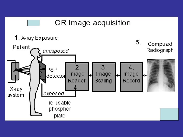
Computed Radiography (CR) is the generic term applied to an imaging system comprised of: ★Photostimulable Storage Phosphor to acquire the x-ray projection image ★ CR Reader to extract the electronic latent image ★ Digital electronics to convert the signals to digital form
• Computed Radiography (CR) uses very similar equipment to conventional radiography except that in place of a film to create the image, an imaging plate (IP) is used. The imaging plate is placed under the object to be examined and the x-ray exposure is made.
X-rays Cassette Conventional radiography Screen-film combination IP only
CR Detector • Photostimulable Storage Phosphor (PSP) • Ba. FBr compound, Eu activated Phosphor Plate Or imaging plate”IP” Barium fluorin bromide Cassette Holder Eu: europium
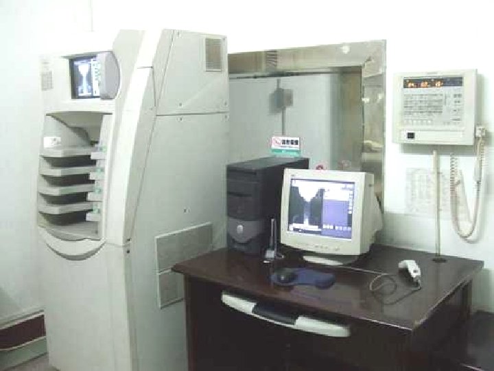
• instead of taking an exposed film into a darkroom for developing in chemical tanks or an automatic film processor, the imaging plate is run through a special laser scanner to read and digitize the image.
Computed Radiography “reader” Information panel Plate stacker
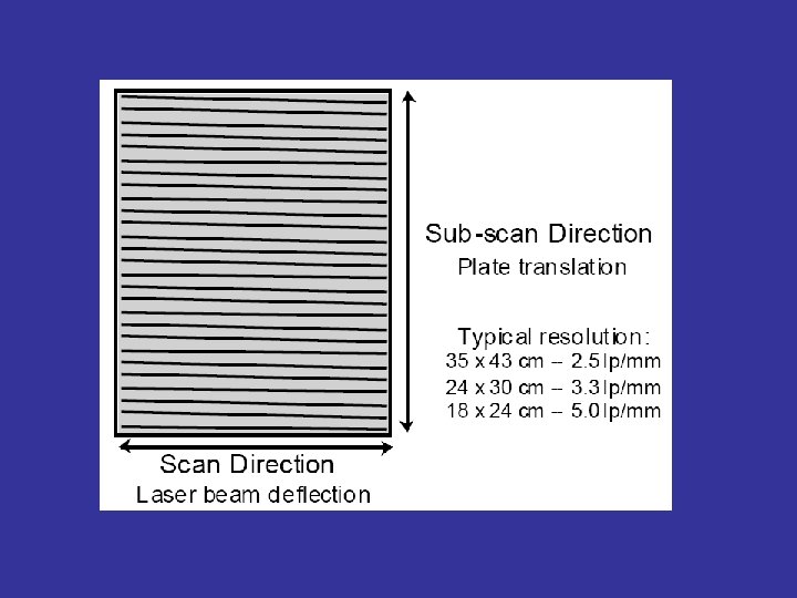
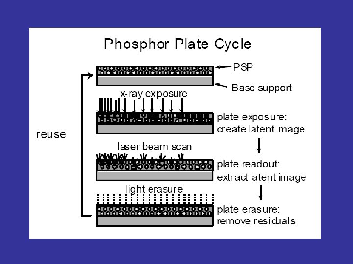
• The CR imaging plate contains photostimulable storage phosphors, which store the radiation level received at each point in local electron energies. When the plate is put through the scanner, the scanning laser beam causes the electrons to relax to lower energy levels, emitting light that is detected by a photo-multiplier tube (PMT), which is then converted to an electronic signal.
photo-multiplier tube
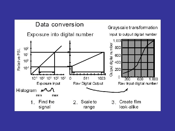
• The electronic signal is then converted to discrete (digital) values and placed into the image processor pixel map. • Imaging plates can theoretically be reused thousands of times if they are handled carefully, although handling under industrial conditions may result in damage after a few hundred uses. An image can be erased by simply exposing the plate to a room-level fluorescent light.
POST-PROCESSING
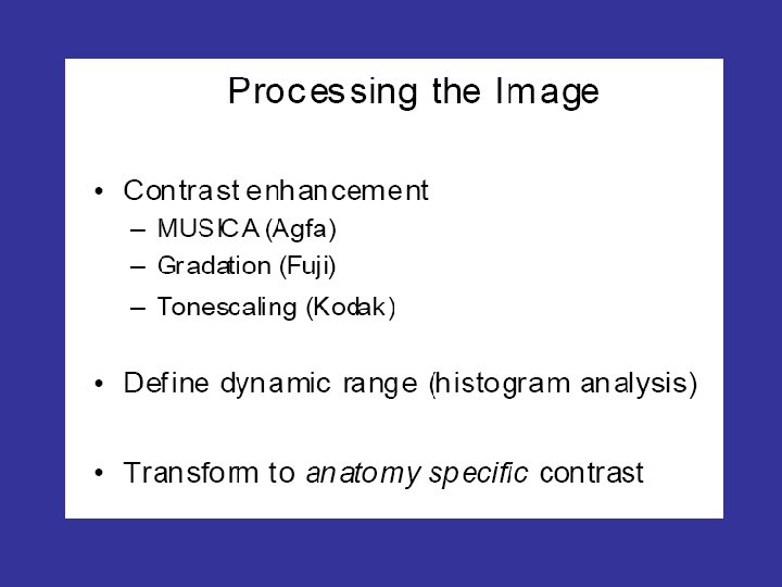
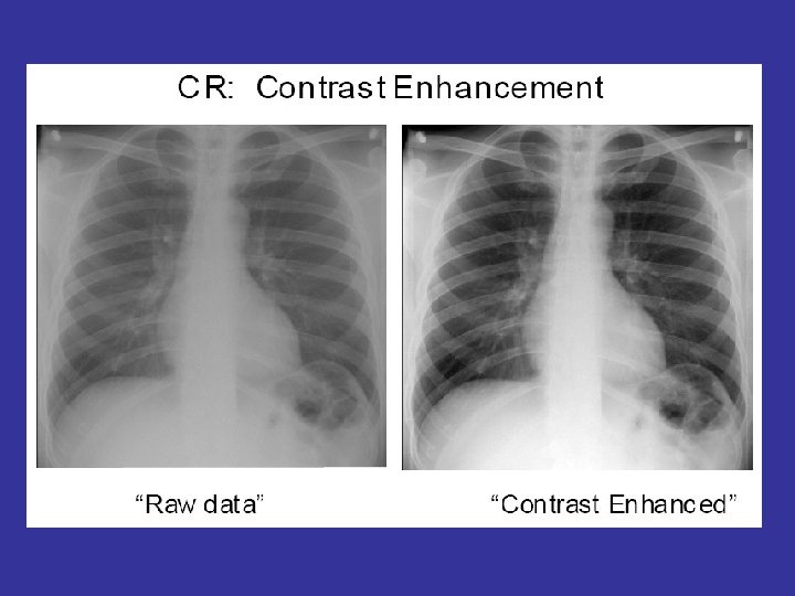
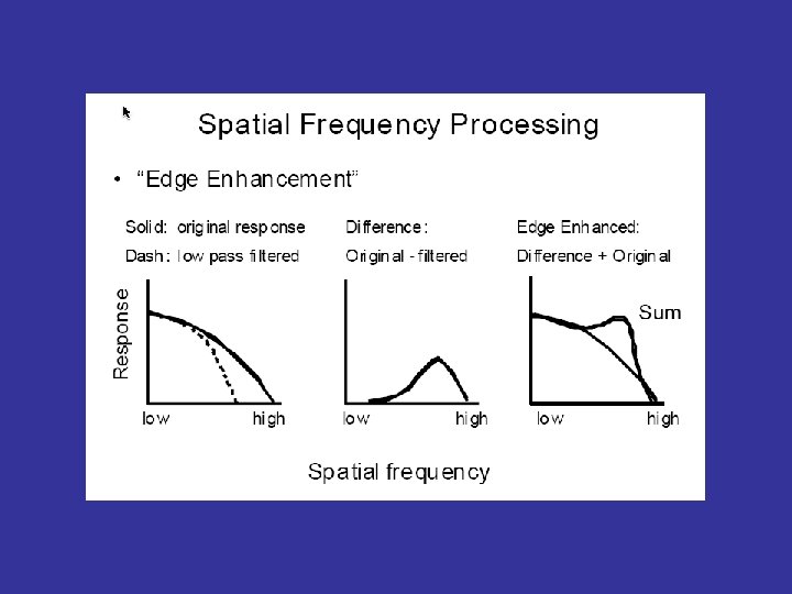
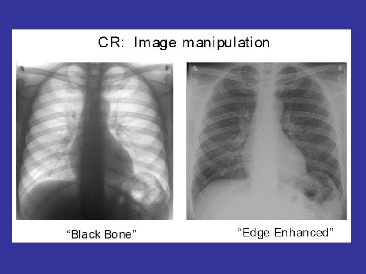
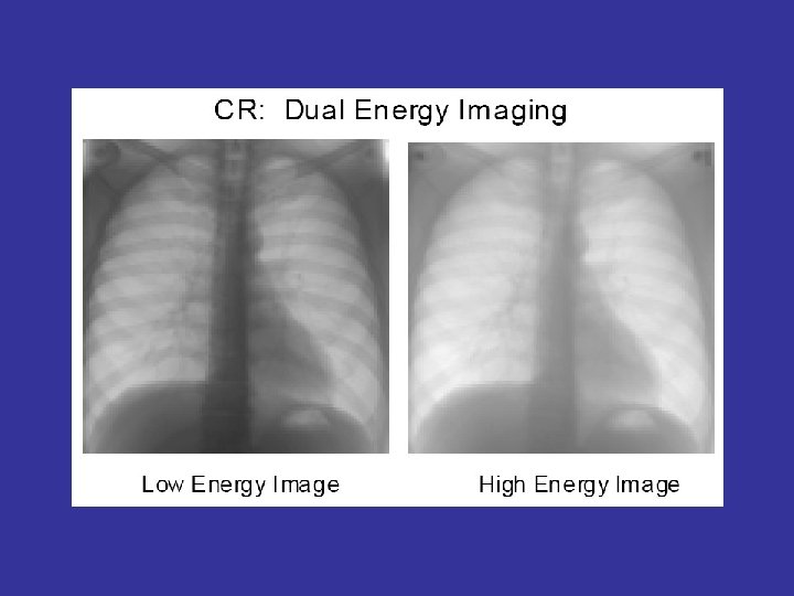
• The digital image can then be viewed and enhanced using software that has functions very similar to other conventional digital image-processing software, such as contrast, brightness, filtration and zoom.
Change the contrast Curve 1 Curve 2
GA GT GC GS RN RT RE 影像A 1. 2 E 1. 6 -0. 14 4 R 0. 5 影像B 0. 9 A 1. 6 -0. 42 4 R 4. 0
GA GT GC GS RN RT RE 影像A 1. 2 D 0. 7 0. 0 3 Q 1. 0 影像B 1. 1 A 0. 8 -0. 01 3 Q 5. 0
GA GT GC GS RN RT RE 影像A 1. 0 F 0. 7 0. 38 4 R 0. 5 影像B 1. 0 A 0. 7 0. 10 4 R 5. 0
GA GT GC GS RN RT RE 影像A 1. 4 0 1. 6 0. 7 4 Q 1. 1 影像B 0. 9 A 0. 8 0. 0 4 Q 5. 0
GA GT GC GS RN RT RE 影像A 0. 63 5 T 0. 5 影像B 0. 9 A 0. 6 0. 20 5 T 5. 0
GA GT GC GS RN RT RE 影像A 0. 9 E 1. 6 -0. 16 4 R 0. 5 影像B 0. 7 A 1. 5 -0. 58 4 R 5. 0 影像C -1. 3 A 1. 5 0. 66 4 R 5. 0
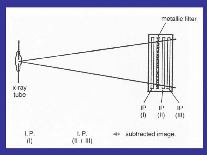
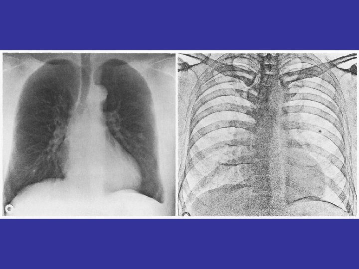
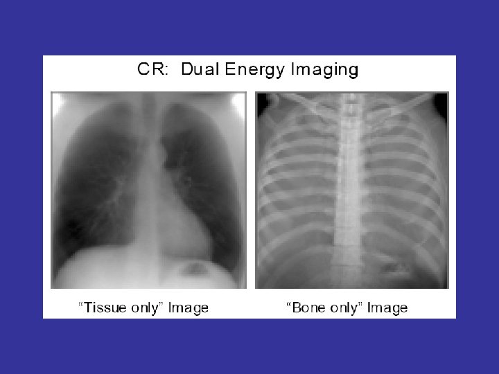
Subtraction
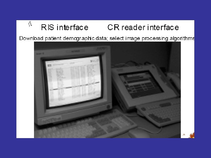
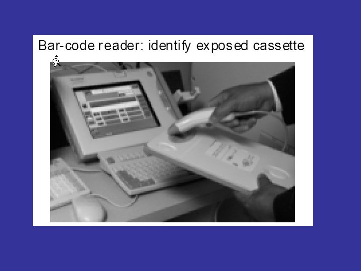
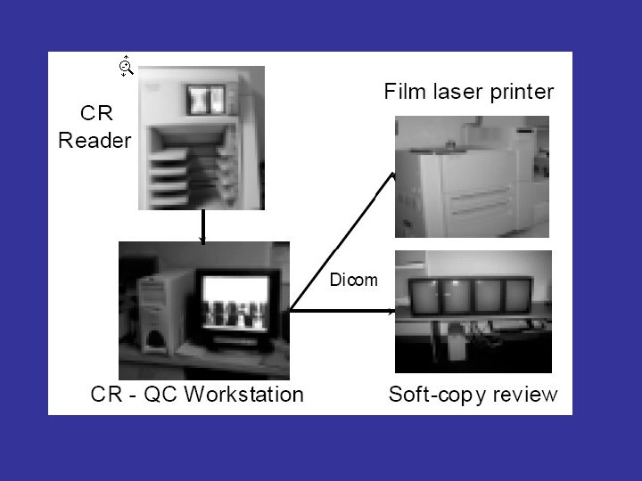
CR Networking • DICOM – Digital Imaging Communications in Medicine – Provides open architecture solutions for modality interfaces, storage/retrieval, and print functions • Technologist QC Workstation • Modality Worklist Input • Processed image output
Medical applications
• Computed Radiography systems are the most common in medical applications because of their low cost. • CR IP's can be used in multiple X-ray sites and the IP's processes through a single reader (scanner), similar to a film processor.
• No silver based film or chemicals; instead a computer is used. • Exposure times are a fraction of typical film exposures, which reduces occupational exposure to radiographers in field applications and is more economical. • Image acquisition is much faster; commonly one minute instead of seven minutes, as with conventional radiography.
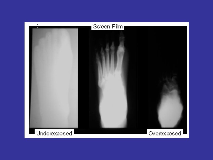
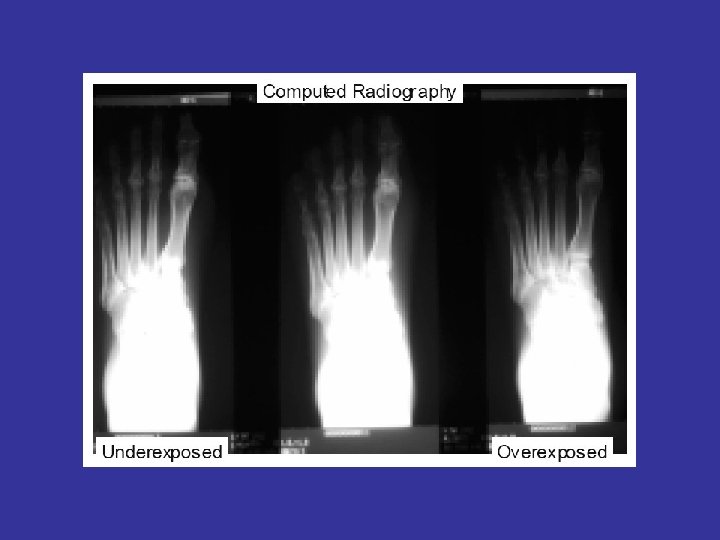
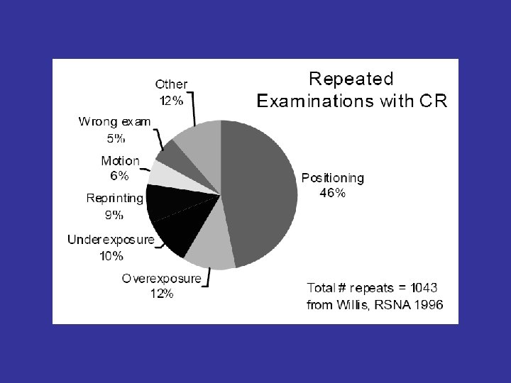
• By adjusting image brightness and/or contrast, a wide range of thicknesses may be examined in one exposure, unlike conventional film based radiography, which may require a different exposure or multiple film speeds in one exposure to cover wide thickness range in a component. Computed radiography often requires fewer reshots due to under- or over-exposure. • Images can be enhanced digitally to aid in interpretation.
• Images can be stored on disk or transmitted for off-site review.
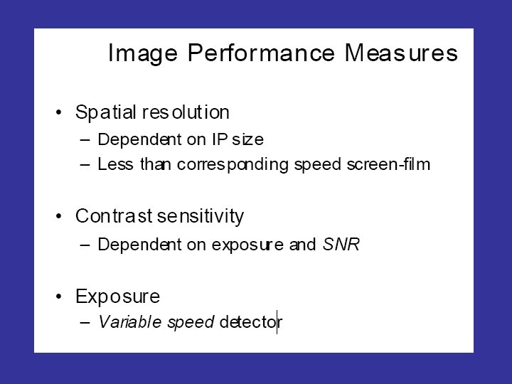
Disadvantages • CR is still not an approved method for most higher quality radiological applications (aerospace), due to the possibility of digital manipulation to the captured image, the inherent geometric unsharpness and resultant lower spatial resolution as compared to film (radiographic) images, SNR (signal vs. noise) issues and sensitivity to scattered radiation
• Imaging plates (IP's) are quite expensive and can be easily damaged, if the system being used requires manual handling of the IP's. Theoretically, IP's may be reused thousands of times, but constant use will always result in damage to the IP and image artifacts.
CR Trends • Lower system costs • Smaller footprint • High throughput systems • Low throughput systems “Table-top” units • Integrated QC workstations for images • DICOM output