The normal cell is confined to a fairly


 hormonal hyperplasia, which increases the functional capacity of a tissue")


demonstrates the normal thickness of the left ventricular wall for")


. – When a")
. – Normal function")


 is a slice of")


 to mature columnar epithelium (so-called Barrett")


shows normal skeletal")
- Slides: 97
The normal cell is confined to a fairly narrow range of function and structure. • genetic programs – metabolism, – differentiation, – specialization; • by constraints of neighboring cells; • by the availability of metabolic substrates.
What if demands changes • handle normal physiologic demands, maintaining a steady state called homeostasis.
In addition to demands environment changes • Cells are constantly exposed to changes in their environment • Cells adapt to acceptable changes in their environment by modifying metabolism or growth pattern •
The main causes of disease that lead to changes in the environ ment of cells
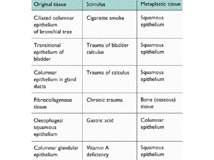
Adaptation to severe stresses and some pathologic stimuli • The adaptive responses – hypertrophy • an increase in the sizes of individual cells, – hyperplasia • increase in the number of cells, – atrophy • decrease in the size and function of cells. – Metaplasia change in the differentiation of cell s
2. 3 Changes in growth of cells.
Adaptation, reversible injury, and cell death can be considered a spectrum • limits of adaptive response to a stimulus are exceeded, or • the cell is exposed to a more injurious agent or severe stress, • a sequence of events follows that leads to cell injury. • Cell injury is reversible up to a certain point, • the stimulus persists or is severe enough from the beginning, the cell reaches a "point of no return" and suffers irreversible cell injury and ultimately cell death.
Example • For instance, in response to increased hemodynamic loads, the heart muscle first becomes enlarged, a form of adaptation. If the blood supply to the myocardium is insufficient to cope with the demand, the muscle becomes reversibly injured and finally undergoes cell death
Cell death, • is one of the most crucial events in the evolution of disease of any tissue or organ. • There are two principal patterns of cell death, necrosis and apoptosis.
Necrosis is the type of cell death that occurs after such abnormal stresses as ischemia and chemical injury, and it is always pathologic.
Apoptosis occurs when a cell dies through activation of an internally controlled suicide program. It is designed to eliminate unwanted cells during embryogenesis and in various physiologic processes, such as involution of hormoneresponsive tissues upon withdrawal of the hormone.
Stages in the cellular response to stress and injurious stimuli.
Summary of tissue response to environmental change. Adaptive responses allow cells to survive in the face of a change in the cellular environment. Failure to adapt is associated with cell damage or cell death.
subcellular alterations. • Stresses of different types may induce other changes • Cells that are exposed to sublethal or chronic stimuli cells may show a variety of subcellular alterations. – Subcellular alterations may operate through Metabolic derangements in cells – may be associated with intracellular accumulations of a number of substances, including proteins, lipids, and carbohydrates. – Calcium is often deposited at sites of cell death, resulting in pathologic calcification. • cell aging is also accompanied by characteristic morphologic and functional changes.
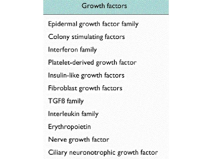
Nature and Severity of Injurious Stimulus Cellular Response Table 1 -1. Cellular Responses to Injury Altered physiologic stimuli: Cellular adaptations: • Increased demand, increased trophic stimulation (e. g. growth factors, hormones) • Hyperplasia, hypertrophy • Decreased nutrients, stimulation • Atrophy • Chronic irritation (chemical or physical) • Metaplasia Reduced oxygen supply; chemical injury; microbial infection Cell injury: • Acute and self-limited • Acute reversible injury • Progessive and severe (including DNA damage) • Irreversible injury → cell death Necrosis Apoptosis • Mild chronic injury • Subcellular alterations in various organelles Metabolic alterations, genetic or acquired Intracellular accumulations; calcifications Prolonged life span with cumulative sublethal injury Cellular aging
The relationships between normal, adapted, reversibly injured, and dead myocardial cells. The cellular adaptation depicted here is hypertrophy, and the type of cell death is ischemic necrosis. In reversibly injured myocardium, generally effects are only functional, without any readily apparent gross or even microscopic changes.
Hyperplasia • Hyperplasia is an increase in the number of cells in an organ or tissue, usually resulting • in increased volume of the organ or tissue. • Hyperplasia takes place if the cellular population is capable of synthesizing DNA, thus permitting mitotic division;
Hyperplasia • hyperplasia and hypertrophy are two distinct processes, • frequently both occur together, • and they may be triggered by the same external stimulus. • For instance, hormone-induced growth in the uterus involves both increased numbers of smooth muscle and epithelial cells and the enlargement of these cells. • by contrast, hypertrophy involves cell enlargement without cell division.
Hyperplasia can be physiologic or pathologic.
Physiologic Hyperplasia • (1) hormonal hyperplasia, which increases the functional capacity of a tissue when needed, • (2) compensatory hyperplasia, which increases tissue mass after damage or partial resection.
Hormonal hyperplasia • is best exemplified by the proliferation of the glandular epithelium of the female breast at puberty and during pregnancy and the physiologic hyperplasia that occurs in the pregnant uterus.
compensatory hyperplasia • remaining tissue grows to make up for partial tissue loss • Live hyperplasia • after unilateral nephrectomy,
Mechanisms of Hyperplasia. • increased local production of growth factors, • increased levels of growth factor receptors on the responding cells, • Turning on many cellular genes, including genes encoding growth factors, receptors for growth factors, and cell cycle regulators, • In hormonal hyperplasia, the hormones may themselves act as growth factors and trigger the transcription of various cellular genes.
Pathologic Hyperplasia • Endometrial hyperplasia is an example of abnormal hormone-induced hyperplasia. • This form of hyperplasia is a common cause of abnormal menstrual bleeding.
Pathologic Hyperplasia • Benign prostatic hyperplasia is another common example of pathologic hyperplasia induced by responses to hormones, androgens. • Although these forms of hyperplasia are abnormal, the process remains controlled, because the hyperplasia regresses if the hormonal stimulation is eliminated. • Responsiveness to normal regulatory control mechanisms that distinguishes benign pathologic hyperplasia's from cancer, in which the growth control mechanisms become defective. • Pathologic hyperplasia, however, constitutes a fertile soil in which cancerous proliferation may eventually arise. • Thus, patients with hyperplasia of the endometrium are at increased risk for developing endometrial cancer
Pathologic Hyperplasia • Hyperplasia is also an important response of connective tissue cells in wound healing, in which proliferating fibro-blasts and blood vessels aid in repair • Under these circumstances, growth factors are responsible for the hyperplasia.
HYPERTROPHY • an increase in the size of cells, – resulting in an increase in the size of the organ. • the hypertrophied organ has no new cells, just larger cells. • The increased size of the cells is due not to cellular swelling but to the synthesis of more structural components. • cells capable of division may respond to stress by undergoing both hyperplasia and hypertrophy, • whereas in nondividing cells (e. g. , myocardial fibers), hypertrophy occurs.
Hypertrophy can be physiologic or pathologic
Physiological Hypertrophy • is caused by increased functional demand or • by specific hormonal stimulation. – The striated muscle cells in both the heart and the skeletal muscles are capable of hypertrophy, – they cannot adequately adapt to increased metabolic demands by mitotic division and production of more cells to share the work.
Hypertrophy • The most common stimulus for hypertrophy of muscle is increased workload. • For example, the bulging muscles of bodybuilders. • chronic hemodynamic overload, resulting from either hypertension or faulty valves.
Hypertrophy • The massive physiologic growth of the uterus during pregnancy is a good example of hormone -induced increase in the size of an organ that results from both hypertrophy and hyperplasia • The cellular hypertrophy is stimulated by estrogenic hormones acting on smooth muscle estrogen receptors, eventually resulting in increased synthesis of smooth muscle proteins and an increase in cell size
Physiologic hypertrophy of the uterus during pregnancy. A, Gross appearance of a normal uterus (right) and a gravid uterus (removed for postpartum bleeding) (left). B, Small spindle-shaped uterine smooth muscle cells from a normal uterus (left) compared with large plump cells in gravid uterus (right).
Hypertrophy of cardiac muscle in response to valve disease
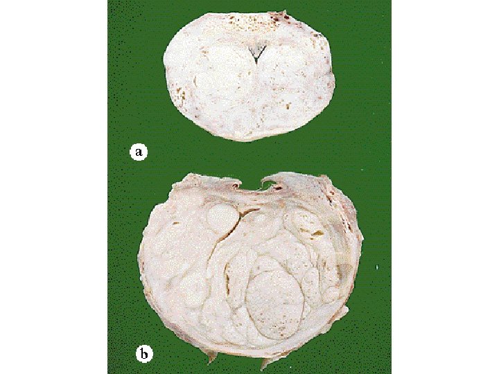
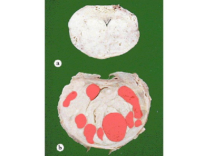
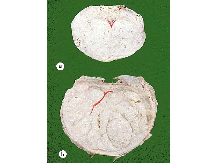
The upper macroscopic specimen (a)demonstrates the normal thickness of the left ventricular wall for comparison with the greatly thickened wall in the lower specimen, taken from a heart in which severe narrowing of the aortic valve caused resistance to systolic ventricular emptying. The increased mass of the left ventricle is due to enlargement of cardiac muscle cells as a result of hypertrophy. This can be seen by comparing the diameter of fibres from the normal heart (b) with those from the diseased heart (c). } Note that the size of nuclei in the hypertrophied cardiac muscle is also increased; it has been found that such nuclei are frequently polyploid (i. e. contain several times the normal quantity of DNA).
Histology of cardiac muscle from a normal heart
Histology of cardiac muscle from a hypertrop hied heart
Hyperplasia and hypertrophy of myometrium in pregnancy. On the left is a normal uterus showing the normal mass of smooth muscle in its wall. On the right is a uterus from a recently pregnant woman, in which the striking increase in mass of smooth muscle is evident. At a cellular level this is due to both hyperplasia and hypertrophy of uterine smooth muscle. +} }}
Mechanisms of Hypertrophy. • induction of a number of genes, which in turn stimulate synthesis of numerous cellular proteins
ATROPHY • Shrinkage in the size of the cell by loss of cell substance is known as atrophy. • When a sufficient number of cells are involved, the entire tissue or organ diminishes in size, or becomes atrophic. •
Atrophy can be physiologic or pathologic.
Physiologic atrophy • The uterus decreases in size shortly after parturition,
Pathologic atrophy • depends on the underlying cause and can be local or generalized.
The common causes of atrophy • Decreased workload (atrophy of disuse). – When a broken limb is immobilized in a plaster cast or – when a patient is restricted to complete bed rest, • The initial rapid decrease in cell size is reversible once activity is resumed. • With more prolonged disuse, skeletal muscle fibers decrease in number as well as in size; • this atrophy can be accompanied by increased bone resorption, leading to osteoporosis of disuse.
The common causes of atrophy • Loss of innervation (denervation atrophy). – Normal function of skeletal muscle is dependent on its nerve supply. – Damage to the nerves leads to rapid atrophy of the muscle fibers supplied by those nerves • Diminished blood supply. – A decrease in blood supply (ischemia) to a tissue as a result of arterial occlusive disease results in atrophy of tissue owing to progressive cell loss. – In late adult life, the brain undergoes progressive atrophy, presumably as atherosclerosis narrows its blood supply
The common causes of atrophy • Loss of endocrine stimulation. – Many endocrine glands, the breast, and the reproductive organs are dependent on endocrine stimulation for normal metabolism and function. – The loss of estrogen stimulation after menopause results in physiologic atrophy of the endometrium, vaginal epithelium, and breast.
The common causes of atrophy • Pressure. • Tissue compression for any length of time can cause atrophy. • An enlarging benign tumor can cause atrophy in the surrounding compressed tissues. – Atrophy in this setting is probably the result of ischemic changes caused by compromise of the blood supply to those tissues by the expanding mass. • Inadequate nutrition. • .
, Atrophy of the brain in an 82 -year-old male with atherosclerotic disease. Atrophy of the brain is due to aging and reduced blood supply. The meninges have been stripped. B, Normal brain of a 36 -year-old male. Note that loss of brain substance narrows the gyri and widens the sulci.
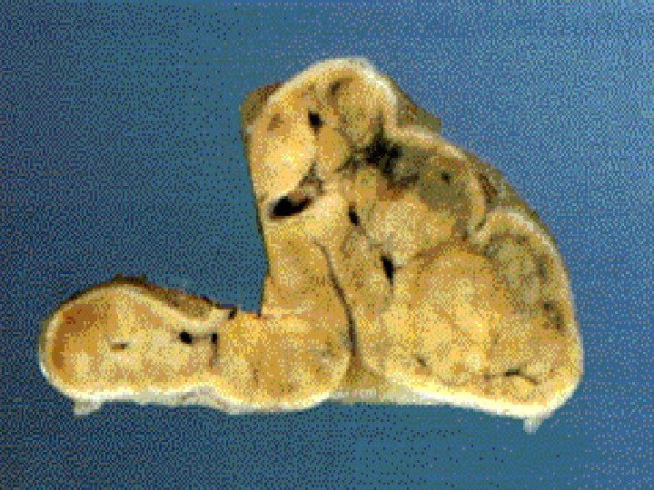
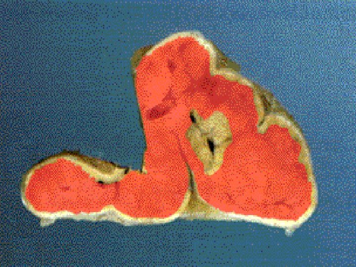
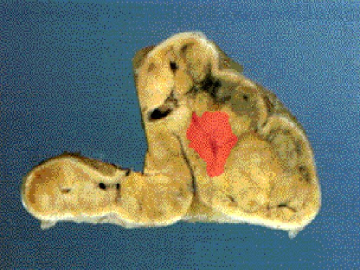
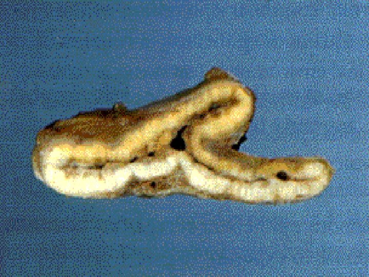
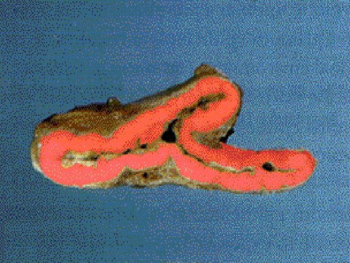
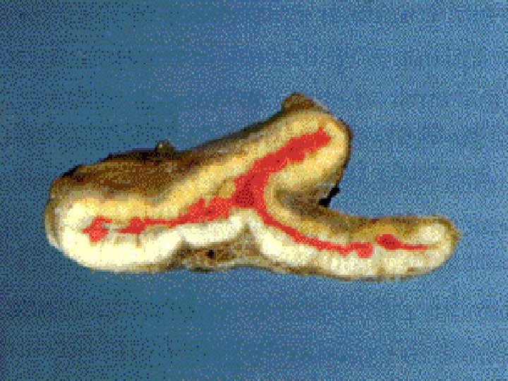
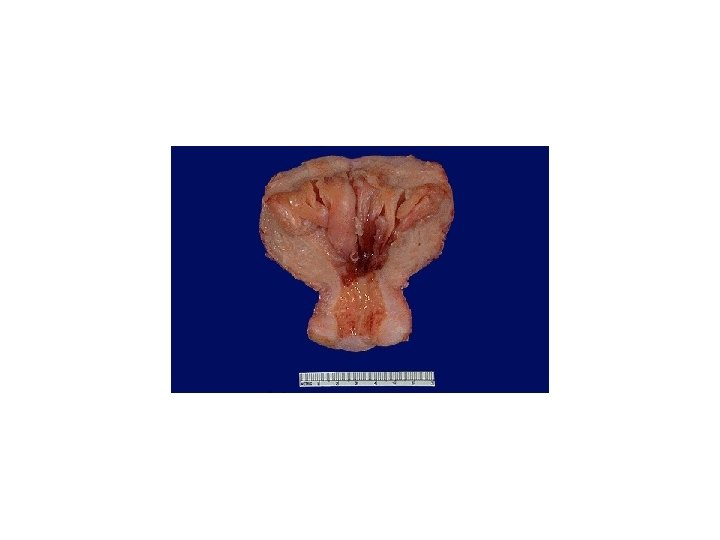
• Atrophy of the adrenal gland. • Specimen (b) is a slice of normal gland, in which yellow cortex can be distinguished from the small amount of grey medulla. • The slice in (a) is from a patient who had surgical ablation of the pituitary gland, leading to lack of ACTH. • This has resulted in reduction in the size of the adrenal gland, specifically affecting the cortex.
A, Schematic diagram of columnar to squamous metaplasia. B,
METAPLASIA • Metaplasia is a reversible change in which one adult cell type (epithelial or mesenchymal) is replaced by another adult cell type. • It may represent an adaptive substitution of cells that are sensitive to stress by cell types better able to withstand the adverse environment.
METAPLASIA • Stones in the excretory ducts of the salivary glands, pancreas, or bile ducts may cause replacement of the normal secretory columnar epithelium by nonfunctioning stratified squamous epithelium • A deficiency of vitamin A (retinoic acid) induces
METAPLASIA • The most common epithelial metaplasia is columnar to squamous as occurs in the respiratory tract in response to chronic irritation. • In the habitual cigarette smoker, the normal ciliated columnar epithelial cells of the trachea and bronchi are often replaced focally or widely by stratified squamous epithelial cells.
Metaplasia • . • Although the metaplastic squamous cells in the respiratory tract, for example, are capable of surviving, an important protective mechanism-mucus secretion-is lost. • Thus, epithelial metaplasia is a two-edged sword and, in most circumstances, represents an undesirable change
Metaplasia • the influences that predispose to metaplasia, if persistent, may induce malignant transformation in metaplastic epithelium. • Thus, the common form of cancer in the respiratory tract is composed of squamous cells, which arise in areas of metaplasia of the normal columnar epithelium into squamous epithelium.
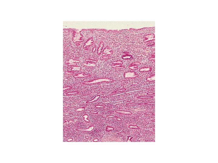
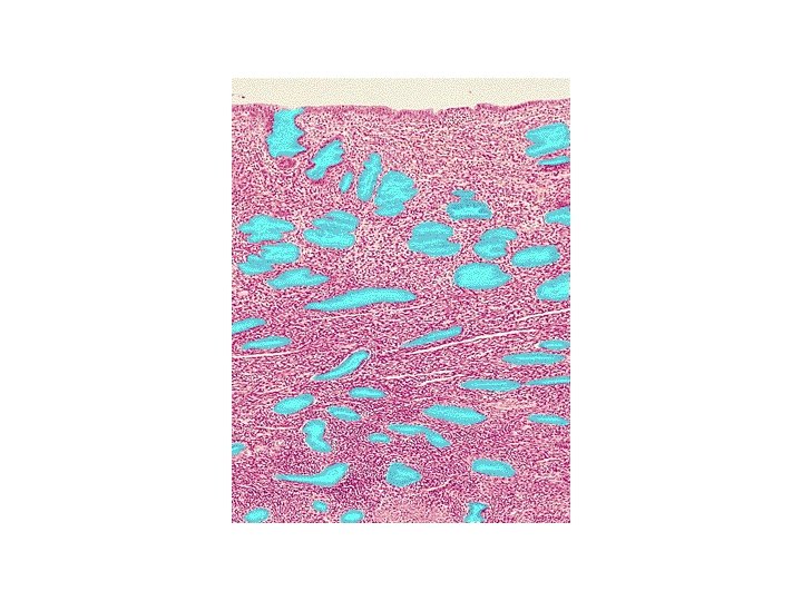
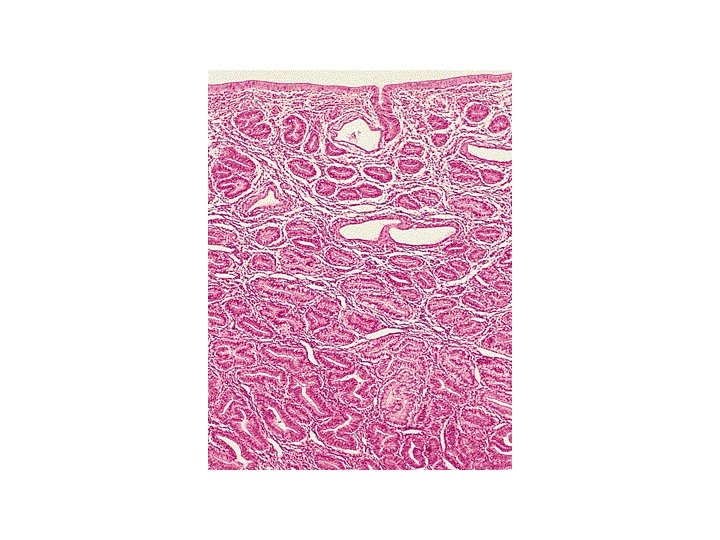
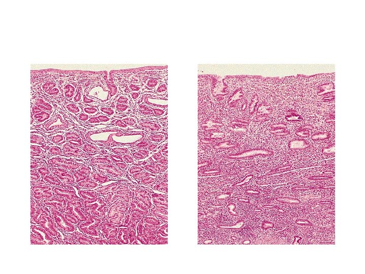
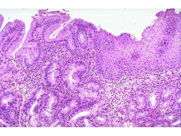
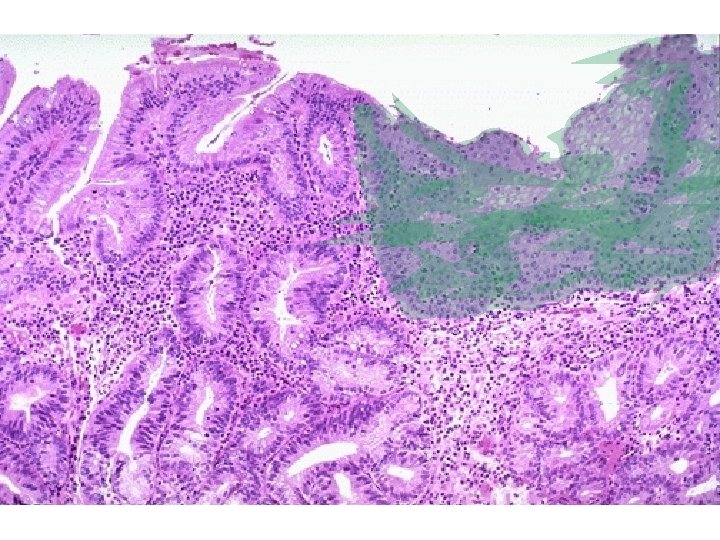
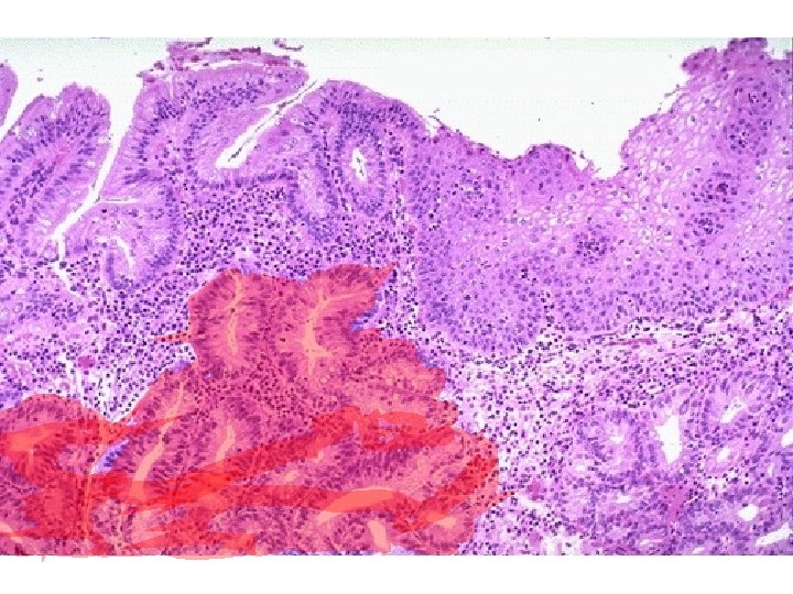
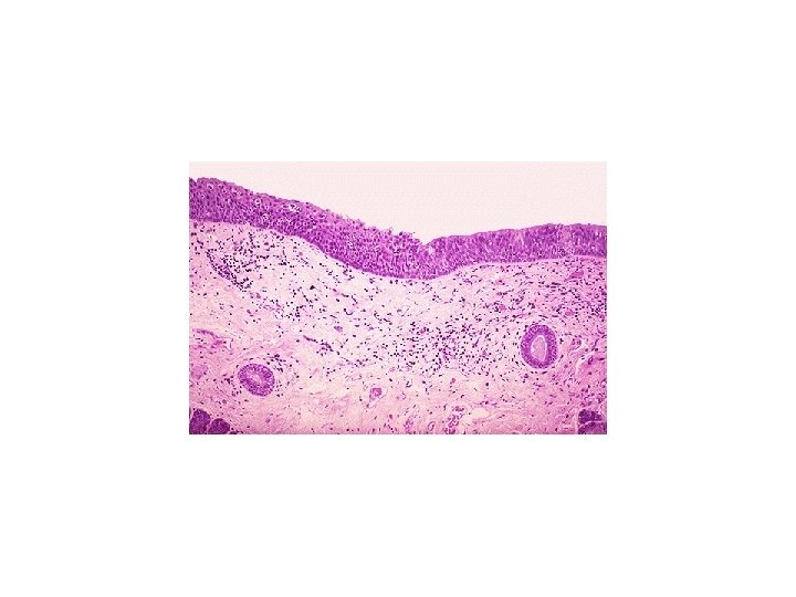
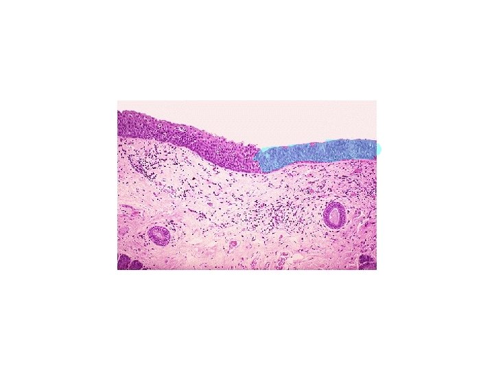
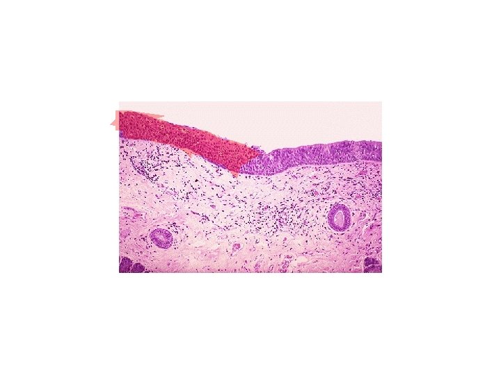
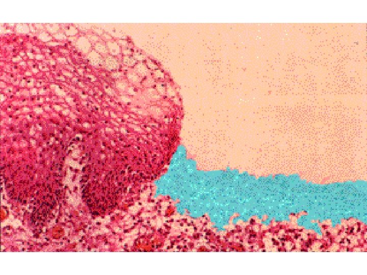
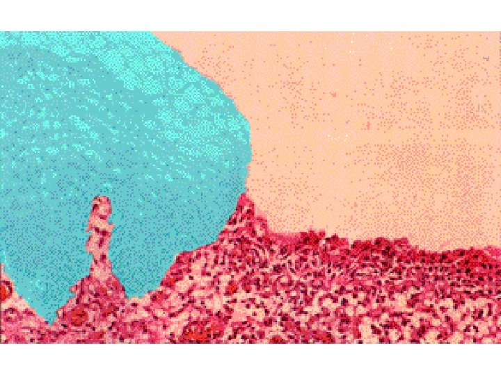
METAPLASIA • Metaplasia from squamous to columnar type may also occur, as in Barrett esophagus, in which the esophageal squamous epithelium is replaced by intestinal-like columnar cells under the influence of refluxed gastric acid • Cancers may arise in these areas, and these are typically glandular (adeno)carcinomas
METAPLASIA • Connective tissue metaplasia is the formation of cartilage, bone, or adipose tissue (mesenchymal tissues) in tissues that normally do not contain these elements. • For example, bone formation in muscle, designated myositis ossificans, occasionally occurs after bone fracture. • This type of metaplasia is less clearly seen as an adaptive response.
Mechanisms of Metaplasia. • Metaplasia does not result from a change in the phenotype of a differentiated cell type; • instead it is the result of a reprogramming of stem cells that are known to exist in normal tissues, or of undifferentiated mesenchymal cells present in connective tissue. • In a metaplastic change, these precursor cells differentiate along a new pathway
Metaplastic transformation of esophageal stratified squamous epithelium (left) to mature columnar epithelium (so-called Barrett metaplasia
Summary of tissue response to environmental change. Adaptive responses allow cells to survive in the face of a change in the cellular environment. Failure to adapt is associated with cell damage or cell death.
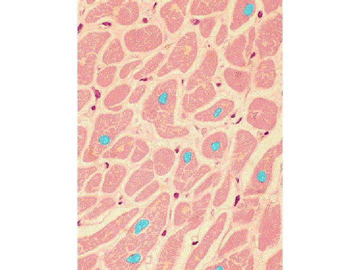
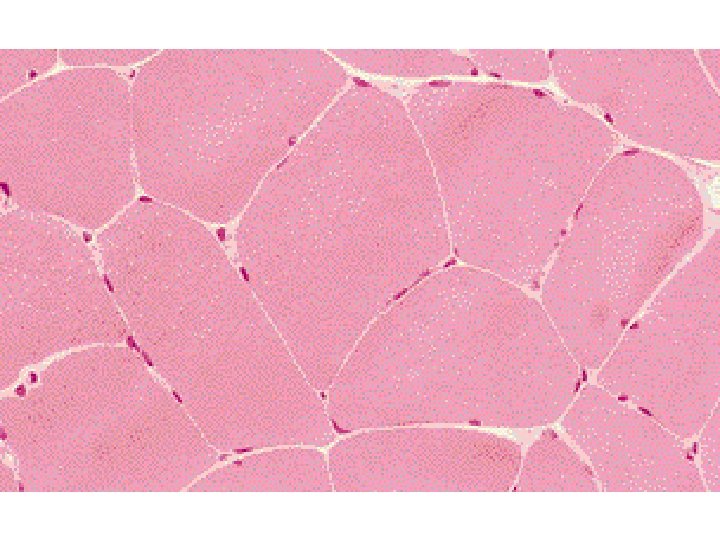
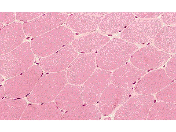
Hypertrophy of skeletal muscle in response } to exercise. Hypertrophy in the absence of hyperplasia is typically seen in muscle where the stimulus is an increased demand for work. Taken at the same magnification, (a) shows muscle fibres in transverse section from the soleus muscle of a normal 50 -year-old male and (b) shows fibres from the same muscle in a veteran marathon runner. Note the dramatic increase in the size of the fibres in response to the demands of marathon running.
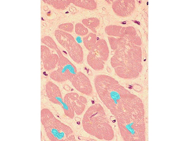
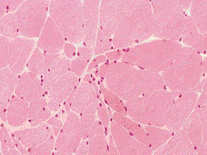
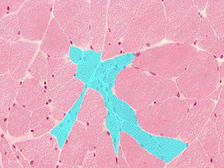
• Atrophy of skeletal muscle with • denervation. • Micrograph (a)shows normal skeletal muscle fibres. In micrograph (b) damage to many of the axons in the main nerve supplying the muscle has caused atrophy of fibres, • Fibres are small and angulated.