Liver diseases I and II The liver is
")


 acute")


 consumption is the leading cause of liver")
: - lipid droplets accumulate in hepatocytes increasing with")
 Hepatitis: 1. Hepatocyte swelling and necrosis (accumulation of fat and")


 shunting of normal substrates away from catabolism")
 induces")


----")
 • NAFLD represents a spectrum of disorders that have")


 is characterized principally by (1) deposition of")


 is")
 • acute, fulminant hepatitis • Chronic hepatitis • Steatohepatitis")


- Slides: 65
(Liver diseases I and II)
• The liver is vulnerable to a wide variety of metabolic, toxic, microbial, circulatory, and neoplastic insults. • The major primary diseases of the liver are viral hepatitis, nonalcoholic fatty liver disease (NAFLD), alcoholic liver disease, and hepatocellular carcinoma (HCC). • Hepatic damage also occurs secondary to some of the most common diseases in humans, such as heart failure, disseminated cancer, and extrahepatic infections. • With the exception of acute liver failure, liver disease is an insidious process in which clinical detection and symptoms of hepatic decompensation may occur weeks, months, or many years after the onset of injury. The ebb and flow of hepatic injury may be imperceptible to the patient and detectable only by abnormal laboratory tests.
Liver Failure • The most severe clinical consequence of liver disease is liver failure: - acute liver failure: the result of sudden and massive hepatic destruction. - chronic liver failure: which follows upon years or decades of insidious, progressive liver injury. - Acute on- chronic liver failure, in which an unrelated acute injury supervenes on a well-compensated late-stage chronic disease or the chronic disease itself has a flare of activity that leads directly to liver failure. 80% to 90% of hepatic functional capacity must be lost before hepatic failure ensues. When the liver can no longer maintain homeostasis, transplantation offers the best hope for survival; the mortality rate in persons with hepatic failure without liver transplantation is about 80%.
Acute Liver Failure • has been referred to as “fulminant liver failure” until recently. • Acute liver failure is defined as an acute liver illness associated with encephalopathy and coagulopathy that occurs within 26 weeks of the initial liver injury in the absence of preexisting liver disease.
• Rarely, there may be diffuse poisoning of liver cells without obvious cell death and parenchymal collapse, such as in diffuse microvesicular steatosis related to fatty liver of pregnancy or idiosyncratic reactions to toxins (e. g. , valproate, tetracycline). In these settings, usually related to primary mitochondrial dysfunction, hepatocytes are unable to perform their usual metabolic functions.
• Pathogenesis: - Acute liver failure is caused by massive hepatic necrosis, most often induced by drugs or toxins. - Accidental or deliberate ingestion of acetaminophen accounts for almost 50% of cases in the United States, while autoimmune hepatitis, other drugs/toxins, and acute hepatitis A and B infections account for rest of cases. In Asia, acute hepatitis B and E predominate. - With acetoaminophen toxicity, the liver ailure occurs within a week of the onset of symptoms, whereas failure due to hepatitis viruses takes longer to develop. - The mechanism of hepatocellular necrosis may be direct toxic damage (as with acetaminophen), but more often is a variable combination of toxicity and immune-mediated hepatocyte destruction (e. g. , hepatitis virus infection).
• Clinical Course: - Acute liver failure manifests first with nausea, vomiting, and jaundice, followed by life threatening encephalopathy, and coagulation defects. - serum liver transaminases are markedly elevated. And liver is initially enlarged due to hepatocyte swelling and edema. - As parenchyma is destroyed, however, the liver shrinks dramatically with decline of serum transaminases. - With unabated progression, multiorgan system failure occurs and, if transplantation is not possible, death ensues
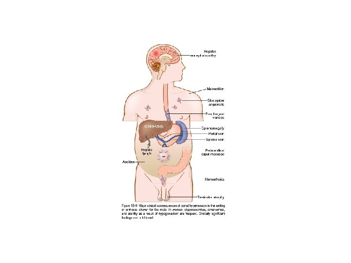
- jaundice and icterus---- retention of bilirubin, - cholestasis----- systemic retention of not only bilirubin but also other solutes eliminated in bile. - Hepatic encephalopathy--- is a spectrum of disturbances in consciousness, ranging from subtle behavioral abnormalities, to marked confusion and stupor, to deep coma and death. Asterixis, a particularly characteristic sign, is manifested as nonrhythmic, rapid extension-flexion movements of the head and extremities. Elevated ammonia levels in blood and the central nervous system correlate with impaired neuronal function and cerebral edema. - Coagulopathy--- Easy bruisability, fatal intracranial bleeding. Due to impaired hepatic synthetic function of vitamin K-dependent and -independent clotting factors. - disseminated intravascular coagulation---- due to decreased hepatic function to remove activated coagulation factors from the circulation. - Portal hypertension----- ascites and hepatic encephalopathy. - Hepatorenal syndrome----- is a form of renal failure occurring in individuals with liver failure in whom there are no intrinsic morphologic or functional causes for kidney dysfunction.
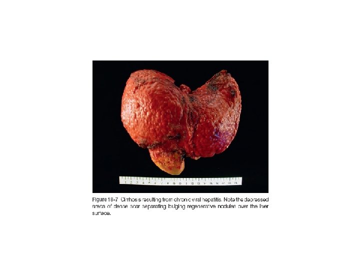
Chronic Liver Failure and Cirrhosis • The leading causes of chronic liver failure worldwide include chronic hepatitis B, chronic hepatitis C, nonalcoholic fatty liver disease, and alcoholic liver disease. • Liver failure in chronic liver disease is most often associated with cirrhosis, a condition marked by the diffuse transformation of the entire liver into regenerative parenchymal nodules surrounded by fibrous bands and variable degrees of vascular (often portosystemic) shunting. • not all cirrhosis leads to chronic liver failure and not all end-stage chronic liver disease is cirrhotic. The Child-Pugh classification of cirrhosis distinguishes between class A (well compensated), B (partially decompensated), and C (decompensated)
• the term cirrhosis implies the presence of severe chronic disease, it is not a specific diagnosis and it lacks clear prognostic implications. • The term cryptogenic cirrhosis is sometimes used to describe cirrhosis when there is no clear cause.
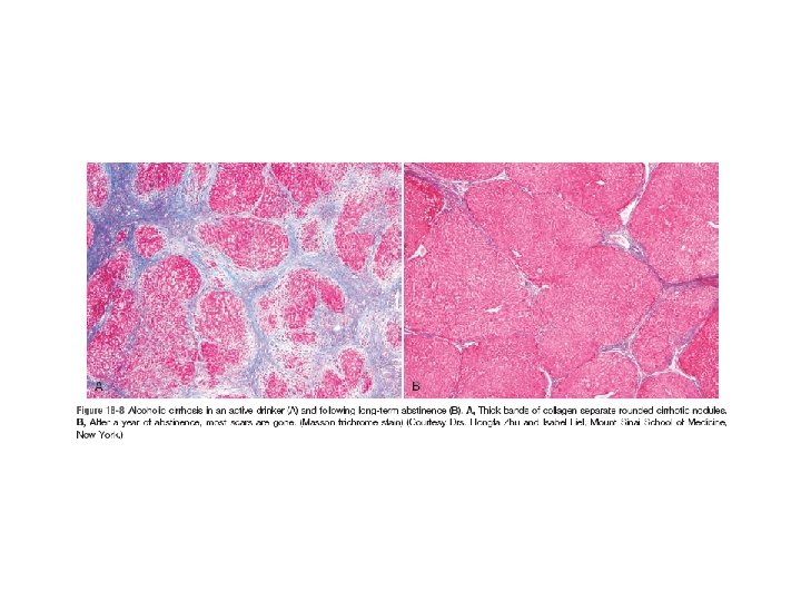
• Although uncommon, regression of fibrosis, albeit rarely, in fully established cirrhosis, does occur; this is another reason why cirrhosis should not be automatically equated with end stage disease. In the past when there were no reliable ways to cure any chronic liver disease, there were no opportunities to see whether cirrhosis could regress. With increasing numbers of effective treatments for cirrhosis-causing conditions, however, we now understand that regression of scars can take place. Scars can become thinner, more densely compacted, and eventually fragment. As fibrous septa break apart, adjacent nodules of regenerating parenchyma coalesce into larger islands. All cirrhotic livers show elements of both progression and regression, the balance determined by the severity and persistence of the underlying disease.
• Clinical Features: About 40% of individuals with cirrhosis are asymptomatic until the most advanced stages of the disease. When symptomatic, they present with nonspecific manifestations: anorexia, weight loss, weakness, and, in advanced disease, symptoms and signs of liver failure discussed earlier. The ultimate causes of death in chronic liver failure, whether cirrhotic or not, include those seen in acute liver failure, and additional grim outcomes, such as development of hepatocellular carcinoma in the context of cirrhosis. Hepatic encephalopathy, bleeding from esophageal varices and bacterial infections ( resulting from damage to mucosal barrier in the gut and Kupffer cell dysfunction) are often the terminal events.
• Impaired estrogen metabolism an consequent hyperestrogenemia in male patients with chronic liver failure can give rise to palmar erythema (a reflection of local vasodilatation) and spider angiomas of the skin. • In men, hyperestrogenemia also leads to hypogonadism and gynecomastia. • Hypogonadism can also occur in women from disruption of hypothalamic-pituitary axis function, either through nutritional deficiencies associated with the chronic liver disease or primary hormonal alterations.
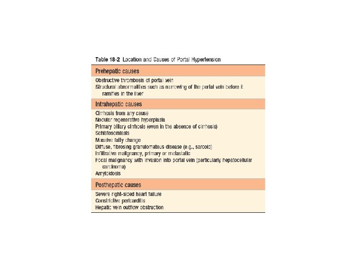
• Portal Hypertension: increased resistance to portal blood flow may develop in a variety of circumstances, which can be divided into prehepatic, intrahepatic, and posthepatic. The dominant intrahepatic cause is cirrhosis, accounting for most cases of portal hypertension. The pathophysiology of portal hypertension is complex and involves resistance to portal flow at the level of sinusoids and an increase in portal flow caused by hyperdynamic circulation. The four major clinical consequences of portal hypertension are: (1) ascites, (2) the formation of portosystemic venous shunts, (3) congestive splenomegaly, (4) hepatic encephalopathy
• Ascites. - The accumulation of excess fluid in the peritoneal cavity. - In 85% of cases, ascites is caused by cirrhosis. - Ascites usually becomes clinically detectable when at least 500 m. L have accumulated. - The fluid is generally serous, having less than 3 gm/d. L of protein (largely albumin), and a serum to ascites albumin gradient of ≥ 1. 1 gm/d. L. - The pathogenesis of ascites is complex, involving the following mechanisms: Sinusoidal hypertension, Percolation of hepatic lymph into the peritoneal cavity and Splanchnic vasodilation and hyperdynamic circulation
• Portosystemic Shunts: With the rise in portal system pressure, the flow is reversed from portal to systemic circulation by dilation of collateral vessels and development of new vessels. Venous bypasses develop wherever the systemic and portal circulation share common capillary beds. Principal sites are veins around and within the rectum (manifest as hemorrhoids), the esophagogastric junction (producing varices), the retroperitoneum, and the falciform ligament of the liver (involving periumbilical and abdominal wall collaterals). Abdominal wall collaterals appear as dilated subcutaneous veins extending from the umbilicus toward the rib margins (caput medusae).
• Splenomegaly: The massive splenomegaly may secondarily induce hematologic abnormalities attributable to hypersplenism, such as thrombocytopenia or even pancytopenia.
• two additional syndromes that occur in chronic liver failure: Hepatopulmonary syndrome: - seen in up to 30% patients with cirrhosis of the liver and portal hypertension. - These patients develop intrapulmonary vascular dilations involving capillary and pre-capillary vessels up to 100 μM in size. The blood flows rapidly through such dilated vessels, giving inadequate time for oxygen diffusion and leading to ventilation-perfusion mismatch and right-to-left shunting, manifesting as hypoxia. - Hypoxia and resultant dyspnea occur preferentially in an upright position rather than in the recumbent position, as gravity exacerbates the ventilation-perfusion mismatch. Portopulmonary hypertension: - refers to pulmonary arterial hypertension arising in liver disease and portal hypertension. - The most common clinical manifestations are dyspnea on exertion and clubbing of the fingers.
Hepatitis Causes: - infectious Hepatitis: viral, Bacterial, Parasitic, and Helminthic - Autoimmune Hepatitis - Drug- and Toxin (Alcohol) - Metabolic
infectious Hepatitis
• Several clinical syndromes may develop following exposure to hepatitis viruses: (1) acute asymptomatic infection with recovery (serologic evidence only) (2) Acute symptomatic hepatitis with recovery, anicteric or icteric-------incubation period, a symptomatic preicteric phase, a symptomatic icteric phase, and convalescence (3) chronic hepatitis, with or without progression to cirrhosis------ HBV, HDV, HCV (m. c)----- increased risk for the development of hepatocellular carinoma. (4) acute liver failure/Fulminant hepatitis with massive to submassive hepatic necrosis----- HAV, HBV, or HDV, HEV (pregnant) (5) The Carrier State
Autoimmune Hepatitis • Autoimmune hepatitis is a chronic, progressive hepatitis with all the features of autoimmune diseases in general: - genetic predisposition, - association with other autoimmune diseases, - presence of autoantibodies, - and therapeutic response to immunosuppression. - There is a female predominance (78%). Triggers for the immune reaction may include viral infections or drug or toxin exposures.
• Autoimmune hepatitis is classified into types 1 and 2, based on the patterns of circulating antibodies: - Type 1: more common in middle-aged to older individuals, is characterized by the presence of antinuclear (ANA), anti–smooth muscle actin (SMA), anti –soluble liver antigen/liver-pancreas antigen (anti. SLA/LP) antibodies, and less commonly, antimitochondrial (AMA) antibodies. - Type 2: usually seen in children and teenagers, the main serologic markers are anti–liver kidney microsome-1 (anti -LKM-1) antibodies, and anti–liver cytosol-1 (ACL-1) antibodies.
• Clinical features: - The disease may be rapidly progressive or indolent, both giving rise eventually to liver failure. acute hepatitis Fulminant hepatitis Chronic hepatitis and cirrhosis - The mortality of patients with severe untreated autoimmune hepatitis is approximately 40% within 6 months of diagnosis and cirrhosis develops in at least 40% of survivors. - diagnosis and intervention are imperative. Immunosuppressive therapy is usually successful, leading to remissions in 80% of patients that permits long term survival. In end stage disease, liver transplantation is indicated.
Drug- and Toxin-Induced Liver Injury As the major drug metabolizing and detoxifying organ in the body, the liver is subject to injury from an enormous array of therapeutic and environmental agents. • Injury may result from: - direct toxicity, - through hepatic conversion of a xenobiotic to an active toxin, - or be produced by immune mechanisms, such as by the drug or a metabolite acting as a hapten to convert a cellular protein into an immunogen. • Exposure to a toxin or therapeutic agent should always be included in the differential diagnosis of any form of liver disease. Reactions may be mild to very serious, including acute liver failure or chronic liver disease. Acetaminophen is the most common hepatotoxin causing acute liver failure. Alcohol is the most common hepatotoxin causing chronic liver disease.
• Drug toxic reactions may be classified as: Predictable reactions: affect all people in a dose dependent fashion------- acetaminophen (suicidal or accidental overdoses of acetoaminophen result in acute liver failure due to effect of toxic metabolite produced by the cytochrome P-450 system) Unpredictable reactions: occurs in rare individuals, depend on idiosyncrasies of the host, particularly the propensity to mount an immune response to the antigenic stimulus or the rate at which the agent can be metabolized ---- halothane Both classes of injury may be immediate or take weeks to months to develop.
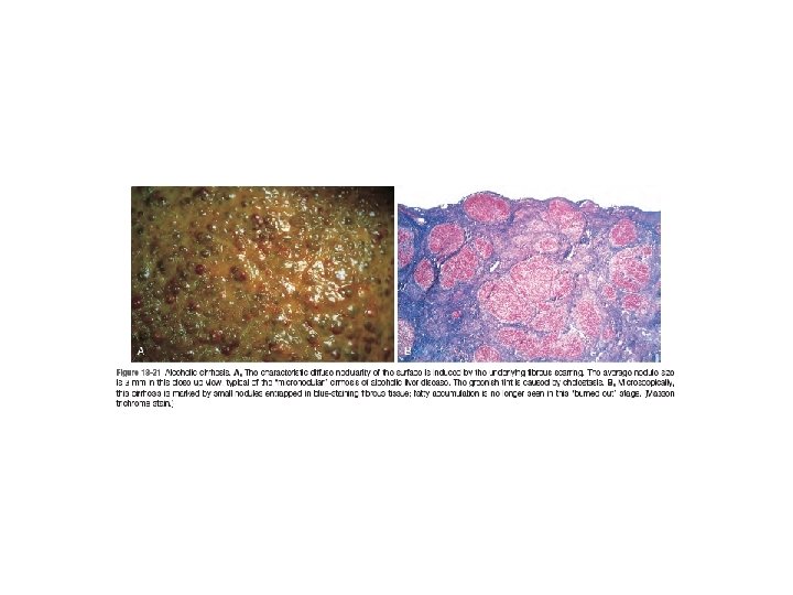
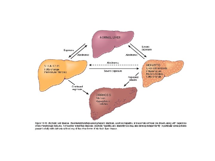
Alcoholic Liver Disease • Excessive alcohol (ethanol) consumption is the leading cause of liver disease in most Western countries. • There are three distinctive, albeit overlapping forms of alcoholic liver injury: (1) hepatocellular steatosis or fatty change, (2) Alcoholic (or steato-) hepatitis, (3) Steatofibrosis including cirrhosis in the late stages of disease.
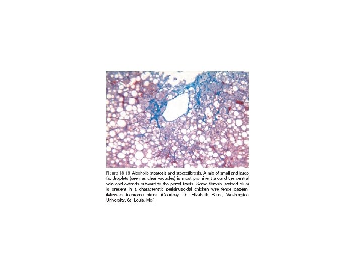
• Hepatic Steatosis (Fatty Liver): - lipid droplets accumulate in hepatocytes increasing with amount and chronicity of alcohol intake. - Fatty change is completely reversible if there is abstention from further intake of alcohol.
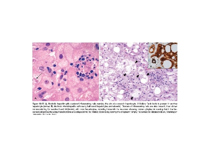
• Alcoholic (Steato-) Hepatitis: 1. Hepatocyte swelling and necrosis (accumulation of fat and water, as well as proteins that are normally exported) 2. Mallory-Denk bodies (intermediate filaments such as keratins 8 and 18 in complex with other proteins such as ubiquitin, characteristic but not specific ) 3. Neutrophilic reaction. They may be more or less admixed with mononuclear cells
• Alcoholic steatofibrosis: Alcoholic hepatitis is often accompanied by prominent activation of sinusoidal stellate cells and portal fibroblasts, giving rise to fibrosis. Early stages of scarring can regress with cessation of alcohol use, but the farther along toward cirrhosis the liver gets, the more vascular derangements prevent a full restoration of normal. Complete regression of alcoholic cirrhosis, while reported, is rare
Pathogenesis: • Short-term ingestion of as much as 80 gm of alcohol over one to several days generally produces mild, reversible hepatic steatosis. • Daily intake of 80 gm or more of ethanol generates significant risk for severe hepatic injury. • Daily ingestion of 160 gm or more for 10 to 20 years is associated more consistently with severe injury. • Only 10% to 15% of alcoholics, however, develop cirrhosis and It may take 10 to 15 years of drinking for the development of cirrhosis. Thus, other factors must also influence the development and severity of alcoholic liver disease. These include: - Gender: Women seem to be more susceptible to hepatic injury than men. - Ethnic and genetic differences: Genetic polymorphisms in detoxifying enzymes and some cytokine promoters may play significant roles and contribute to ethnic differences. ALDH*2, a variant of aldehydedehydrogenase (ALDH), found in 50% of Asians, has a very low activity. Individuals homozygous for ALDH*2 are unable to oxidize acetaldehyde and do not tolerate alcohol, leading to alcohol intolerance characterized by upper body flushing and, variably, nausea or lethargy. - Comorbid conditions: Iron overload , HCV and HBV infection------ increased severity of liver disease.
• Exposure to alcohol causes steatosis, dysfunction of mitochondrial and cellular membranes, hypoxia, and oxidative stress.
• Hepatocellular steatosis results from (1) shunting of normal substrates away from catabolism and toward lipid biosynthesis, as a result of increased generation of reduced NADH by the two major enzymes of alcohol metabolism, alcohol dehydrogenase and acetaldehyde dehydrogenase; (2) Impaired assembly and secretion of lipoproteins; (3) Increased peripheral catabolism of fat, thus releasing free fatty acids into the circulation.
• - - alcoholic hepatitis: Acetaldehyde (the major intermediate metabolite of alcohol) induces lipid peroxidation and acetaldehydeprotein adduct formation, further disrupting cytoskeletal and membrane function. Cytochrome P-450 metabolism produces reactive oxygen species (ROS) that react with cellular proteins, damage membranes, and alter hepatocellular function. The induction of cytochrome P-450 enzymes in the liver by alcohol increases alcohol catabolism in the endoplasmic reticulum and enhances the conversion of other drugs (e. g. , acetaminophen) to toxic metabolites. alcohol impairs hepatic metabolism of methionine, which decreases glutathione levels, thereby sensitizing the liver to oxidative injury. Alcohol causes the release of bacterial endotoxin from the gut into the portal circulation, inducing inflammatory responses in the liver. alcohol stimulates the release of endothelins from sinusoidal endothelial cells, causing vasoconstriction and contraction of activated myofibroblastic stellate cells, leading to a decrease in hepatic sinusoidal perfusion.
Clinical Features: Hepatic steatosis: may cause hepatomegaly, with mild elevation of serum bilirubin and alkaline phosphatase levels. Severe hepatic dysfunction is unusual. Alcohol withdrawal and the provision of an adequate diet are sufficient treatment. - alcoholic hepatitis: tends to appear acutely, usually following a bout of heavy drinking. Symptoms and laboratory manifestations may range from minimal to those that mimic acute liver failure. Between these two extremes are the nonspecific symptoms of malaise, anorexia, weight loss, upper abdominal discomfort, and tender hepatomegaly, and the laboratory findings of hyperbilirubinemia, elevated serum aminotransferases and alkaline phosphatase, and often a neutrophilic leukocytosis. With proper nutrition and total cessation of alcohol consumption, the alcoholic hepatitis may clear slowly. However, in some patients, the hepatitis persists, despite abstinence, and progresses to cirrhosis. - In contrast to other chronic liver diseases where serum ALT tends to be higher than serum AST, serum AST levels tend to be higher than serum ALT levels in a 2: 1 ratio or higher in alcoholic liver disease. This can be helpful in differential diagnosis of chronic liver injury when adequate history is not available. • -
• The manifestations of alcoholic cirrhosis are similar to those of other forms of cirrhosis may be clinically silent, discovered only at autopsy or when stress such as infection or trauma tips the balance toward hepatic insufficiency. • In the end-stage alcoholic the proximate causes of death are (1) hepatic coma, (2) massive gastrointestinal hemorrhage, (3) intercurrent infection (to which these patients are predisposed), (4) hepatorenal syndrome following a bout of alcoholic hepatitis, (5) hepatocellular carcinoma (the risk of developing this tumor in alcoholic cirrhosis is 1% to 6% of cases annually).
Metabolic Liver Disease • either acquired or inherited. - non-alcoholic fatty liver disease (NAFLD)---- m. c - hemochromatosis, - Wilson disease, - α 1 -antitrypsin deficiency - neonatal hepatitis
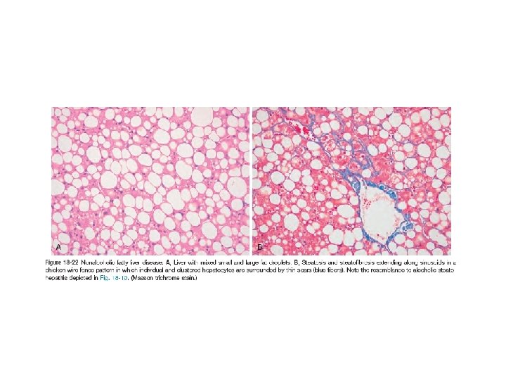
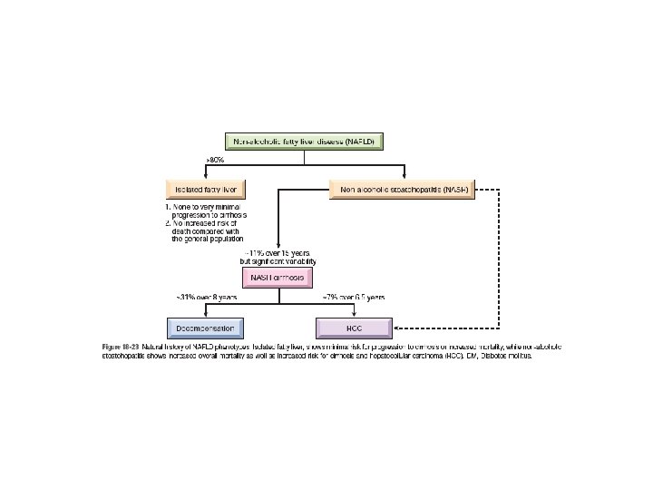
Nonalcoholic Fatty Liver Disease (NAFLD) • NAFLD represents a spectrum of disorders that have in common the presence of hepatic steatosis (fatty liver) in individuals who do not consume alcohol or do so in very small quantities (less than 20 g of ethanol/week)------ Pathologic steatosis is defined as involving more than 5% of hepatocytes • The term “nonalcoholic steatohepatitis” (NASH) is often used to denote overt clinical features of liver injury, such as elevated serum transaminases, but the designation NAFLD is preferred, with steatohepatitis reserved for histologic features of hepatocyte injury already described in the section on alcoholic liver disease. • NAFLD are most consistently associated with the metabolic syndrome----obesity, type 2 diabetes mellitus , dyslipidemia, and hypertension. • Greater than 90% of previously described “cryptogenic cirrhosis” (i. e. , cirrhosis of unknown cause) is now thought to represent such “burned out” NAFLD.
• Pathogenesis: - Insulin resistance gives rise to hepatic steatosis----- obesity - Hepatocellular oxidative injury resulting in liver cell necrosis and the inflammatory reactions to it. In individuals with established insulin resistance and metabolic syndrome, the visceral adipose tissue not only increases, but also becomes dysfunctional, with reduced production of the lipid hormone, adiponectin, and increased production of inflammatory cytokines such as TNF-α and IL-6. These changes in turn promote hepatocyte apoptosis. Fat laden cells are highly sensitive to lipid peroxidation products generated by oxidative stress which can damage mitochondrial and plasma membranes, causing apoptosis. Kupffer cell production of TNF-α and TGF-β activate stellate cells directly leading to deposition of scar tissue.
• Clinical features: - Individuals with simple steatosis are generally asymptomatic. Clinical presentation is often related to other signs and symptoms of the metabolic syndrome, in particular insulin resistance or diabetes mellitus. - Serum AST and ALT are elevated in about 90% of patients with NASH. Despite the enzyme elevations, patients may be asymptomatic. Others have general symptoms such as fatigue or right-sided abdominal discomfort caused by hepatomegaly - The goal of treating individuals with NASH is to reverse the steatosis and prevent cirrhosis by correcting the underlying risk factors, such as obesity and hyperlipidemia, and to treat insulin resistance. - NASH also increases the risk of hepatocellular carcinoma as do other metabolic diseases. Imaging studies may reveal at accumulation in the liver. However, liver biopsy is the most reliable diagnostic tool for NAFLD and NASH, and for assessment of scarring.
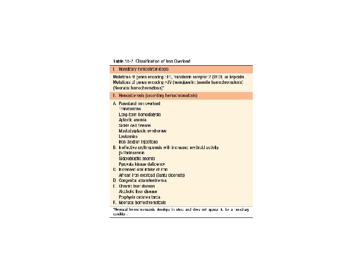
Hemochromatosis • Hemochromatosis is caused by excessive iron absorption, most of which is deposited in parenchymal organs such as the liver and pancreas, followed by heart, joints, and endocrine organs. • Fully developed cases exhibit (1) micronodular cirrhosis in all patients; (2) diabetes mellitus in 75% to 80% of patients; (3) abnormal skin pigmentation in 75% to 80% of patients. Iron accumulation in hereditary forms is lifelong but the injury caused by excessive iron is slow and progressive; hence symptoms usually first appear in the fourth to fifth decades of life in men and later in women since menstrual bleeding counterbalances the accumulation until menopause------ hemochromatosis affects more males than females (ratio of 5 to 7 : 1).
• Pathogenesis: In hereditary hemochromatosis, regulation of intestinal absorption of dietary iron is abnormal, leading to net iron accumulation of 0. 5 to 1 gm/year. The disease manifests itself typically after 20 gm of stored iron have accumulated (the total body iron pool ranges from 2 to 6 gm in normal adults; about 0. 5 gm is stored in the liver, 98% of which is in hepatocytes). The main regulator of iron absorption is the protein hepcidin, encoded by the HAMP gene and secreted by the liver. Therefore, hepcidin lowers plasma iron levels. Conversely, a deficiency in hepcidin causes iron overload. Transcription of hepcidin is increased by inflammatory cytokines and iron, and decreased by iron deficiency, hypoxia, and ineffective erythropoiesis. The adult form of hemochromatosis is almost always caused by mutations of HFE gene on Ch 6 (regulate hepcidin level). Mutations of HAMP gene cause severe juvenile hemochromatosis. Excessive iron appears to be directly toxic to tissues------- inflammation is characteristically absent.
• Severe hemochromatosis (hereditary or secondary) is characterized principally by (1) deposition of hemosiderin in the following organs (in decreasing order of severity) the liver, pancreas, myocardium, pituitary gland, adrenal gland, thyroid and parathyroid glands, joints, and skin; (2) cirrhosis; (3) Pancreatic fibrosis. Biochemical determination of hepatic tissue iron concentration has been the gold standard for quantitating hepatic iron content. In normal individuals, the iron content of liver tissue is less than 1000 μg per gram dry weight of liver. Adult patients with hereditary hemochromatosis exhibit more than 10, 000 μg iron per gram dry weight; hepatic iron concentrations in excess of 22, 000 μg per gram dry weight are associated with the development of fibrosis and cirrhosis. However, with newly available genetic testing for these diseases, quantitative assessment of tissue iron content is no longer necessary for confirmation of a suspected diagnosis.
• Clinical Features: The principal manifestations of classic hemochromatosis include : - hepatomegaly, - Abdominal pain, - abnormal skin pigmentation, -deranged glucose homeostasis or diabetes mellitus - cardiac dysfunction (arrhythmias, cardiomyopathy) - atypical arthritis. - hypogonadism (e. g. , amenorrhea in the female, impotence and loss of libido in the male). Death may result from cirrhosis or cardiac disease. A significant cause of death is hepatocellular carcinoma; the risk is 200 -fold greater than in the general population.
• Currently most patients with hemochromatosis are diagnosed in the subclinical, precirrhotic stage due to routine serum iron measurements (as part of other diagnostic workup). • Treatment by regular phlebotomy steadily depletes tissue iron stores. With treatment, life expectancy is normal. • Screening of family members of probands is important. • Screening involves : - demonstration of very high levels of serum iron and ferritin, - exclusion of secondary causes of iron overload, - liver biopsy if indicated.
Wilson Disease • Wilson disease is an autosomal recessive disorder caused by mutation of the ATP 7 B gene on Ch 13, resulting in impaired copper excretion into bile and a failure to incorporate copper into ceruloplasmin and inhibits ceruloplasmin secretion into the blood. • This disorder is marked by the accumulation of toxic levels of copper in many tissues and organs, principally the liver, brain, and eye, in addition to decrease in circulating ceruloplasmin. Concomitantly, urinary excretion of copper markedly increases from its normal miniscule levels.
• Normally, 40% to 60% of ingested copper (2 to 5 mg/day) is absorbed in the duodenum and proximal small intestine, and is transported to the portal circulation complexed with albumin and histidine. • Free copper dissociates and is taken up by hepatocytes. In the liver copper binds to an α 2 -globulin (apoceruloplasmin) to form ceruloplasmin, which is secreted into the blood. Ceruloplasmin accounts for 90% to 95% of plasma copper. • Excess copper is transported into the bile. • Circulating ceruloplasmin is eventually endocytosed by the liver, and degraded within lysosomes, after which the released copper is excreted into bile. • This degradation/excretion pathway is the primary route for copper elimination. The estimated total body copper is only 50 to 150 mg.
Morphology: • Fatty change (steatosis) • acute, fulminant hepatitis • Chronic hepatitis • Steatohepatitis • Cirrhosis • chronic obstructive cholestasis • Toxic injury to the brain primarily affects the basal ganglia, Nearly all patients with neurologic involvement develop eye lesions called Kayser-Fleischer rings, green to brown deposits of copper in Descemet membrane in the limbus of the cornea. • demonstration of hepatic copper content in excess of 250 μg per gram dry weight is most helpful for making a diagnosis. the vast range of genetic alterations in Wilson disease means that genetic testing is not yet a primary diagnostic modality.
• Clinical Features: • the disorder usually manifests in affected individuals between 6 and 40 years of age. • acute or chronic liver disease. • Neurologic involvement presents as movement disorders (tremor, poor coordination) or rigid dystonia (spastic dystonia, mask like facies, rigidity and gait disturbances); these symptoms may be confused with Parkinsonism. • psychiatric symptoms such as depression, phobias, compulsive behavior, and labile mood. • Hemolytic anemia may occur due to toxicity of copper to red cell membranes.
• The biochemical diagnosis of Wilson disease is based on : - a decrease in serum ceruloplasmin, - an increase in hepatic copper content (the most sensitive and accurate test), - and increased urinary excretion of copper (the most specific screening test). Serum copper levels are of no diagnostic value, since they may be low, normal, or elevated, depending on the stage of evolution of the disease. early recognition and longterm copper chelation therapy (with D penicillamine or Trientine) or zinc-based therapy (which blocks uptake of copper in the gut) has dramatically altered the usual progressive downhill course.
α 1 -Antitrypsin Deficiency • α 1 -Antitrypsin deficiency is an autosomal recessive disorder of protein folding marked by very low levels of circulating α 1 -Antitrypsin (α 1 AT). The major function of this protein is the inhibition of proteases, which are normally released from neutrophils at sites of inflammation. • α 1 AT deficiency is the most commonly diagnosed inherited hepatic disorder in infants and children. • α 1 AT deficiency leads: - Pulmonary emphysema - Cutaneous panniculits - Liver disease: neonatal hepatitis without or with cholestasis and fibrosis Childhood cirrhosis chronic hepatitis steatosis
α 1 AT is plasma glycoprotein synthesized predominantly by hepatocytes. It is a member of the serine protease inhibitor (Pi) family. The gene, located on chromosome 14. The most common genotype is Pi. MM, occurring in 90% of individuals (the “wild-type”). Some deficiency variants, including: - the Pi. S variant, result in a moderate reduction in serum concentrations of α 1 AT without clinical manifestations. - Pi. ZZ protein have circulating α 1 AT levels that are only 10% of normal---- m. c - Pi. MZ heterozygotes have intermediate plasma levels of α 1 AT - Rare variants termed Pi-null have no detectable serum α 1 AT. • With most allelic variants, the protein is synthesized and secreted normally. Deficiency variants show a selective defect in migration of protein from endoplasmic reticulum to Golgi apparatus; this is particularly characteristic of the Pi. Z polypeptide, The mutant polypeptide (α 1 AT-Z) is abnormally folded (protein misfolding) and polymerized, creating endoplasmic reticulum stress and triggering the unfolded protein response, a signaling cascade that may lead to apoptosis. All individuals with the Pi. ZZ genotype accumulate α 1 AT-Z in the endoplasmic reticulum of hepatocytes, but only 10% to 15% of Pi. ZZ individuals develop overt clinical liver disease. Other genetic factors or environmental factors are thus posited to play a role in the development of liver disease.
• Clinical Features. - Neonatal hepatitis with cholestatic jaundice appears in 10% to 20% of newborns with the deficiency. - In adolescence, presenting symptoms may be related to hepatitis, cirrhosis or pulmonary disease. - Alternatively, the disease may remain silent until cirrhosis appears in middle to later life. - Hepatocellular carcinoma develops in 2% to 3% of Pi. ZZ adults, usually, but not always, in the setting of cirrhosis. The treatment, indeed the cure, for severe hepatic disease is orthotopic liver transplantation. patients with pulmonary disease the single most important preventive measure is avoidance of cigarette smoking, because smoking markedly accelerates emphysema and the destructive lung disease associated with α 1 AT deficiency.