Inoculation to Medium The yeast are inoculated with


 Identification of Fungi • Direct microscopic image of the fungus in clinical specimen")


 2. Systemic Miycosis 3. Other Fungi")


 Ectothrix:")

")
 Clinical Identification: Absolute diagnosis of Trichophytosis must be done by laboratory inspection")


 Clinical Identification: Absolute diagnosis of Microsporiosis must be done by laboratory inspection")
 and")


, is characterised by exudative dermatitis which is observed in")


 Clinical Diagnosis: Clinical findings are mostly observed in the respiratory and digestion")


,")


 fungus Blastomyces dermatitidis •")


 or systemic disease characterized by Histoplasma capsulatum, a dimorphic")


 Niger or bird seed agar melanin production : C. neoformans")


- Slides: 94
Inoculation to Medium • The yeast are inoculated with the aid of the inoculum to the surface of the inoculum prepared from the clinical or necropsy material as it is in the same bacteria. • In fungi isolation, a sterile bisturia is formed at 5 points on the surface of the nutrient plantation. Later, small pieces of lesion tissue, skin scrapings or furrows are lightly soaked in the agar in these positive areas. • If the samples are immersed directly in the agar, splitting, breakage may occur in the agar during long-term incubations • Yeast are passively ingested into the new medium as it is in the same bacteria.
Subculture of Fungi If the fungi column is sporulating; Fungus colonies usually start to produce spores from the center to the periphery, and are often sports that give a colonial characteristic color. A substance is first dipped into the surface of a sterile agar portion to make it slightly damp and sticky, then used to collect spores from a column. If the fungi species is a fast-breeding fungus, the inoculum is inoculated immediately below the surface of the agar, at the exact center point on a new plate surface. If the fungus forms small, slow-breeding colonies, the agar plate is divided into four portions and each section is separately planted.
Subculture of Fungi If the fungi column is not sporulating; • In the fungus colon, the aging and death phase begins with the hypha in the center of the colon. For this reason, the passage of the hyphen around the colon is required. • A small agar block (5 mm 2) in the center is cut off on the medium to be used for passage with a sterile blister. Using the same bisturia, a similar sized and shaped agar is cut out from the side of the column to be passaged to include fungal hyphae. • This agar block is carefully placed on the medium to be passaged in such a way that the mushroom part is above. The interrupted hypha will be regenerated and will grow towards the periphery from the block surface.
Microscopic Examination of Fungal Colonies • Investigation with Dissecting microscope • preparation of lamel preparation with LPCB (Lactophenol Cotton Blue ) (wet mount method) • Adhesive tape method • Lam Culture Technique
c-) Identification of Fungi • Direct microscopic image of the fungus in clinical specimen • Colony morphology and pigmentation type • Microscopic image of macroconidia (fruiting heads) and spores in fungal colon • Yeast morphology and budding • Biochemical tests for yeast and less frequent and frequent fungi • Effect specific tests such as germ tube test for Candida albicans • Specific serological tests • Status of contaminants!!!
Safety Conditions for Mycology • Most of the fungi that cause disease in animals are also pathogenic to humans. • Particular care should be taken when considering materials and cultures that are thought to contain pathogenic fungi!!! • Particular care should be taken against pathogens that are easily aerosolized, such as Coccidioides immitis, which form highly infective arthrospores at 25 and 37 ° C. • Dimorphic fungi such as Cryptococcus neoformans and Blastomyces dermatitidis cause very serious disease in humans. • Ideally, all mycologic examinations should be done in the biosafety cabinet.
Treatment and Prevention
• Mammalian cells are lack of enzymes that destroyes fungal cell Wall polysaccharides. Because of this, fungi can not be eradicated by the host defence mechanisms. • Memeli hücreleri, mantarların hücre duvarı polisakkaridlerini parçalayan enzimlere sahip değildir. Bu nedenle mantarlar, hayvanın konakçının defans mekanizmalarıyla eradike edilemezler. • Since mammalian and fungal cells are eukaryotic, both of them have the same cell structure and also look like each other biochemically. • • Hem memeliler ve hem de mantarlar ökaryotik organizmalar olduklarından, her ikisindeki hücresel yapı, biyokimyasal olarak birbirine benzerdir. All eukaryotic cells’ cell membrane have sterols; in fungi these are ergosterol and in mammalian cells these are cholestrol. Thus, the invasive fungi elemination leads to severe side effects in their hosts. • Bütün ökaryotik hücrelerin, hücre membranları steroller içerir; mantarlarda bu ergosterol iken, memelilerde ise kolesteroldür. Dolayısıyla, invaze olan mantar etkenini bozacak maddeler konakçıda da ciddi yan etkilere neden olabilmektedir. • Although the first chemotherapeutic agent is an antifungal (oral iodids), the developing of these agents were slow in comparison to antibacterial agents. • Her ne kadar ilk kemoterapotik ajan 1903 yılında kullanılan bir anti-mikotik (oral iodidler) iken, bu ajanların geliştirilmesi anti-bakteriyel ajanlara göre yavaş olmuştur. • It was difficult to inhibit the invasive organism while protecting the host. This situation slows down the new drug developments. • Konakçıya minimal zarar vererek invaze olan organizmayı inhibe etmek için, gerekli selektif toksisitenin ökaryotik hücreler için oluşturulması güç bir hedef olmuştur. Bu da yeni ilaç geliştirme çalışmalarını yavaşlatmıştır.
• Flucanozole is used for the AIDS patients to treat the cryptococcosis. • Flukanazol bugün cryptococcosis’li AIDS hastalarının tedavisinde tercih edilen ilaçtır. Spinal sıvıyı (BOS) penetre ettiği için idealdir. • Azolles leads to the inhibition of ergesterol synthesis. • Azollerin genel etki mekanizması hücre duvarı sentezini etkileyen ergesterol sentezinin inhibisyonudur. Oral uygulama, düşük toksisite önemli dezavantajlarıdır. • Ketoconazole, Fluconazole, Itraconazole, Voriconazole, Posaconazole • Griseofulvin, is used in severe skin and nail infections. It has a very slow effect. Orally routed. And the mechanism of its effect is due to the accumalation on stratum corneum layer and to penetrate the tissue in order to prevent the fungal invasian. • Şiddetli deri ve tırnak infeksiyonlarında kullanılan, oldukça yavaş etkili bir ilaçtır. Oral yolla uygulanır. Etkisi, stratum corneum tabakasında birikmesi ve buradan da dokuya geçerek daha ileri fungal penetrasyonu ve üremeyiş engelleyecek şekilde bariyer oluşturması prensibine dayanır. • 5 -fluorosytosine, inhibits the RNA synthesis, mostly used in criptococcosis treatment. Orally routed. • Alilamines, Terbinafine (lamisil). Used in dermathophyte infections. • Echinocandins (caspofungin): New antifungal agent approved by FDA.
General Characteristics of Fungal Infections
• Although the dermatophytes are known to be obligate parasites, most of the pathogenic fungi are living in the environment as saprophyte or have a close relation with both humans and animals commensally • Dermatofitlerin birkaçının obligat (zorunlu) parazitler olduğu düşünülse de patojenik mantarların çoğu çevrede saprofit olarak yaygındır ya da hayvan ve insanlarla ilişkili komensaller olarak bulunmaktadır. • Most of the fungi are opportunist pathogens and the factors that lead to developing of infections are: – Antibiotic usage in a long period. Thus the change in normal microbiata of host – Immunsupression – Simultaneıus infections • Mantarların çoğu fırsatçı patojenler olup mantar infeksiyonlarının şekillenmesinde rol oynayan predispoze edici faktörler: – Uzun süreli antibiyotik kullanımı sonucu konakçının normal mikrobiyotasının değişmesi – İmmunosupresyon – Eş zamanlı infeksiyonlar
• Skin and mucous membrane injuries or the loss of skin integrity • Deri ve muköz membranlarda yaralanmalar veya deri bütünlüğünün bozulması • Continous moist areas on skin • Deride sürekli nemli bölgelerin bulunması • Exposure to high infective dose of spores like «brooder pneumonia» observed in chicks caused by Aspegillus fumigatus • Ya da civcivlerde görülen “brooder pneumonia” da Aspergillus fumigatus sporlarında olduğu şekilde yüksek dozda infektif doza maruz kalmak • No epidemies are observed in fungal infections except «ringworm» that appear suddenly • Mantar hastalıkları «ringworm» enfeksiyonları dışında genellikle epidemiler şeklinde görülmez • In fungal infections there are no exo or endotoxins but in food of animals sometimes toxix metabolites can be developed by the fungi while developing on these food • Mantar infeksiyonlarında ekzotoksin ve endotoksin sentezlenmemektedir, ancak hayvan yemlerinde mantar üremesi sırasında daha önceden oluşan toksik metabolik ürünlere bağlı mikotoksikozisler şekillenebilmektedir.
1. Dermathophytes (Cutan Mycosis) 2. Systemic Miycosis 3. Other Fungi
Dermathophytes
1. • • • Cutan Mycosis Trichophyton Genus Microsporium Genus Epidermophyton Genus 2. Dermatophilosis
• Dermathophytes are close related fungi that uses keratin to reproduce • • Dermatofitler üremek için keratini kullanan yakın ilişkili mantarlar bütünüdür. They make infections on superficial regions like, stratum corneum, nails, hairs of both animals and humans • Derinin dış stratum corneum tabakası, tırnaklar, pençe ve insan ve hayvanların saç ve kılları gibi yüzeysel alanlarda infeksiyon oluştururlar. • Classical lesions are circular lesions called «Ringworm» • • Klasik lezyonlar “Ringworm” olarak adlandırılan dairesel lezyonlardır. Conventionally dermathophytes are classified as «Fungi Imperfecti» , nevertheless some of them are classified as Ascomycetes because of their known sexual reproduction • Geleneksel olarak dermatofitler “Fungi Imperfecti” sınıfında gösterilirken bazıları için seksüel aşama tespit edilmiş olup Ascomycetes grubunda sınıflandırılmıştır. • There are more than 38 dermathophyte species • • 38’den fazla dermatofit türü bulunmaktadır. The ones that effect animals are classified in the genus Microsporum and Trichophyton • Hayvanları etkileyenler Microsporum ya da Trichophyton genusunda yer almaktadır.
Frequent Dermathophyte Infections 1. Tinea capitis 2. Tinea pedis 3. Tinea corporis 4. Tinea cruris 5. Tinea barbea 6. Tinea ungium
Trichophyton Genus
• In animals, trichophytones lead to dermathomycoses particularly observed on skin, hair and nails • Trikofiton İnfeksiyonları (Trikofitozis), hayvanlarda, Trikofiton cinsine ait mantarlar tarafından özellikle deri, kıl ve tırnakların keratinize kısımlarında oluşan bir dermatomikozistir. • Some of them are zoonotic • • Trikofiton cinsi çok geniş türe sahiptir ve bazıları zoonoz karakterlidir. On solid agars the coloies can be cotton, granular, puffy, mucoid shaped and in different colour • • Katı besiyerinde üreyen kolonileri kadife, pamuk, granüler, kabarık, mukoid görünümde ve çeşitli renklerde olabilmektedir. The macroconidiums are oval, lemon, cigar or cylindirical and contains 2 -12 cells. They are rarely observed as a group • • Makrokonidiumlar, oval, limon, puro ya da silindirik biçimlidir ve 2 -12 hücrelidir. Tek tek bulunurlar, nadiren gruplar halindedir. The microconidiums are one cell, spherical or oval shaped. They can be found on hyphae one by one or as clusters • Mikrokonidiumlar, tek hücreli, yuvarlak, oval ya da armut biçimlidir. Hifa boyunca ya tek bulunurlar ya da kümeler halinde yer alırlar. • They do not give flourescence under the wood lamb light!!!! • Trikofitonlar, Wood Lambası altında fluoresans vermezler !
Trichophytones can be classied in two types according to the hair invasion 1) Ectothrix: The fungal arthropores can be found out of the hairs not inside • • T. mentagrophytes T. equinum T. verrucosum T. rubrum 2) Endothrix: The fungal arthropores can be found inside of the hair in parallel or irregularly • T. tonsurans • T. violaceum
Epidemiology • These kind of infections can be found all over the earth • Trikofiton’lardan ileri gelen dermatofitozislere dünyanın her yerinde sıkça rastlanmaktadır. • Trichophytosis can be spread directly by contact or indirectly between animals • Trikofitozis, direkt temas veya indirekt yollarla bir hayvandan diğer hayvana kolaylıkla bulaşır. • The infection is more contogious in especially in winter and in the crowded, dirty and moist barns • Özellikle kış aylarında kalabalık, pis ve rutubetli ahırlarda bulaşma daha çabuk şekillenir. • Mostly the young animals are effected • Genellikle genç hayvanlarda daha çok görülmektedir.
Important Pathogenic Species • • • Trichophyton Trichophyton Trichophyton equinum ( Horse, Dog ) rubrum ( Cow, Dog ) gallinae ( Chicken, Turkey, Dog ) soudanese ( Dog, Cat, Monkey ) megninii ( Horse, Cow, Dog ) violaceum ( Cow ) verrucosum ( Cow, Sheep, Horse, Dog ) concentricum ( Cow, Dog ) mentagrophytes ( Dog, Cat, Cow, Horse )
Identificaiton 1) Clinical Identification: Absolute diagnosis of Trichophytosis must be done by laboratory inspection because it can be clinically misdiagnosed with other skin diseases, insect bites, bacterial infections. 2) Laboratory Inspection: Microscopy: Skin scrapings and hair samples must be taken from the outside of the lesion. Samples must be put on a clear slide and inspected with %10 KOH on microscope. Arthrospores, hyphae with branches and septums are seeked.
Culture: SDA is optimal. Samples are sticked into the different parts of the agar. Petri dishes are incubated for 2 weeks at 25 C. The macroscopic and microscopic morphology of colonies can be inspected.
Hair Perforation Test • To discriminate the T. mentagrophytes and T. rubrum • T. Mentagrophytes can invade to hair tissue and make conical perforation – Hair sample is taken from a child – This hair autoclaved at 121°C for 15 min to sterilized it – These steril hair samples are left on the 3 -5 day subculture of the tested dermathophyte and incubated at 25°C – On the 7 th day the hair samples are stained with LCB for the inspection of perforation
Treatment: Topical antifungals, Thiabendazole, Miconazole, Ecoconazole, Ketoconazole, İtraconazole, Limesulphur solution, 5 % sodium hypochlorite solution can be used topically. Systemic antifugals can be used if topical treatment doen not work. For example; ketoconazole, clotrimazole, itraconazole, terbinafine. Mostly terbinafine is the most efficient. Nowadays, Griseofulvin isn’t used because of its acute toxicity Prevention – Control • T. verrucosum (LTF-130 strain) • Live vaccine. Contains conidia and hyphal elements. Used for both prophylaxis and curation.
Microsporum Genus
• It is a dermathomycoses caused by Microsporum species in both animal and humans’ hair and skin • Mikrosporum İnfeksiyonları (Mikrosporozis), insan ve hayvanlarda, Mikrosporum cinsine ait mantarlar tarafından kıl ve deride oluşan bir dermatomikozistir. • The arthrospores are smaller than the Trichophytone’s. They can surround the hair like a package • Mikrosporum cinsine ait mantarların artrosporları, Trikofiton cinsine ait mantarların artrosporlarından daha küçüktür ve kılların etrafında mozaik görünümlü paketler oluştururlar. • The morphology of colonies are thin, granullar or cotton shaped and with different colours • • Katı besiyerinde üreyen kolonileri ince, granüllü, kadife veya pamuk görünümlü ve çeşitli renklerde olabilmektedir. In microscopy big, thin and thick walled, multi compatment (3 -15 cells) and shuttle shaped macroconidiums can be inspected • Mikroskop altında incelemelerde büyük, ince veya kalın duvarlı, çok bölmeli ( 3 -15 hücreli) ve mekik şeklinde makrokonidiumlara rastlanmaktadır. • Microconidiums can be observed as spherical, oval and unicellular on the hyphae one by one • • Mikrokonidiumlar, tek hücreli, yuvarlak, oval ya da armut biçimlidir. Hifalar üzerinde saplı ve tek hücreler tarzında yer alırlar. Microsporum species give yellow-green fluoresence under the wood lamp!!! • Mikrosporum’lar, Wood Lambası altında parlak sarı-yeşil renkli fluoresans verirler !
Epidemiology • Can be seen all over the World • Mikrosporum’dan ileri gelen dermatofitozislere dünyanın her yerinde sıkça rastlanmaktadır. • Spread by direct contact or indirectly • Mikrosporozis, direkt temas veya indirekt yollarla bir hayvandan diğer hayvana kolaylıkla bulaşır. • The infection is more contogious in especially in winter and in the crowded, dirty and moist barns • Özellikle kış aylarında kalabalık, pis ve rutubetli ahırlarda bulaşma daha çabuk şekillenir. • Mostly the young animals are effected • Genellikle genç hayvanlarda daha çok görülmektedir.
Important Pathogenic Species • • • Microsporum Microsporum Microsporum canis ( Dog, Cat, Horse, Rabbit, Rodents ) nanum ( Dog, Pig ) cookei ( Dog, Cat, Guinea pig ) gypseum ( Dog, Cat, Horse, Rodent ) audouinii ( Dog, Monkey, Rodent ) distordum ( Dog, Monkey ) persicolor ( Human, Dog, Rat ) ferrugineum ( Human, Animal ) vanbreuseghemii ( Human, Animal )
Identification 1) Clinical Identification: Absolute diagnosis of Microsporiosis must be done by laboratory inspection because it can be clinically misdiagnosed with other skin diseases, insect bites, bacterial infections.
Wood’s Lamb inspection: • While growing, M. canis, M. distortum, M. audouini (human) and M. ferrugineum (human) can produce some metabolites which also give green fluorescence by Wood’s Lamb UV light (366 nm) • Suspected M. canis infections can be diagnosed • The infected sites are generally face, front paws and abdominal areas • However half of the M. canis infections doesn’t give fluorescence, because of this future laboratory inspection must be performed • Topical ointments lead to false positive results
Laboratory Inspection Skin scrapings and hair samples must be taken from the outside of the lesion. 1) Microscopy: Arthrospores, hyphae with branches and septums can be onserved. 2) Culture: SDA is optimal. Samples are sticked into the different parts of the agar. Petri dishes are incubated for 2 weeks at 25 C. The macroscopic and microscopic morphology of colonies can be inspected. Treatment: Topical and systemic treatment is performed for 10 days with antifungal agents. Itraconazole ( Anorexia risk is low in cats ) Terbinafine Ketoconazole Thiabendazole Miconazole Griseofulvin (Isn’t used because of its acute toxicity) (In Siamese, Himalayan, Abyssinian cats myelosupression can be observed)
Epidermophyton Genus
• First article of Epidermophyton was published in 1870 by Harz, about a Tinea cruris case • Epidermophyton cinsine ait ilk bilimsel bildiri Harz tarafından 1870 yılında Tinea cruris vakasından izole edilerek yapılmıştır. • In the beginning it was called Acrothecium floccosum; then in 1923 Ota and Langeron named it as Epidermophyton floccosum • Acrothecium floccosum olarak bildirilen etken, 1923 yılında Ota ve Langeron tarafından Epidermophyton floccosum olarak yeniden isimlendirilmiştir • In Epidermophyton genus there are 2 species described – Epidermophyton floccosum (Mostly in gogs) – Epidermophyton stockdaleae (Non pathogen)
• Usually isolated from the cases of tinea corporis, tinea pedis, tinea cruris and tinea ungium • There is no need for spesific medium for the isolation of agent. SDA and 25 C is optimal. • In solid media they develop expanded hyphae like other dermathophytes. Macroconidias are long and thin shaped with 1 -9 septums. They do not include microcodias • The most important virulence factor is its proteinase enzyme that it produces at 37 C
Treatment: Topical and systemic antifungals, Thiabendazole, Miconazole, Ecoconazole, Ketoconazole, İtraconazole, Clotrimazole, Terbinafine • The most effective one is Terbinafine which can be used both topically and systematically
Dermatophilosis Genus
• Dermatophilosis (Mycotic Dermatitis), is characterised by exudative dermatitis which is observed in leg, head and neck skin of animals Etken Dermatophilus congolensis. – Aerobic, Motile spores (They have flagella to move independently. When it finds suitable environmental conditions spores will develop branched filaments) – Mostly seen in hot and moistured climate – They can not be found in the environment. Only observed on the skin and hair
• The lesions are mostly observed on the back, head, neck and the both sides of the bodies of horses, cows, sheeps and goats • There are papules, vesicles, oedema and suppuration which can also be misdiagnosed as skin infections • Between knee joint and nail coroner of sheeps there can be found 24 cm lesions called “Strawberry Foot Rot”
Treatment – Protection • The treatment of new infection is easy and sometimes animal can recover spontaneously. However treatment of the chronic infections are very difficult • Pomat iodure, iodophorm ointment, %5 salicylic acid and topical antifungal agents can be used • If necessary Penisilin-Streptomisin can be used • For the foot lesions zinc sulphate or copper sulphate can be used • Maintaining the skin of animals dry is the most important prevention strategy. Insect and arthropode control must be performed
Dermatofitlerin genel yaşam dönemleri
Aspergillosis
• Aspergillus species lead to respiratory system infections and sometimes they can also rarely cause systemic infections • Aspergillozis, Aspergillus türleri tarafından oluşturulan, genellikle solunum sistemine yerleşen ve bazen de generalize (sistemik) durum gösteren bir mantar hastalığıdır. • Main species that causes infections in animals are: Aspergillus fumigatus the major agent. Beside this; – Aspergillus niger, – Aspergillus flavus, – Aspergillus terreus, – Aspergillus nidulans § A. fumigatus and A. flavus have endotoxin. A. flavus also synthesised a very potent toxin called aflatoxin.
Epidemiology • The intake of Aspergillus spores by inhalation leads to the frequent observation of respiratory system infections • Spores can be observed in soil, decayed food and plants. Inhalation of these spores by the animals that are fed in these environments lead to Aspergillosis • Sporlara toprakta, çürümüş veya çürümekte olan gıdalarda, bitkilerde oldukça sık rastlanmaktadır. Böyle yerlerde yaşayan ya da beslenen hayvanlarda solunum yolu ile sporların alınması sonucunda Aspergillozis meydana gelir. • In the tissue and pathologic materials the conidiums, coniophores and micelial elements can be seen • • Dokularda veya patolojik maddelerde genellikle konidiumlara, konidiofor ve miselyal elementlere rastlanır. All animals are susceptible. In-appropriate caring and nutrition rules are the predisposition factors for the disease • Hastalığa hemen her hayvanda rastlanmaktadır. Kötü bakım-besleme koşulları ve hijyenik olmayan ortamlar predispose edici faktörlerdendir. • Transmission from animal to animal is very rare in comparison to the other fungal infections • Hayvandan hayvana bulaşma, diğer mantar infeksiyonlarına göre daha nadir rastlanmaktadır.
Diagnosis 1) Clinical Diagnosis: Clinical findings are mostly observed in the respiratory and digestion systems. In some animals abortions dur to the fungi can also be observed. Aspergillus infections are frequently seen in the poultry. Acute Aspergillosis are observed in young animals, Chronic Aspergillosis is seen in adults. - Culture: Tissue with lesions and other materials can be cultured on SDA with antibiotic and incubated at 25 C. The colonies can be evaluated for the both macro and micro morphological characteristics. - In order to diagnose Aspergillus species the conidiophore, vesicle, sterigma and conidial chain must be evaluated. The head of the conidia must be investigated for its shape and colour; the structure of ascospores; the sequence number of sterigma; the length of conidiophofore and the size of the conidium is important.
- Microscopy: The materials are stained and investigated for the presence of conidophores, conidiums, vesicle, sterigma and hyphae Treatment Topical antifungal agents, Amphotericin-B , Enilconazole, Miconazole , Terbinafine Systemic antifungal agents, • Amphotericin-B ( 1. 5 mg/kg, 5 days) • Fluconazole ( 5 mg/kg, 7 days ) • İtraconazole ( 5 -15 mg/kg, 21 days ) • Ketoconazole ( 10 -30 mg/kg, 21 days) • Voriconazole ( 5 -10 mg/kg, 7 days)
Dimorphic Fungi
• Dimorphic fungi have two different reproduction types: – Fungus: In nature as a saprophyte or at 25 -30 C on agar cultures while incubation – Yeast or yeast like: In animal tissues or at 37 C on specific enrichment agar cultures • Fungal or micelial form is the stabil form in comparison to these forms • Mantar ya da miselyal form bu iki form arasındaki daha stabil formdur. • These fungi can cause deep or systemic mycoses in human and animals • Bu mantarlar insan ve hayvanlarda derin ya da sistemik mikozislere neden olurlar.
Diseases that are caused by dimorphic fungi Dimorphic Fungus Hosts Disease Lesion Site Geographical distribution Sporothrix schenckii Horse, Dog, Cat, Human Sporotriciosis Subcutaneous nodulles, Rarely systemic All over the world Blastomyces dermatitidis Dog, human North American Blastomycoses Primarly dog Lung, skin and other organ metastases USA, Africa, Asia and Europe Histoplasma capsulatum Dog, Cat, Human Histoplasmosis Primarly lungs Secondarly intestines Sporadic in the world Equide Epizootic Lenfangitis Lymphatic system, lymph nodulles and systemic Africa, Asia, France, Italy, Rusia, Egypt Coccidiomycosis Primarly lungs sekondarly bones and other organs USA, Mexica, South America Histoplasma farciminosum Coccidioides immitis Dog, Human
Sporotrichosis
Sporotrichosis, The chronic-granulamotous inflamation and ulceration of leg skin and lymph vessel • Caused by Sporotrichum schenckii (Dimorphic) • In the pathological material and slides prepared from the tissues it can be seen as long, spherical, cigar shaped and yeast like cells like bud • The parasitic form of S. schenckii, in in-vitro environment, tiamine, biotine and aminoasides must be added, also incubated at 37 C • At 25°C colonies formed in 3 -5 days and look like first white-cream then dark-skin like shaped • At 37°C the yeast like, S typed, soft and cream colour colonies will occure
Epidemiology • Sporotrichum schenckii, can be found in nature – Soil, water, fertilizer (gübre), decayed plant and the oral and gastrointestinal mucosa of rats – Penetration to the body is from the portantres of skin – Moisture and heat is important in the initial of disease. At 30 C and higher temperatures the rate of the occurence of the disease will be very less Clinical Findings • In equide spores that penetrate from the microscopic portantres of the skin will form the lesions found in the skin and lymphoid tissue under the skin. Sometimes they can metastase to the inner organs – Mallein test can be performed for the differntiation from Malleus in equide • Usually the nodulles under the chest skin will get bigger, hardened and became ulserative. The hair at that site will fall and pus will leak from the wounds
Treatment and Control – Amphotericin-B, Griseofulvin and Sodium – Potasium Iodure – Hygenic precautions must be ruled – The legs of animals must be controlled routinely and prvented from the wound occurences
Blastomycosis
Blastomycosis, The chronic-granulamotous and suppurative infection caused by dimorphic (diphasic) fungus Blastomyces dermatitidis • Blastomyces dermatitidis, is in mycelial form when incubated at 22 - 25 ˚C and yeast like form at 37 ˚C • In living organisms the body temperature is 36 -37 ˚C so in the body and pathological material the fungus can be found as yeastlike form • In skin, lungs, bones, neural system, urogenital system and other orgns the lesions can be occured
• Blastomyces dermatitidis infections can be observed in dogs lived in Canada and USA. Agent can reproduce in the nature and the spores can be inhalated by air • There is no spread between living organisms. All living organism will take the agent individually from outside and infection will occur • There are two clinical forms: Skin Blastomycosis and Systemic Blastomycosis • Systemic Blastomycosis, occures when the agent invade into primer tissue sites. Lung, liver, kidney, splen and related organs are the tissue that the lesions are observed • Skin Blastomycosis, rarely from skin wound but mostly by hematogen ways are the main routes that the disease agent causes skin lesions. Skin and under skin the lymph nodes are effected and the abscess, furuncules will occur
Identification • Clinical identification, B. dermatitis lesions can be confused with other related lesions similar to lesions of many bacterial and viral agents. • Necropsy is found in the animals, in the skin and subcutaneous tissues, in the lungs and other internal organs, numerous nodules. Lesothoracic lung, liver, spleen, kidney, lymph ovules, skin lesions are sent to the relevant laboratory for laboratory diagnosis. • Culture: Antibiotic SDA or brain-heart infusion agar is applied from the lesioned tissue, organs and other materials and left to incubate at 25 ˚C and 37 ˚C for about 10 -15 days. Macro and micro morphology of breeding fungus colonies are examined. • Microscopy: Lenticular materials are first treated with 10% KOH or Lactophenol Cotton Blue, and cross-lamellar examination is performed. Under the microscope, large, round, thick-walled, granular and some buds are visible.
• Treatment and Preservation: There is no known effective therapeutic agent in the treatment of the disease. Generally, Amphotericin-B is administered to the animals while the injured is being treated with surgical interventions. - The general hygienic conditions must be observed and the sick animals and healthy animals must be kept away from each other
Histoplasmosis
Histoplasmosis, is a localized (pulmonary) or systemic disease characterized by Histoplasma capsulatum, a dimorphic fungus in humans and animals. is a localized (pulmonary) or systemic disease characterized by Histoplasma capsulatum, a dimorphic fungus, in humans and animals. • • Dogs and pets are the most sensitive animals. • Histoplasma capsulatum, is mycelial form when incubated at 22 - 25 ˚C; When cultivated at 37 ˚C, it grows in yeast-like form. • • The colonies produced at 25 C in Sabouraud dextrose agar are preceded by white-pink, then brown, forming aerial mycelium like cotton. • In pathological materials, especially candelabra dyed preparations, small, oval, yeast, mononuclear, and sometimes polymorphonuclear cells are affected. • In cultures made from pathological materials, white and aerial hyphae (Type. A) and other brown colored (Type-B) colonies are formed.
Epidemiology • H. Capsulatum, exists in saprophytic form to soil. The presence of poultry feces in contaminated areas and in the perches has a good development environment. • • There is no animal or animal contamination. Individuals taking active sports are infected at the end. • It has been reported that the root of the chickens is isolated. • The environments in which the wounds occur are reservoirs for humans and animals. • This disease is more common in specially trained dogs for hunting trails (crawling, digging). Apart from this, there are similar rates in cats, pigs and sheep. • The agent is localized in some regions and produces endemic disease.
Clinical Symptoms • Infection is difficult to diagnose because of a latent and chronic course in animals. Some of the symptoms that arise are not specific enough to describe the disease. Laboratory Results • Microscopy: Dyeed and unpainted preparations prepared from animal materials are found in yeast form. Mononuclear and sometimes polymorphonuclear structures are found in preparations prepared from blood, bone marrow and lymph nodules. • Culture : Antibiotic SDA is removed from the receiving material by incubation at 25 °C and 2 °C for 2 weeks at 37 °C. Micro and macro morphologies of breeding colonies are examined. When necessary, the yeast form is re-sown to allow for conversion to a mycelial form.
Coccidioidomycosis
Coccidioidomycosis, is a chronic, noncommunicable disease that is usually localized to the respiratory system in humans and animals. • The agent has a dimorphic character, it is Coccidioides immitis. • C. immitis, is very resistant to drying. It can protect its vitality for long periods on environmental conditions • It develops easily in antibiotic SDA medium and at 25° C in 35 days.
Epidemiology • C. immitis and effective spores are more common in the soil • In dry and windy weather, spores that are involved in the air are removed by the breathing air and become localized to the lungs. • There is no animal or animal contamination. • The possibility of infection from the portents in the vicinity is very rare and no digestive system infections have been found. • Infection is mostly dog, cattle, horse, cat, pig and sheep.
Clinical Symptoms • Many of the C. immitis infections are often overlooked because they are seen in latent or subclinical forms • It is almost impossible to diagnose precisely without laboratory analysis even in the case of clinical trials. • The most obvious symptom is cough, which is similar to tuberculosis and other pulmonary infections.
Laboratory Results Microscopy: Lenticular examination is first performed with 10% KOH or Lactophenol Cotton Blue on the lesional material. Under the microscope, it is examined in terms of thick walled and refractile spherules. • Culture : Sterilization of antibiotics and antibiotics from clinical materials is performed instead of SDA fattening and incubation is allowed at 25 C and 37 C. Reproduced colonies within 3 -5 days are examined in terms of C. immitis culture formation, but care should be taken that spores are not scattered around and not taken up with breathing air.
Patogenic Yeast
Patojenik Mayalar; • Candida albicans • Cryptococcus neoformans • Malassezia pachydermatis • Geotrichium candidum
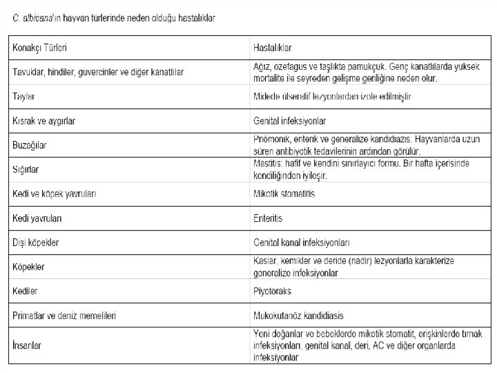
Candidiasis
Candidiasis, is infectious in Candida species generally in the digestive tract and mucous membranes, predominantly in C. albicans in the vast majority of humans and animals (90%). • C. albicans, is located commensally in the mouth, esophagus, stomach, intestinal tract, subcutaneous tissues, lung, breast tissue and genital tract. affective lesions are encountered. • Most of the leading infections are of endogenous origin and predisposing causes such as immunosuppression, long-term antibiotic therapy and inadequate care-feeding are the reasons for the infection. • Infection, acute in young animals; is a chronic course in adult animals.
Clinical Symptoms • In poultry, clinical signs are hardly observable. • The cases are usually sporadic and rarely endemic. Upper respiratory tract and digestive system are affected the most. • In acute cases, yellow-white lesions are loose in the mucosa of the necropsy coats; In chronic cases, it is observed on the surface of the mucosa and a structure covered with necrotic membrane like towel. • Mycoses from C. albicans are rarely encountered in ruminants and pigs. In pigs cases, abortus, mastitis, mycotic stomatitis, pneumonia and rumenitis are usually observed. • In dogs and cats skin, otitis, intestinal candidiasis, genital candidiasis
Laboratory Diagnosis • For the diagnosis of the disease, depending on the location of localization, mouth, esophagus, stomach, cow, milk, uterine flow, skin scraping etc. materials should be sent to the appropriate laboratory conditions. • • Direct Microscopy Skin and mucosa excavations are treated with 10% KOH and examined under a microscope. Oval-budding yeast-like cells and short mycelial structures are observed. Lactophenol is examined by cotton bluish or Gram stain on the frother where it is prepared from uterine fluids and milk. Culture Sabouraud Dextrose Agar is planted with both antibiotics and antibiotics from the laboratory materials. The petri dishes are allowed to incubate at 25 ° C and 37 ° C for 3 -5 days. Some Candida species are inhibited by cycloheximide. Plates are cultivated with a small inoculum volume as in the same bacteria.
Identification Colony morphology • • C. albicans colonies usually form within 3 -5 days. They form colonies of 4 -5 mm in diameter, with a sweet, fruity smell that is bright, high-convex in white or creamy color. Microscopic morphology • A small piece of Lactophenol Cotton Blue can be prepared from a single colon, or pre -fixed colonies can be stained with Gram Stain or Methylene Blue. • C. albicans bloody agar and Sabouraud Dextrose Agar to form thin-walled, budding cells. Demonstration of Germ Tubes • • A small inoculum prepared from breeding colonies is inoculated into 0. 5 ml of sheep, cattle, rabbit or human serum and incubated for 2 -3 hours at 37 °C. The prepared mixture is examined in a drop phase contrast microscope or light microscope. The appearance of small tubers that protrude out of some yeast cells is characteristic of C. albicans.
Treatment and Protection • Candidiasis is largely due to predisposing factors. For this reason, predisposing factors must be removed first. • Nystatin can be added to copper sulphate and baits for preservative drinking water. • Some studies have shown that formic acid applications may be beneficial to feeds. • Amphotericin is primarily used in animals to be treated. In addition, topical applications can be performed on lesioned areas.
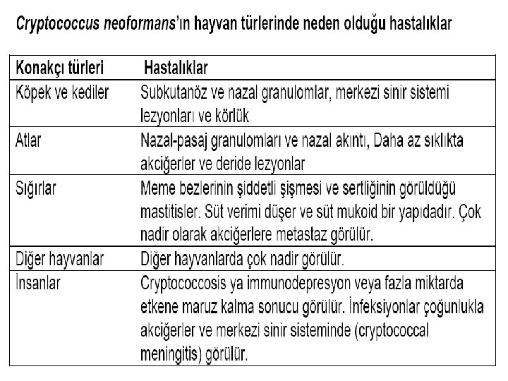
Cryptococcosis
• Cryptococcosis, is a subacute and chronic disease characterized by Cryptococcus neoformans in humans and animals. Within 19 Cryptococcus species only Cryptococcus neoformans are pathogenic for humans and animals. • Thin-walled budding yeast that varies from spherical to oval and varies in diameter from 2. 5 to 20 μm. • • Cells are surrounded by a polysaccharide capsule in a mucoid structure with varying thickness, and this capsule is larger in animal tissues. The young cells are single and bud with a thin neck from the main cell. • Cryptococcus neoformans is a member of the Fungi Imperfecti class. • Cryptococcosis (European blastomycosis, torulosis) is a subacute or chronic infection involving the central nervous system, respiratory system and eye.
Epizotology • C. neoformans are very common on earth. Fruit juices, milk, soil, healthy animals were found in the skin, mucous membranes and intestinal tract. . • Due to the high content of creatine in the pigeon's ovary, it is present in excess in the stool and can survive for longer than 1 year in pigeon feces. • Creatinine can be used by C. neoformans, while inhibiting many other microorganisms.
Pathogenesis • The infectious infection is usually through respiration, firstly localization in the nasal cavity or paranasal sinuses, and then transmission to the brain and brain membranes. • The infection of the brain membranes is known as tuberculous meningitis. • Occasionally, subcutaneous granulomas occur in the disease, mostly in the cervical or pedal regions. • C. neoformans can affect any mammal, but cryptococcosis is more common in cats, dogs, cattle, horses, and humans. . • Antifagocytic and immunosuppressive capsules play a role in the active virus. • Cryptococcal lesions macroscopically resemble myxomatous neoplasms. These include capsular slime, yeast cells, some inflammatory cells, histiocytes, epitheloids and giant cells.
Laboratory Diagnosis • Cryptococcus should be studied very carefully when working with materials thought to contain C. neoformans (ideally in the biosafety cabinet) because it can cause serious illnesses in the causative organ. • Cerebrospinal fluid, lesions or exudates, milk taken from animal with mastitis, biopsy specimens and tissues Microscopy • The preparation can be prepared from cerebrospinal fluid or clean exudates and examined by India ink or nigrosin staining. With these dyes, the capsule can be shown characteristically. • Histological sections of tissue biopsies taken from the lesions can be stained with PAS-hematoxylin stain. With this staining, yeast cells will be dyed instead of capsules. The capsule will be observed as an empty area around the cell • In Mayer's mucicarmine stain, the yeast wall and capsules are painted red, which is determinant for C. neoformans. • LPCB or nigrosine staining displays spherical, capsule-encapsulated budding cells.
• Culture C. neoformans does not contain bloody agar and cycloheximide, but Sabouraud Dextrose is very good in agar. • Cultures are incubated aerobically for up to 2 weeks at 37 ° C. • Capsule breeding can be increased by incubation at 37 ° C in 5% CO 2 environment in agar. • While saprophytic cryptococcus species can not grow at 37 ° C, C. neoformans grows easily at temperatures up to 40 ° C. • The colony recurrence is not observed until about 2 weeks of incubation. • Colonies tend to mucoid as they become S-type, moist, bright and aging. It is initially white, and the latter forms a yellowish shadow. At 25 ° C and 37 ° C, mucoid yeast colonies are formed and are separated from the dimorphic fungi by this breeding shape.
Biochemical tests • b) Niger or bird seed agar melanin production : C. neoformans is one of the few Cryptococcus species that use creatinine in media containing diphenolic and polyphenolic compounds and that produce melanin pigmented (brown) colonies. The media are intensively cultivated and incubated at 37 ° C aerobically for at least 1 week. The dark brown pigment occurs around the breeding colon first and then on the entire medium. c) Biochemical profile : Biochemical profile of isolate API 20 C and Uni-Yeast-Tek commercial systems is determined for accurate diagnosis. Mouse inoculation Mice are inoculated intraperitoneally. If they do not die by themselves, euthanasia is administered 2 weeks later and there are gelatinous lesions in the abdominal cavity and lungs. C. neoformans is the only Cryptococcus species that is pathogenic to mice. Immunological tests Lam latex agglutination test kits have been developed for the determination of antigen in serum and cerebrospinal fluid. • Indirect FA tests are used to detect antibodies. Antibodies may not always be displayed as they may be combined with circulating antigen.
Mycotoxins & Mycotoxicosis
Mycotoxin, are toxic substances or metabolites that are synthesized by various pathogenic fungi species and cause intoxications of latent, acute or chronic character when taken by humans and animals. Mycotoxins taken several times and in large quantities, usually cause acute mycotoxicosis. In some cases there may be no clinical signs and latent mycotoxicosis may occur. However, most cases of mycotoxicosis occur as chronic mycotoxicosis. In order for pathogenic fungi to synthesize toxin, it is necessary to bear this feature in genetic character. In addition, toxin synthesis may not be observed when there are no optimal conditions for reproduction.
• General Properties of Mycotoxins They are secondary fungi metabolites, produced by different types of mushrooms, that bring about a wide variety of toxic effects. They do not have antigenic properties, they do not gain immunity. • It is heat resistant and is active even at low concentrations. • Carcinogenic, mutagenic, teratogenic and immunosuppressive. • The events are mostly seasonal and sporadic, and are associated with certain feedstuffs or meralands. • Inter-individual transmission is not the issue. • Antibiotic treatment is ineffective. • The healing process varies depending on the type and amount of mycotoxin taken and the duration of consumption of the contaminant. • Diagnosis is made only by showing the presence of toxin in the suspected bait or in the tissues, secretions and excretions of the sick animal. • Most have a specific target organ or tissue. • Target organs help to diagnose the typical lesions.
According to Tissue and Organs Mycotoxin 1. 2. 3. 4. 5. 6. 7. Hepatotoxin Nephrotoxin Neurotoxin Myototoxin Dermatotoxin Genitotoxin Alimenter toxin In addition, other important effects of mycotoxins are, 1. 2. 3. 4. Carcinogenic Effect Mutagenic Effect Teratogenic Effect Immunosuppression
Aflatoxicosis • In humans and animals, an acute or chronic mycotoxicosis caused by Aflatoxins. • The aflatoxin word originates from Aspergillus flavus. However, Aflatoxins are also synthesized by some species of Aspergillus and Penicillium fungi. • The aflatoxins are called B 1, B 2, G 1, G 2, M 1, M 2 • Hepatotoxic, Teratogenic, Mutagenic and Carcinogenic. • In animals, according to breeding direction, loss of yields. • Immunosuppression and nervous system disorders in young people occur clinically, • Stored grain is frequently isolated in feed, vegetables and fruits. • There are many cases of aflatoxicosis in cattle, pigs, horses, poultry, dogs, rats and fish.
Ergotism, is an intoxication caused by the alkaloids of Sclerotium, a resistant form of Claviceps purpura, a fungus of the class Ascomycetes. • The agent lives as parasites on wheat, barley, rye. The mushroom effect that develops and matures here passes to the durable form towards the winter, namely Sclerotium. • The result of getting ridiculous or overdose is acute or chronic ergotism. • Acute form occurs with nervous findings. In chronic form, gangrenous findings are seen.
Mycotoxicosis of Fusarium • Fusarium graminearum • Plant and soil are common. Many species are saprophytes but some produce mycotoxins. • Zearalenone • Oestrogenic effect frequently in farm animals, hyperaemia and edema in vulva, swelling in breast glands and infertility stands out.
Okratoksikozis Octratoxin, which is synthesized by some Aspergillus and Penicillium species, is a mycotoxicozis. • Aspergillus ochraceus and Penicillium viridicatum are the two most important species. • There are 3 different types of ocratoxin (A, B, C). The most effective of these is ocratoxin-A. • Pathological disorders are usually found in farm animals. • Nephrotoxic and Hepatotoxic. • Asymptomatic clinical manifestations such as degenerative renal disorders, weight loss, polydipsia and polyuria are encountered.
Facial Eczema • Pithomyces chartarum • The Sporidesm ! • Hepatotoksikozis • Photosensitisation