Department of Pathology Faculty of veterinary medicine VIRAL


")


 MACRO * Buccal lesions : -Vesicle or bullae on anterior 2/3 of")


 ………Buccal cavity")

 Clinical Signs (Fever(aphthus fever), Off food, Salivation,")

 Type (ii) Tropism : Epitheliotropic. (iii) R. O. I *")
 Lesions : Cutanous & Systemic lesions. (vi) Inclusions : I/C inclusion bodies")

 MACRO (ii) MICRO * (Pock lesions) in areas devoid of")
 quit previous")
 quit previous")
 quit previous")
 quit previous")
 High power quit previous")
 quit previous")

 Index")


. Isolation And")


 4 - Parainfluenza III virus (I/C) DEF.")
 4 - Parainfluenza III virus (I/C) PATHO")

 (I/N+I/C) DEF. Cause R.")
 (I/N+I/C) (i) Bran like")

 Erosion in muzzle,")


 MACRO (ii) MICRO 9")


 or LUNG OR PULMONARY ADENOMATOSIS DEF. Pathogen esis Viral disease")

 The cattle")
 OR Fuzzy lambs: Def.")
 DEF. Cause R. O. I Lesions")
 DEF. Cause R. O. I Viral")


 Canine measles Hard pad disease ( I/N")
 DEF. Viral disease")
 quit previous")
 DEF. Viral disease of canine Ch’Ch’")


 quit previous")

- Slides: 165
Department of Pathology Faculty of veterinary medicine
VIRAL DISEASES
Introduction
* Viruses can be classified into two classes DNA and RNA viruses. * Most viruses stimulate lymphocytes, however, equine encephalomyelitis stimulate neutrophils firstly and then lymphocytes. * Most viruses result in coagulative necrosis, however, equine encephalomyelitis result in liquifactive necrosis.
Virus and tissue reaction
* Some viruses resulting in lysis and necrosis of cells----------------Herpes v. *Some viruses resulting in Proliferation of cells-------------------Pox, Leukosis. *Some viruses resulting in Fusion of cells and syncytial formation-------Paramyxovirus. *Some viruses resulting in Apoptosis of cells---------------------Rift valley fever. *Some viruses resulting in Transformation of cells-----------------Lung adenomatosis. *Some viruses resulting in Inclusion bodies which either – --------intranuclear ( I/N )------Herpes. ---------intracytoplasmic ( I/C )------Pox. ---------I/N and I/C--------Paramyxovirus.
Virus and infection
* Acute infection: Virus is eliminated by host immune response. * Chronic or persist infection: Virus is not eliminated and can be isolated for long time. * Latent infection: Virus is apparently eliminated and reappears under stress factors as herpes.
Immunotolerant animal: *Animal get infected by virus and considered it as self antigen(no antibodies) *Animal appeared normal but sheds virus to other animals As in BVD infection.
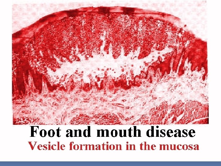
Viral diseases characterized by vesicle formation
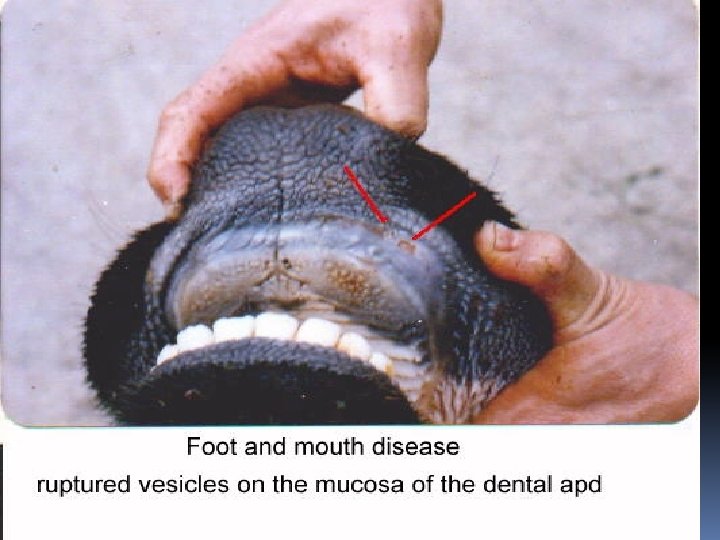
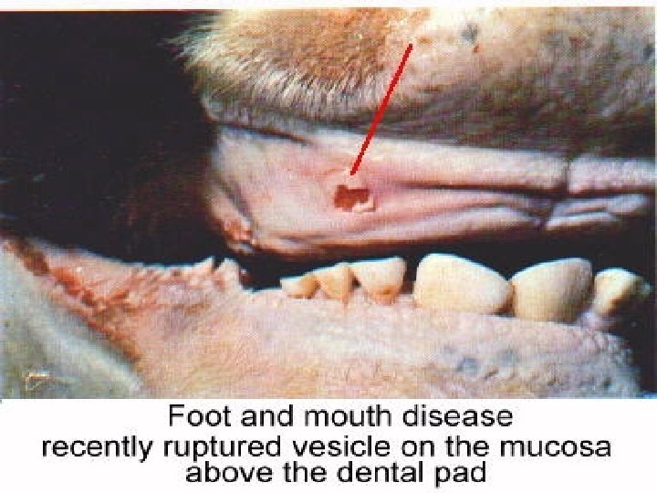
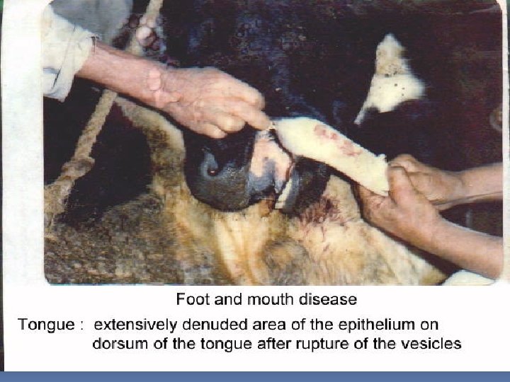
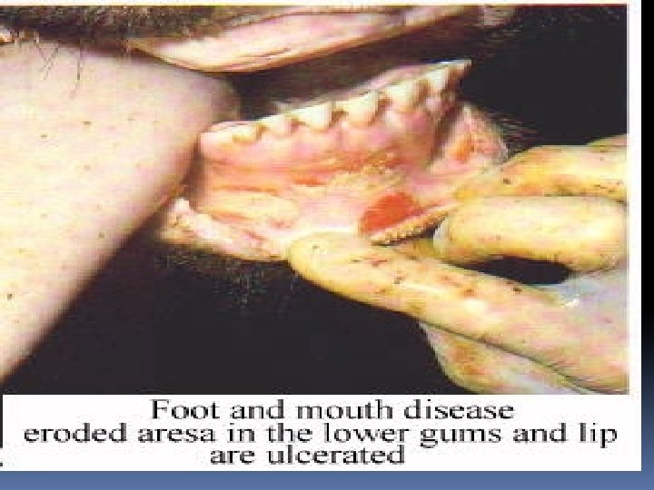
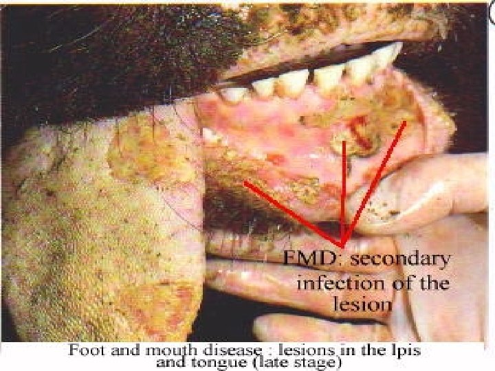
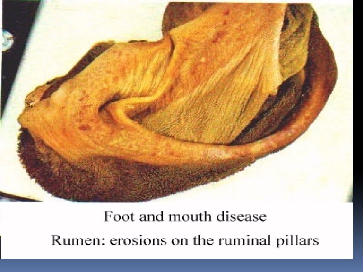
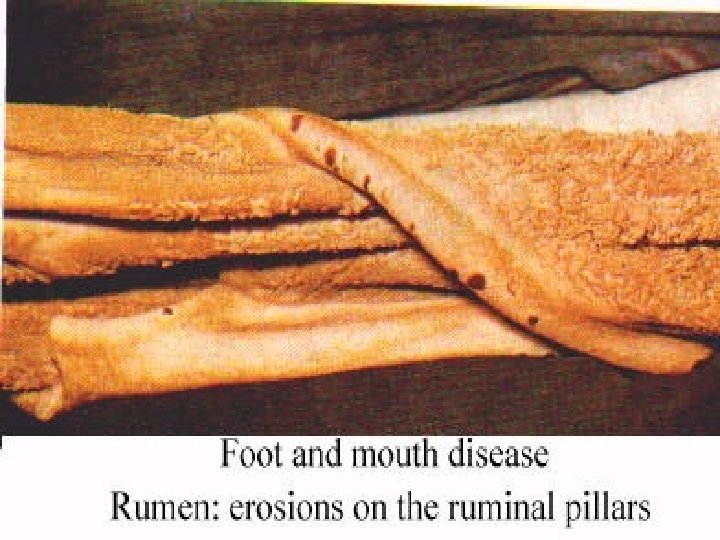
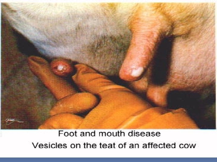
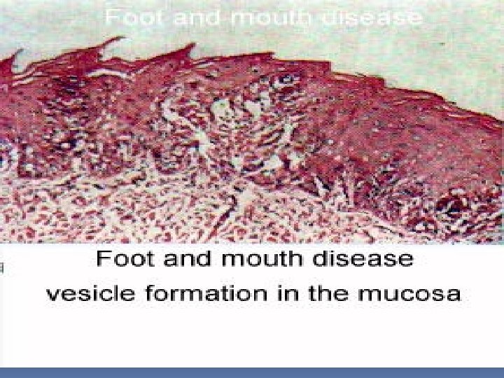
Foot and mouth disease F. M. D Aphthus fever Aphthus ruminitis DEF. * Viral disease of cloven footed animals; cattle, sheep, camel, and may pig. * Not affect horse. * Ch’ch’ : Vesicle formation on the buccal cavity, interdigital space, rumen, reticulum, omasum, and udder. Cause Picornaviridae(epitheliotropic)R. N. A R. O. I Ingestion – Inhalation Pathogenesis Virus ( via R. O. I )…………………. . Buccal cavity ( str. Spinosum ) ……………………Ballooning degeneration(hydropic)……………Vesicle( aphthus )or bullae ………………………. Erosion ……………. . Ulcer……………………. . Virus to circulation ………………………………. . Viremia ………………. G. I. T, Interdigital space & udder.
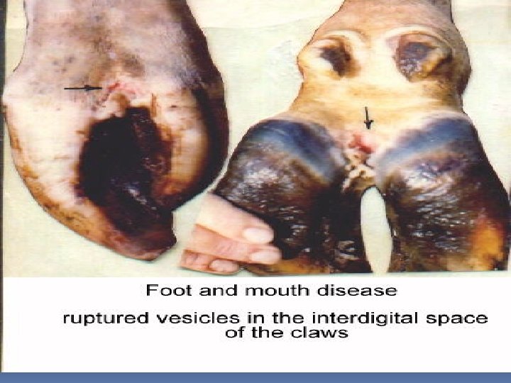
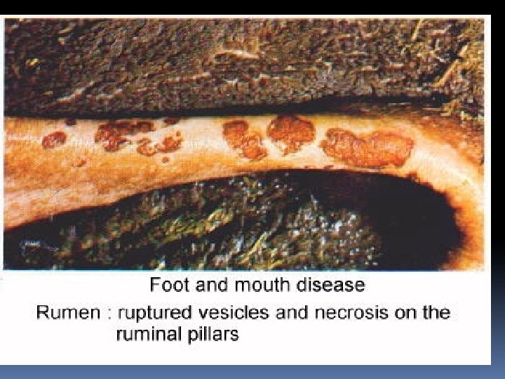
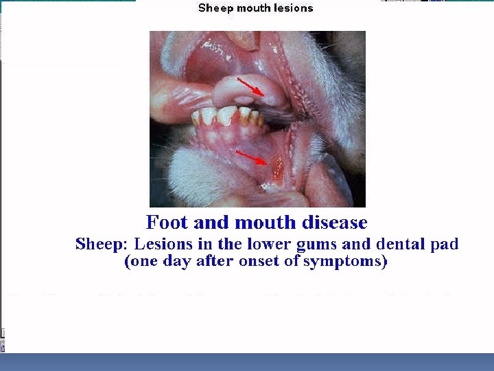
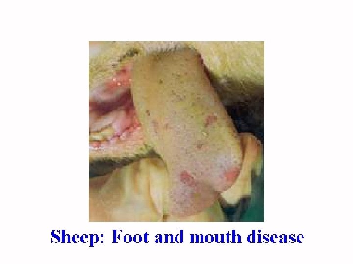
Lesions (i) MACRO * Buccal lesions : -Vesicle or bullae on anterior 2/3 of the dorsum of tongue. -Dental pad and buccal cavity are also affected. * Foot lesions : -Affect interdigital space ………………. . Lamness. -In complicated cases the claws may slaughed. * G. I. T lesions : -Vesicles, Necrosis & Erosions in oesophagus, rumen APHTHUS RUMINITIS. , reticulum, omasum, and intestine. -Peticheal H. on abomasums, intestine, and subendocardium. * Udder lesions : Vesicle on Teat extend to Mamary ts …………………. Mastitis. (ii) MICRO *Epidermis -Ballooning (hydropic Degenerations) -Vesicle(serous fluid, epithelial cells, inflame. cells(mostly lymphocytes & few neutrophil……………………. . Erosion …………………………Ulcer * Dermis Dermatitis i) Congestion ii) Inflammatory cells ( lymphocytes + neutrophils) iii) Perivascular cuffing
Lesions in bucal cavity
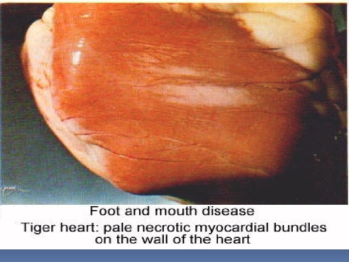
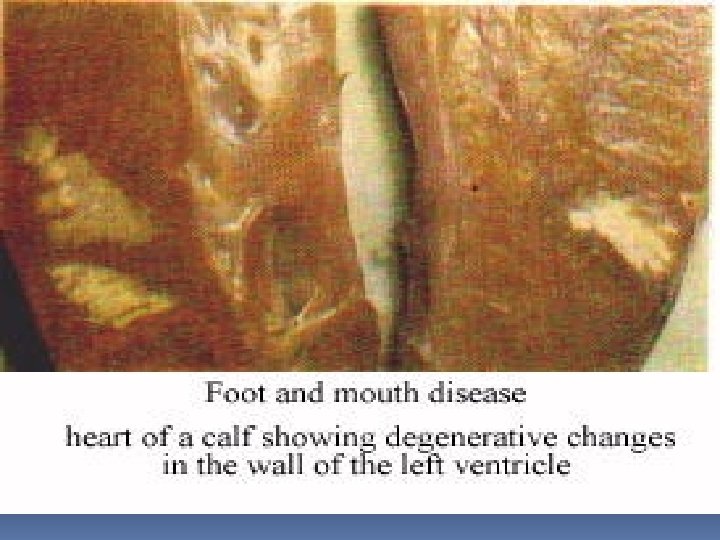
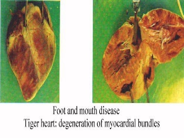
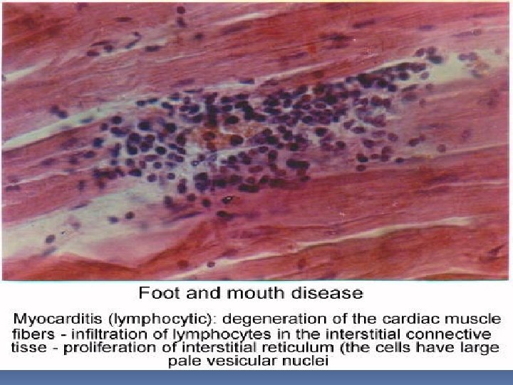
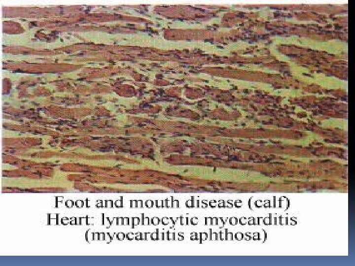
Malignant F. M. D DEF. : *Per acute F. M. D of young calves end with rapid death due to (ACUTE GENERAL VENOUS CONGESTION ). *Ch’ch’ non suppurative myocarditis. Cause : R. N. A virus. R. O. I : Ingestion & Inhalation. Pathogenesis : Virus via R. O. I……Viremia …. Heart muscles & Skeletal muscles. Lesions : (i) MACRO : Greyish necrotic foci on the wall of left ventricle take TIGER appearance SO known as TIGER HEART. (The same lesions are seen in skeletal Ms. ) ii)MICRO : Zenker’s necrosis + Inflammatory cells ( Lymphocytes + Macrophages ). (The same lesions are seen in skeletal Ms. ) ***F. M. D In SHEEP : Vesicle on teat, vulva, & dental pad////// Milder than in cattle//// F. M. D In Lambs & Pigllets Usually DIE.
Non-suppurative myocarditis
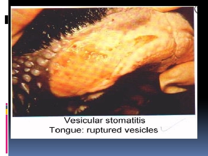
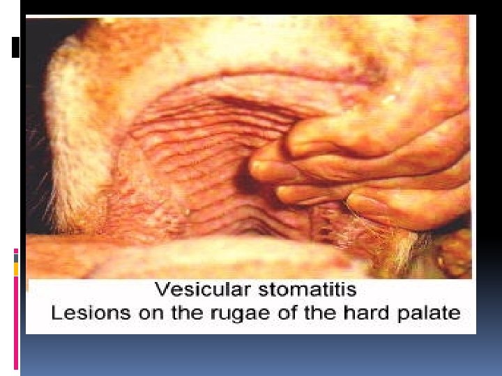
Vesicular stomatitis DEF. : * Viral disease affect mainly horses. * May affect cattle and pig. * Ch’ch’ vesicle on the buccal cavity, Interdigital space and udder * Seasonal disease as it occurs in summer coz of mosquito. * Enzootic disease as it occurs in U. S. A Cause: Rhabdoviridae genus (aphthous) e’ 7 Ag types(A, O, C, sat 1, sat 2, sat 3, As. IA 1) e’out cross protection between them R. O. I Mosquito bite wound contamination Vesicular exanthema Def. : * Viral disease affect mainly pigs. * Ch’ch’ vesicle on the buccal cavity, Interdigital space And udder Cause: Calciviridae R. O. I: Ingestion
Vesicular stomatitis Vesicular exanthema Pathogenesis Virus ( via R. O. I ) ………Buccal cavity ( str. Spinosum)………. . spongiosis ………………. Vesicle ( aphthus ) or bullae… Erosion…………Ulcer. Virus to circulation ……… Viremia …… ………………G. I. T, Interdigital space & udder. Lesions: As F. M. D ( Buccal, Foot, and Udder Lesions ONLY) N. B (HYDROPIC DEGENERATION REPLACED BY SPONGIOSIS IN HORSE VESICULAR STOMATITIS)
F. M. D Vesicular stomatitis Vesicular exanthema ANIMALS Cloven footed Horse, Cattle, and Pigs FATALITY Fatal in young Not fatal MYOCARDIUM Affected IN YOUNG Not affected IN EGYPT Present Not present
DIAGNOSIS OF FMD, Vesicular stomatitis&Vesicular exanthema: (1) Clinical Signs (Fever(aphthus fever), Off food, Salivation, Smaking of mouth, vesicles on its sites. (2) Post mortem lesions(Macro and Micro). (3) Isolation And Idenification Of Virus. (4) Diffrential diagnosis from other vesicular diseases. Control measures : (1)Complete eradication of infected animal. (2) Attenuated vaccine for only 1 virus to avoid cross immunity.
Pox. Viridae virus
GENERAL Characters : (i) Type (ii) Tropism : Epitheliotropic. (iii) R. O. I * Cow & Buffalo ……. mild affection. …. Affect teat & udder causing pock lesions. Microscopic lesions as those of sheep. * Camel …………Fatal in Young ………… Local in adult. Ch’Ch’ Pock lesions. : D. N. A virus with 6 genera; Orthopox, Capripox, Lapripox, Suipox, Avipox, and Parapox. : Insect bite, Wound contamination (contact ), Inhalation, Ingestion, and Coitus. (iv) Animal susceptiple : * Affect most animals except dogs. * Horse pox------ 3 forms; Leg, Buccal, and Genital. Ch’Ch’ Pock lesions. * Human … (i) Pseudo cow pox ( MILKER’S NODULES ) Transmitted to milkers from teat cow pox through milking. (ii) Small pox ( VARIOLA ) Fatal as it has systemic reaction + skin affections. Human vaccinized against small pox.
(v) Lesions : Cutanous & Systemic lesions. (vi) Inclusions : I/C inclusion bodies ( GUARNIERI BODIES ) (viii) Pathogenesis of sheep pox lesions In order : (i) ROSEOLA St. ……. . Red line of dilated vessel. (ii) MACULA St. ……. Edema & Inflammatoty exudates. (iii) PAPULE St. ………Proliferations of cells. (iv) VESICULAR St. ……Vesicle formation. (v) Pastule St. (vi) UMBLICATED St. ( POCK LESION )……………. . vesicles ruptured giving ulcer with elevated borders. (vii) ENCRUSTATION St……. . Crust with scab formation. (viii) HEALING St. …………. . Healing of deep pitted ulcer.
SHEEP POX DEF. * Viral disease of SHEEP and goat * Ch’ch’ cutaneous eruption (pock lesions) in areas devoid of wool (lips, nostril, cheek, inner thigh&scrotum). *Also affect pharynx, trachea, lung, Rumen & kidney which increase mortality. ORF SCABY MOUTH CONTAGIOUS ECTHYMA CONTAGIOUS LABIAL DERMATITIS CONTAGIOUS PASTULAR DERMATITIS ULCERATIVE VULVITIS ULCERATIVE DERMATOSIS INFECTIOUS BALANOPOSTHITIS LIP & LEG ULCERATION * Viral disease of SHEEP, GOAT • Viral diseaseof SHEEP & & human. GOAT. * Ch’ch’ Papules, Vesicle formation &Pastules on (oralcavity, oral commisure & lips) covered with thick scab. *Ch’ch’ ulcer formation on ( vulva, glanspenis, lip& leg) Cause Pox virus Para pox Pox virus R. O. I DIRECT CONTACT INGESTION DIRECT CONTACT COITUS Pathogenesis Roseola, Macula, Papule, Vesicle, Pock lesion, Crust& Healing. _____________
SHEEP POX Lesions (i) MACRO (ii) MICRO * (Pock lesions) in areas devoid of wool (lips, nostril, cheek, inner, Thigh &scrotum) *Also affect pharynx, trachea, lung, rumen&kidney which increase mortality. Epidermis : * Hydropic deg. &spongiosis in prickle cell layer. * Neutrophils exocytosis in prickle cell blayer. * I/C EOSINOPHILIC inclusion body ( GUARNIERI BODIES ) Dermis : * Dermatitis i)Congestion. ii)Lymphocytes. iii)Perivascular cuffing. * Pathognomonic lesions SHEEP POX CELL * It’s virus infected Macrophages appeared e’ -margination of chromatin, -prominent nucleoli - I/C inclusion * Formed in deep dermis. ORF SCABY MOUTH CONTAGIOUS ECTHYMA CONTAGIOUS LABIAL DERMATITIS CONTAGIOUS PASTULAR DERMATITIS • Ch’ch’ Papules, Vesicl formation &Pastules on (oral cavity, oral commisure & lips) covered with thick scab. • Formation of papillomatosis growth on lips covered with brown crust. Epidermis : * I/C BASOPHILIC inclusion body ( GUARNIERI BODIES ) Dermis : * Dermatitis i)Congestion. ii)Lymphocytes. iii)Perivascular cuffing. *Pathognomonic lesions PSEUDO CARCINOMATOUS HYPERPLASIA Downward proliferation of epidermal cells into dermis ULCERATIVE VULVITIS ULCERATIVE DERMATOSIS INFECTIOUS BALANOPOSTHITIS LIP & LEG ULCERATION Ulcer formation on vulva, glanspenis, lip& leg covered with brown scab. Epidermis : * Destruction of epidermal layer into dermis( ULCERATION ). * I/C inclusion body ( GUARNIERI BODIES ) Dermis : Dermatitis i)Congestion. ii)Lymphocytes. iii)Perivascular cuffing.
Sheep pox - under tail (elevated papule) quit previous
Sheep pox (scab formation) quit previous
Cow pox (udder) quit previous
Cow pox (udder) quit previous
Avian pox (intracytoplasmic inclusions) High power quit previous
Avian pox (intracytoplasmic inclusions) quit previous
Orf quit previous
Contagious echthyma (Orf) Index
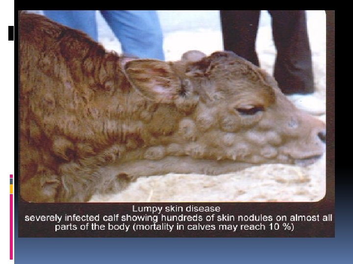
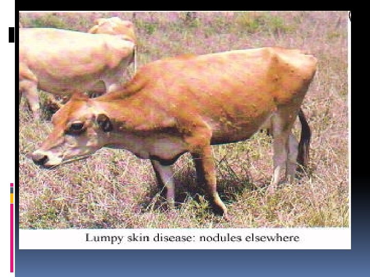
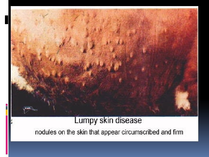
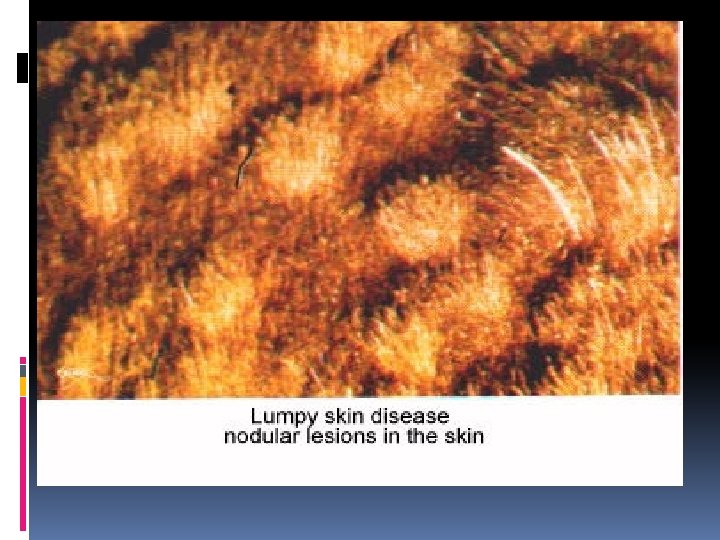
LUMPY SKIN DISEASE L. S. D DEF. * Viral disease of CATTLE & BUFFALOS. * Ch’ch’ nodule formation on Nose, eyelid &conjunctiva. scrotum&glanspenis in males. udder&vulva in females. * Also Ch’Ch’ LYMPHADENOPATHY. Cause Capri pox R. O. I INSECT BITE Pathogenesis Lesions (i) MACRO (ii) MICRO Bovine Papular stomatitis * Viral disease of cattle. * Ch’ch’ : Papules on; Buccal mucosa, Muzzle, Gum, Dental pad, and Lips. Para pox virus Vasculitis ………. Thrombosis ………Ischemia ………. . Necrosis *Erythema------ Papule------- expand slowly and similar to COIN ( necrosed center ). * Intra dermal LUMPS ( Hard indurated nodules ) *SIT FAST: Necrosed nodules with sequestration ………. Deep ulcer. * Erythema begins. • Papule------- On Buccal mucosa, Muzzle, Gum, Dental pad, and Lips Epidermis : * Vacuolar degeneration and Necrosis. * I/C EOSINOPHILIC inclusion ( GUARNIERI BODIES ) Dermis : a) Epidermis : i) Hyperplasia in epidermal cells. ii) Vacuolar degeneration iii) I/C inclusions. I ) Lymphocytic infiltration. Ii) Vascular congestion. Iii) Perivascular cuffing b) Dermis : Dermatitis----I ) Lymphocytic infiltration. Ii) Vascular congestion. Iii) Perivascular cuffing. * Dermatitis * Pathognomonic lesions - Vasculitis - Thrombosis -I/C inclusion inside macrophage
Lumpy skin disease quit previous
Lumpy skin disease quit previous
DIAGNOSIS OF pox viridae: Clinical Signs. Post mortem lesions(Macro and Micro). Isolation And Idenification Of Virus. Diffrential diagnosis from other epitheliotropic diseases.
VIRAL DISEASE OF CATTLE
Affect Respiratory System
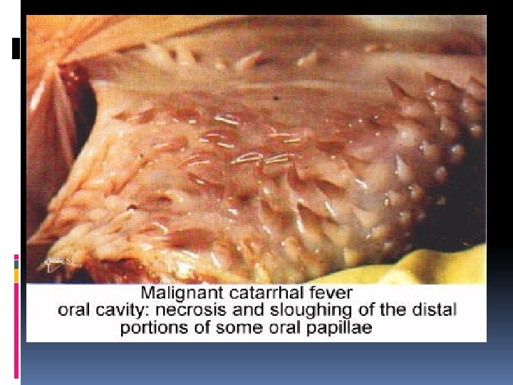
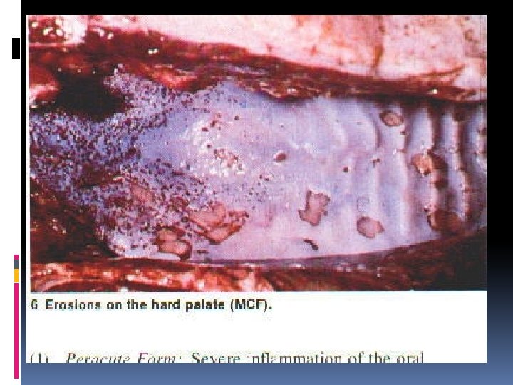
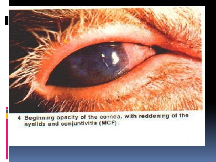
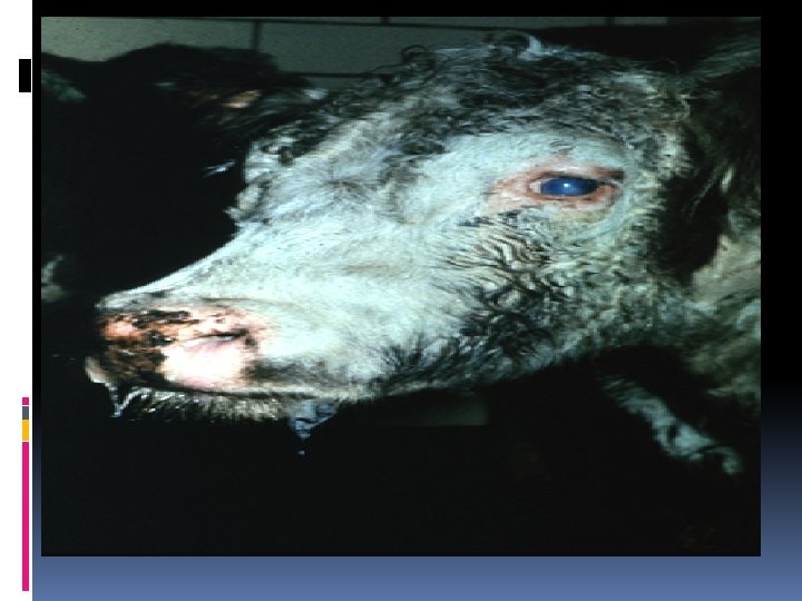
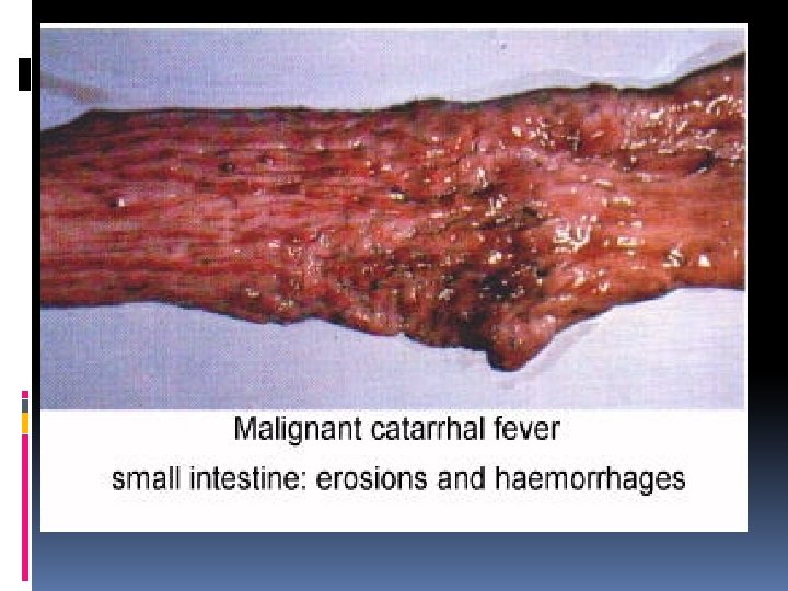
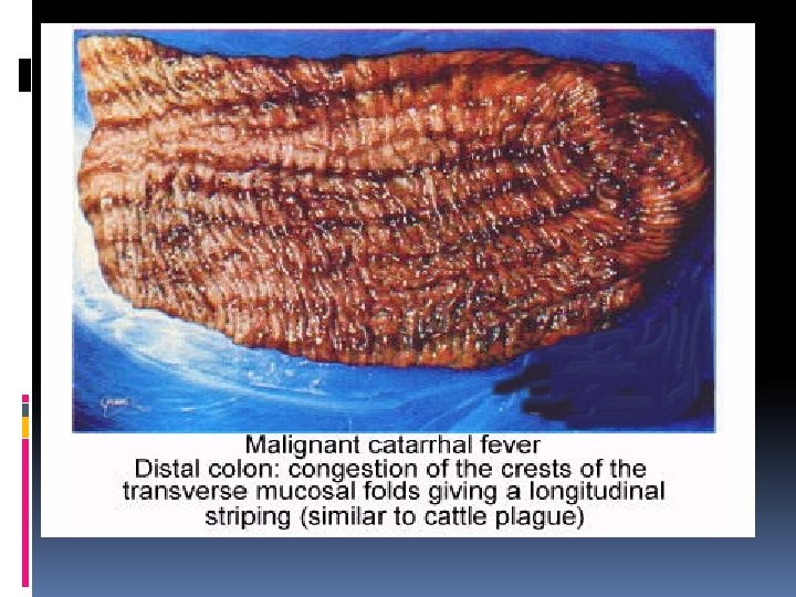
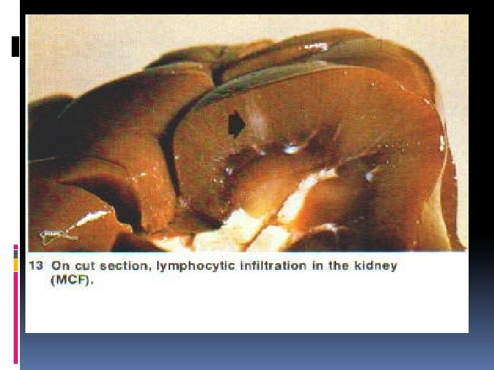
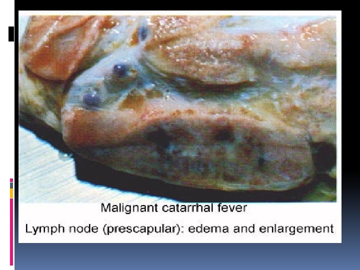
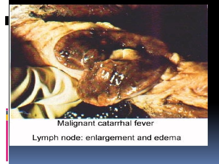
1 - Bovine Malignant OF Cattarhal Fever VIRAL DISEASE CATTLE B. M. C. V Malignant Head Cattarhal Fever M. H. C. F (I/N) Viral disease of cattle &buffaloes ch’ch’: (i)Erosion in upper respiratory tract (ii) Keratoconjunctivitis. DEF. (iii) Encephalitis. (iv) Erosive stomatitis & gastroenteritis. (v) Lymphadenitis. (vi) Cutaneous exanthema. D. N. A Herpes virus Cause (African(wildbeets)&sheep strain) R. O. I Ihalation + Ingestion
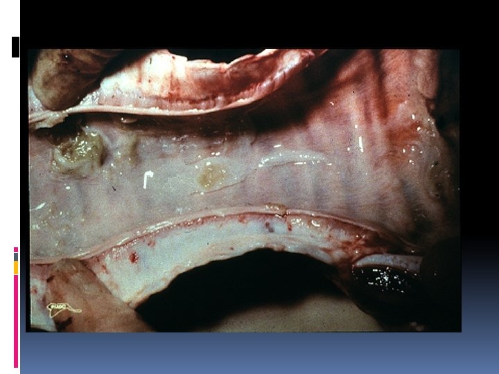
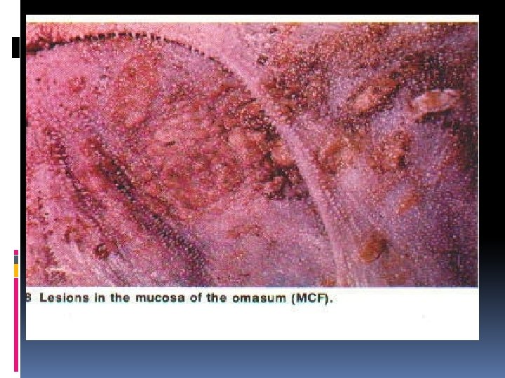
1 - Bovine Malignant Cattarhal Fever B. M. C. V Malignant Head Cattarhal Fever M. H. C. F (I/N) (i) Diphteretic membranes in nasal mucosa, pharynx, trachea&bronchi. Lesio (ii) Keratoconjunctivitis, corneal opacity& edema of eyelid. ns (iii) Encephalitis, Gliosis, myelitis& perivascular cuffing. (iv) Erosion &ulcers along oral mucosa, omasum &intestine. (v) Liver enlarged & mottled. (i) (vi) Kidney shows Interstitial nephritis. MAC (VII) L. N ENLARGED, EDEMATOUS & HEMORRHAGIC (PATHOGNOMONIC) RO (viii) In most cases, s/c blood vessels are tortuous in thorax and abdomen As well as fibrinous poly arthritis. (ix) Cutanous vesicular & popular exanthema. (i) Necrosis of epithelium along respiratory and gastrointestinal tract. (ii) Congested blood vessels. MICR (iii) Inflammatory cells mostly lymphocytes. O (iv) Vasculitis with fibrinoid necrosis of most bl. vs particularly brain ( PATHOGNOMONIC in M. H. C. F only not present in I. B. R) (v) (I/N) inclusion body Called ( COWDRY TYPE A ) stained RED with PHLOXIN TARTRAZIN stain.
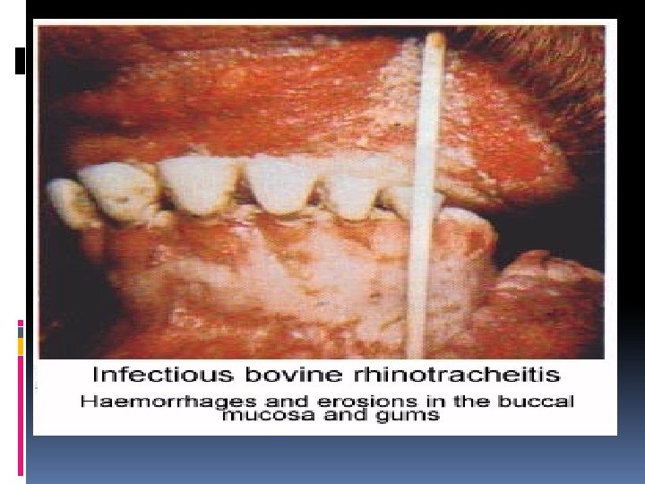
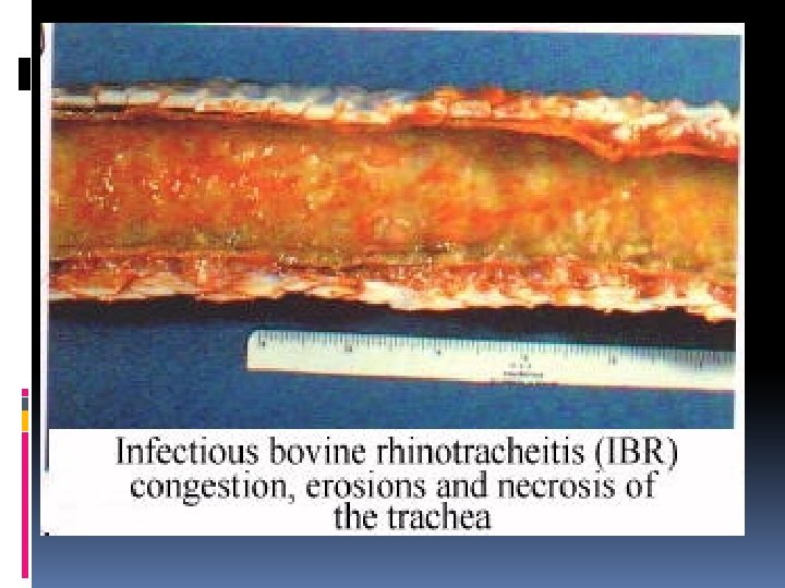
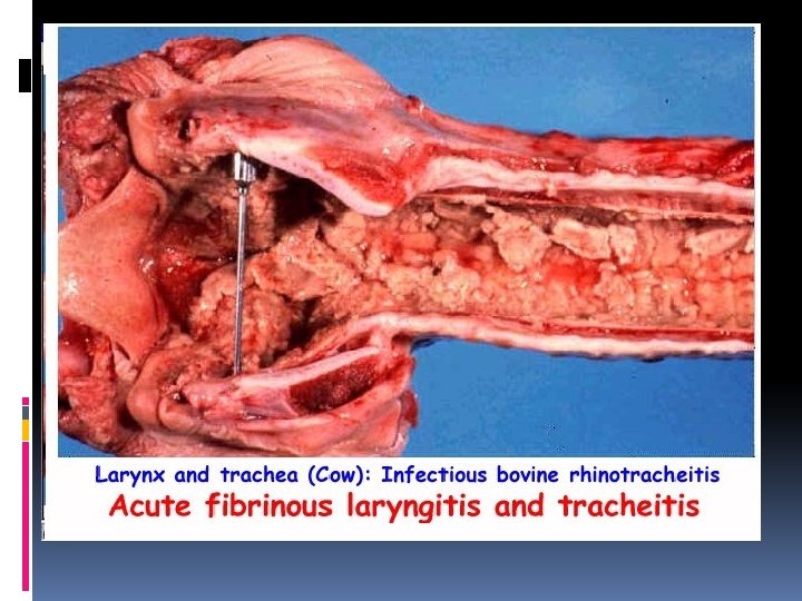
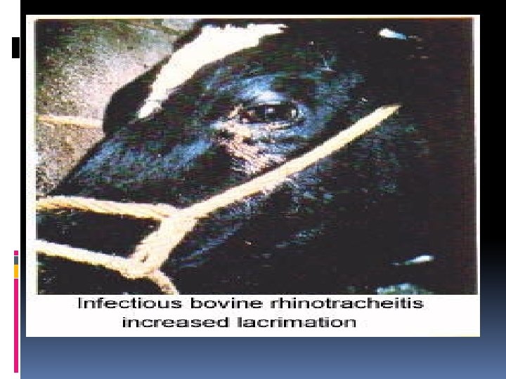
2 - Infectious Bovine Rhinotrachiitis I. B. R Red Nose Disease I. P. B & I. P. V & C. V. V (I/N) Viral disease of cattle & buffaloes ch’ch’: (i) Rhinotrachiitis. (ii) Keratoconjunctivitis. (iii) Encephalitis. DEF. (iv) Balano Posthitis (I. P. B) (Infectios Pastular Balanoposthitis) (v) Vulvo vaginitis (I. P. V) (Infectious Pastular Vulvovaginitis) (vi) Abortion if at late pregnancy. D. N. A Herpes v. Cause (3 subtypes; resp. , genital, encephalic) Inhalation + Sexual intercourse (C. V. V) R. O. I (Coital Vesicular Vulvovaginitis)
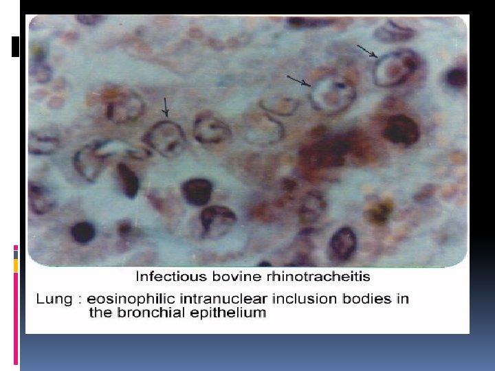
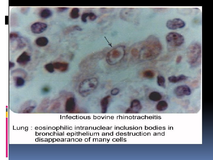
2 - Infectious Bovine Rhinotrachiitis Red Nose Disease I. P. B & I. P. V & C. V. V (I/N) I. B. R 3 FORMS OR 3 Subtypes (i) Respiratory form(subtype): (a)Erosive rhinitis(RED NOSE), Trachiitis& Bronchitis with diphtheritic membranes. Lesions (b)Keratoconjunctivitis & corneal Opacity DUE TO transmission of Virus via trigeminal nerve. (e) Encephalitis, Gliosis & perivascular cuffing. IBR in young calves is fatal & Ch’Ch’ resp. distress, diarrhea&end by convulsion&death. (i) MACRO (ii) Genital form or subtype : (a) Vulvo Vaginitis : Inflam. , vesicles&pustules of vulva & vagina. (b) Balano Posthitis : Inflam. , vesicles&pustules of penis & prepuse. (c) Mastitis. (iii) Abortion form : Infection in late pregnancy ……………. mamification & still birth; Intra uterine fetal death 72 hrs before abortion & aborted fetous showed ………………… Necrotic hepatitis & I/N inclusion body Surrounded by hallo zone (i) Necrosis of epithelium along respiratory and gastrointestinal tract. (ii) Congested blood vessels. MICRO (iii) Inflammatory cells mostly lymphocytes (v) (I/N) inclusion body Called ( COWDRY TYPE A ) stained RED with PHLOXIN TARTRAZIN stain.
3 - Bovine respiratory syncytial virus (I/C) 4 - Parainfluenza III virus (I/C) DEF. Viral disease of cattle ch’ch’: Interstitial pneumonia accompany by syncytial cells. Cause R. N. A viruses ( Pneumo virus-paramyxoviridae ) R. O. I Inhalation
3 - Bovine respiratory syncytial virus (I/C) 4 - Parainfluenza III virus (I/C) PATHO Virus stimulate bronchi and alveolar epithelium result in Bronchointerstitial GENESI pneumonia with syncytial formation S: (ONLY IN BOVINE RESPIRATORY SYNCYTIAL VIRUS) Lesions Lung is atlectatic and consolidated with interstitial pneumonia. (IN BOTH VIRAL INFECTION) (i) interstitial and subpleural emphysema. MACRO (ONLY IN BOVINE RESPIRATORY SYNCYTIAL VIRUS) (ii) MICRO * Bronchiolitis with cells proliferation esult in syncytial cells contained(I/C) inclusion. • Hyperplasia of pneumocyte type II (Lung epithelization). * Accumulation of lymphocyte and plasma cells • Bronchioles&alveoli filled with exudates accomp. by neutrophils lymphocytes¯ophages *Bronchial epithelium is hyperplastic , vacuolated&contain I/C inclusion. * Hyperplasia of pneumocyte type II with squamous metaplasia and rare syncytial cells.
Affect Digestive System
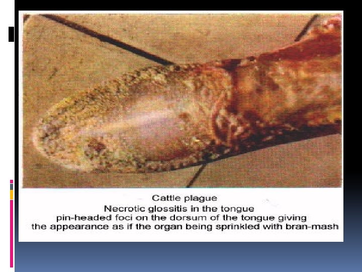
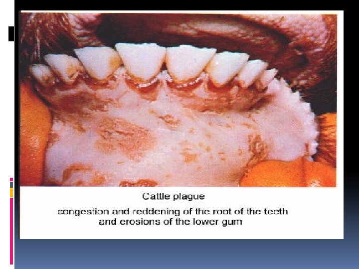
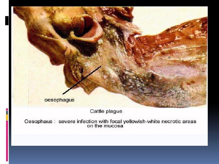
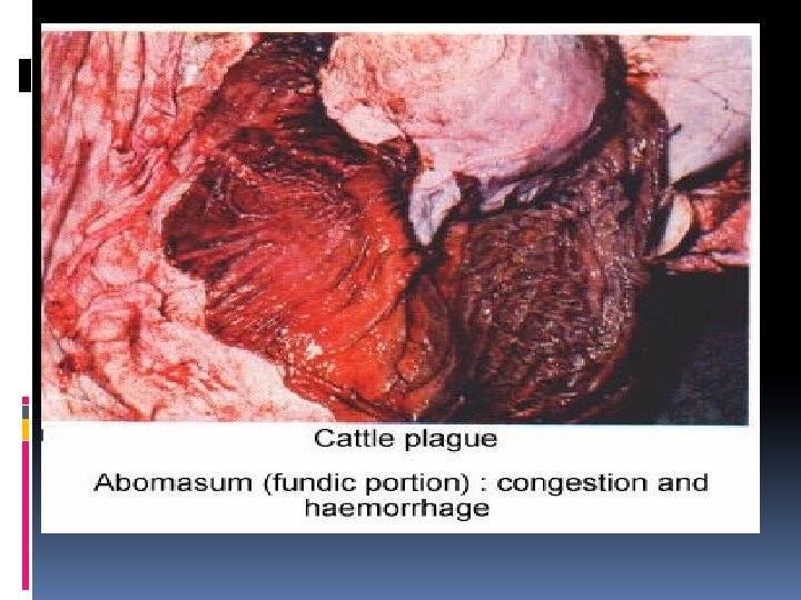
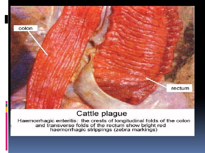
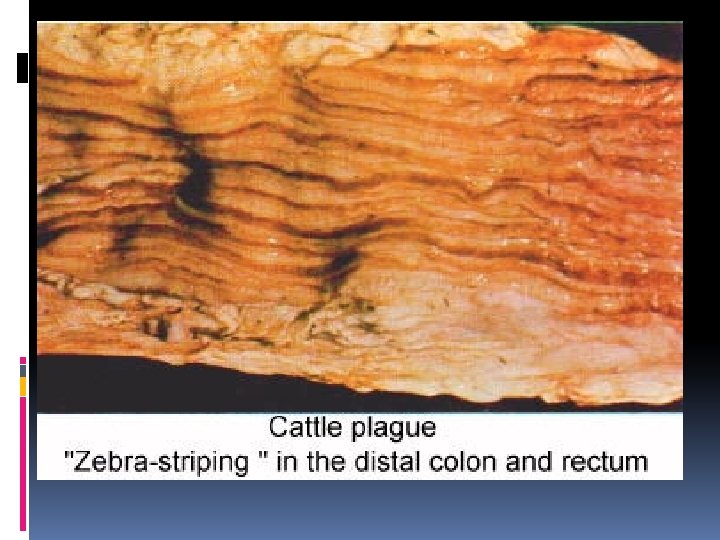
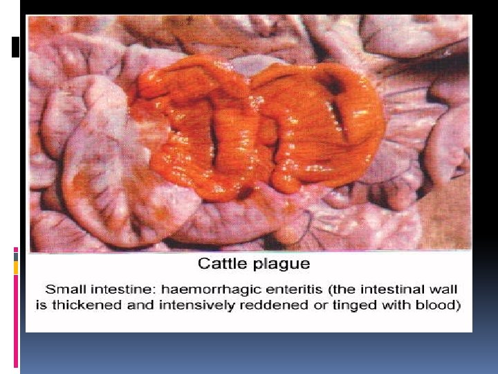
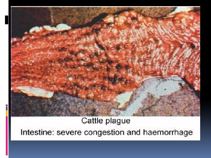
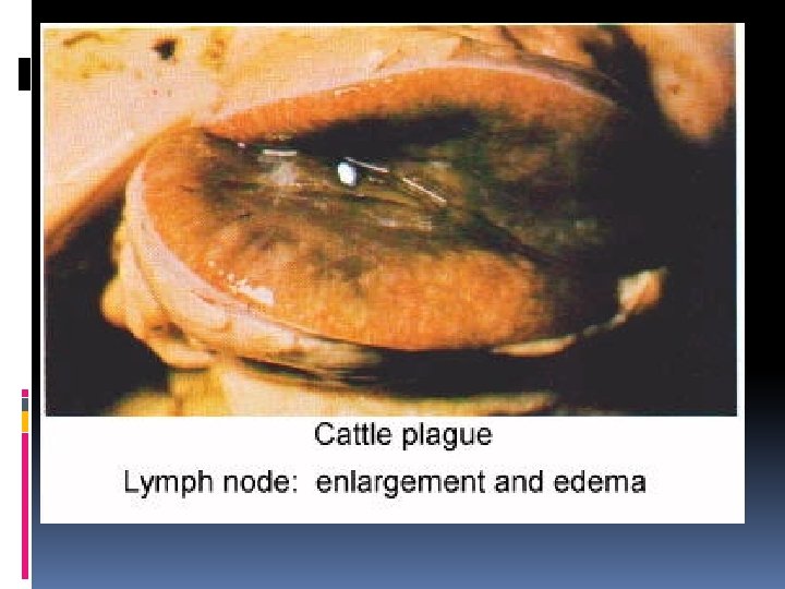
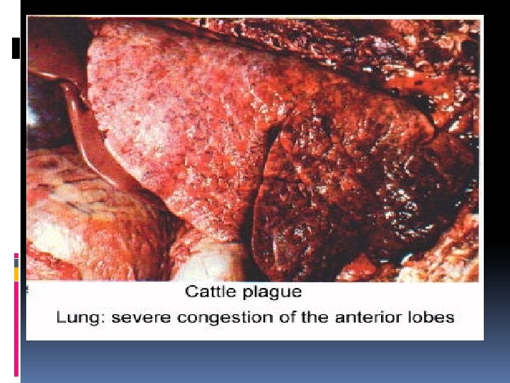
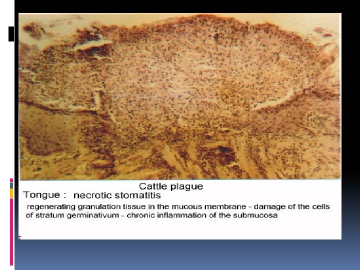
5 - Rinder Pest R. P Cattle plague. (C. P) (I/N+I/C) DEF. Cause R. O. I Viral disease of cattle and buffaloes ch’ch’: (i) Necrosis & ulceration of G. I. T. (ii) Degeneration of lymphoid tissues ( L. N + Spleen ). Paramyxoviridae Ingestion OR Inhalation Pathog Ingestion …………………. Regional L. N and tonsils where it multiplies enesis : Viremia & secondary multiplication in G. I. T, upper respiratory, and lymphoid tissues.
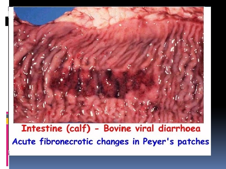
5 - Rinder Pest R. P Cattle plague. (C. P) (I/N+I/C) (i) Bran like Necrosis & ulceration in Buccal cavity at ventral aspect of tongue, tongue tip, dental pad, gum, oral comissure & inner aspect of lips. Lesions (ii) Necrosis & ulceration in pharynx & esophagus. (iii) Congestion, edema & necrosis in abomasal folds. ( Pathognomonic ) (iv) Congestion at ilio cecal & Caecocolic junction( Zebra marking ) ( Pathognomonic) (i) (v) Necrosis of mucosa of ilium & large intestine w’ get invaginated in necrotic lymphoid ts. (Infundibulum OR Herniation) ( PATHOGNOMONIC ) MACRO (vi) Necrosis & ulceration in payer’s patches. (vii) Lymphoid tissues : L. N …………………Enlarged & hemorrhagic Spleen ……………. Enlarged Then Atrophied (viii) Hemorrhage in larynx, trachea, bladder &subendocardium. (i) Syncytia formation( fusion of necrotic epithelium) + Multinucleated cells w’ (ii) contain I/N + I/C inclusions(PATHOGNOMONIC) (ii) Lymphoid tissues showing necrosis of lymphoid follicles w’ appeared as craters. MICRO (iii) Lamina propria of small and large intestine show----congestion, edema& lymphocytic infiltrations.
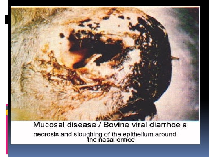
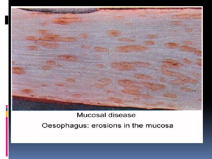
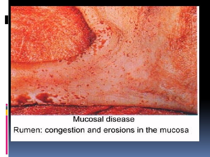
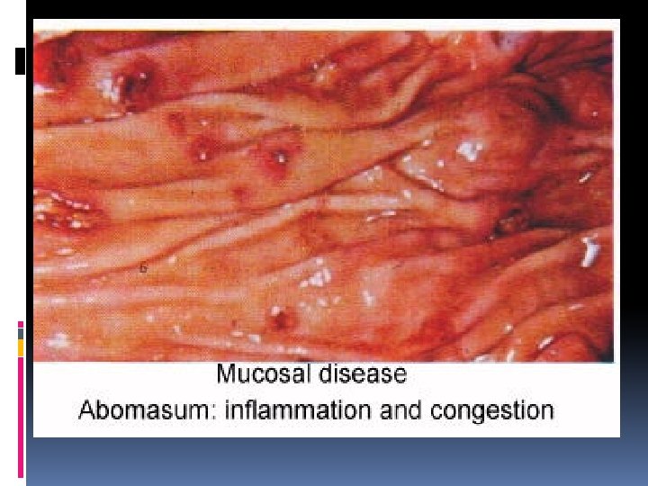
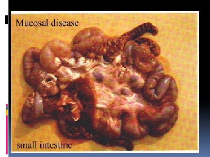
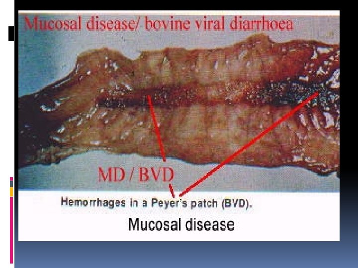
6 - Bovine Viral Diarrhea B. V. D Mucosal Disease DEF. Cause R. O. I Viral disease of cattle and buffaloes ch’ch’: (i) G. I. T lesions & diarrhea. . (ii) Leucopenia (immunosupression). (iii) Still birth fetous with C. N. S lesions. Flaviviridae Ingestion OR Inhalation Pathog There are 2 strains of BVD; enesis : Cytopathic strain ( CP-BVD ), Non cytopathic strain ( NCP-BVD ) Cattle may infected with each of cp-BVD & Ncp-BVD and give the infection to fetous At 1 st 120 d----- Abortion, mummification, Resorption, teratogenic effect > 120 d------ Abortion OR Healthy calve. Some cattle may infected with NCP-BVD and didn’t show signs and pass virus to fetous during 1 ST 120 ds………………Persistant infected calf ( PI calf ) , immune tolerant calf, or chronic shedders. If PI calf infected with CP-BVD-----Show mucosal disease and usually end fatally.
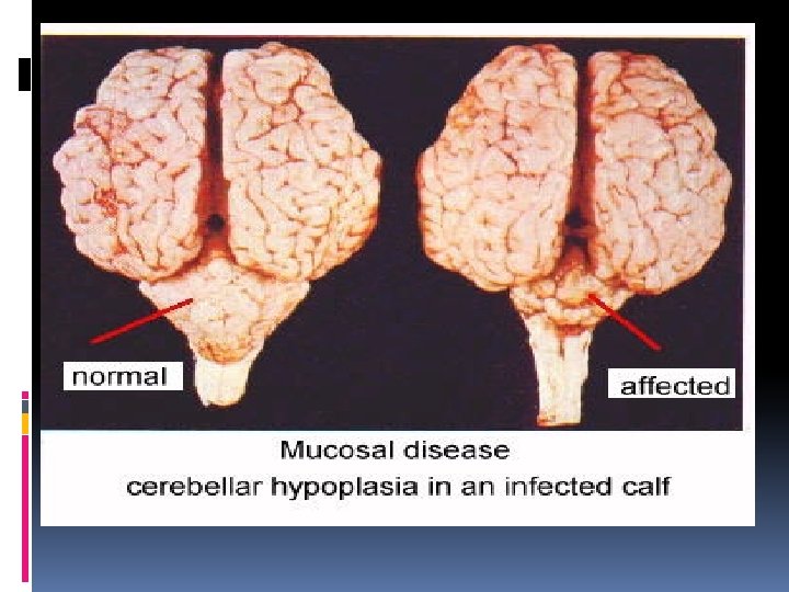
6 - Bovine Viral Diarrhea B. V. D Mucosal Disease i) Erosion in muzzle, nose, larynx, and pharynx. (ii) Linear erosion along oesophagus. (iii) Erosion along rumen, omasum, and abomasums. (i) (iv) Congestion & edema along abomasums &small intestine. MACRO (v) Necrosis in pyres patches. (vi) Zebra marking in intestine. (vii) Destructed lymphoid tissues. (viii) Erosions in skin & interdigital space( lameness). (ix) Foeti suffered from mummification, abortion with or without teratogenic characters as : *cerebral or cerebellar hypoplasia*porencephaly- hydranencephaly*microphthalmia- cataract*optic neuritis *thymus hypoplasia. • Necrosis of epithelium along G. I. T with; congested bl. vs. , and lymphocytic (ii) infiltrations. * Submucosal bl. vs. of G. I. T show vasculitis. MICRO * Lymphoid tissues ( L. N, spleen, thymus, and pyer patches ) showing necrosis. * Aborted fetous : *Brain of foeti show demylination. *Depletion of the lymphoid tissues (L. N &Spleen)
BVD RP OESOPHAGUS Linear erosion Necrotic foci SKIN LESIONS Interdigital spasce Absent FOETAL LESIONS CHRONIC FORM Present(Cerebral) Absent Present Absent VASCULITIS Present Absent INCLUSIONS Absent Present
DEF. 7 - Corona and Rota viruses Viral disease of cattle& most domestic animals ch’ch’: DIARRHEA Cause Coronaviridae & Rotaviridae R. O. I Ingestion Pathogenesis : Ingestion …………. . Intestine where viruses replicate in enterocytes …………………resulting in damage and exofoliation of cells………………………followed by villous atrophy. Lesions * Villous atrophy and villi appeared club shaped, blunt, and fused to each others ( mucosa become flat or cuboidal ) MICRO *Hyperplasia & Hypertrophy Of Crypts. * Lamina propria is heavy infiltrated by Macrophages & lymphocytes. IN CORONA VIRUS ONLY NOT IN ROTA: * Colon mucosa become flat or cuboidal. * Mesentric l. N & payer patches destructed.
Affect Lymphoid Tissues
DEF. Cause R. O. I 8 - Bovine ephemeral fever 9 - Bovine Leukosis Three day sickness Viral disease of cattle and Viral disease of cattle Ch’Ch’ : enlargement of buffaloes Ch’Ch’ : lymph nodes. Fever for three days, lamness, and enlargement of lymph nodes. R. N. A viruses ( Rhabdoviridae ) Mosquitoes Virus by mosquitoes Viremia …………. . Localized in; Muscles, Joints, Spleen, and Pathog Lymph nodes enesis R. N. A viruses ( Retroviridae ) Ingestion OR Congenital *The animal reacted with virus in four different ways; 1. It doesn’t infected due to genetic characters. 2. It is permenant infected with antibodies ( Latent infected ). 3 - It is permenant infected with Benign lymphosarcoma. 4 - It is permenant infected with malignant lymphosarcoma *Also there are SPORADIC and ENZOOTIC leucosis. 1 - THE ENZOOTIC LEUKOSIS: * Usually affect cattle from 4 -8 years. * Ch’Ch’: Fatal lymphosarcoma. 2 - THE SPORADIC LEUKOSIS : * May be JUVENILE cal f-------- < 5 m. * Thymic -------------------- < 2 y. 1. OR * Cutaneous ------------------ 1 -3 y.
8 - Bovine ephemeral fever Three day sickness Lesions (i) MACRO (ii) MICRO 9 - Bovine Leukosis ENZOOTIC LEUCOSIS : i) Edema & hemorrhages all over i) Enlargement of superficial and body. mesenteric lymph nodes. ii) Enlargement of spleen and ii) Abomasum contain nodules and lymph nodes ulcers. iii) Interstitial lung emphysema. iii) Heart, spleen, and kidney are iv) Polyserositis in joints, tendon& greatly enlarged. peritoneum SPORADIC LEUCOSIS : CALF LYMPHOMA-------Enlargement of lymph nodes. THYMUS LYMPHOMA----Enlargement of thymus. CUTANOUS LYMPHOMA----Cutanous plaques. Inflammation of ; Joint, tendon, and muscles Ch’Ch’ : Uniform lymphocytic infiltration in i) Congested blood vessels. affected organs ii) Inflammatory cells mostly lymphocyte
Leukosis
VIRAL DISEASE OF SHEEP
1 -JAAGSIEKTE (driving sickness) or LUNG OR PULMONARY ADENOMATOSIS DEF. Pathogen esis Viral disease of sheep ch’ch’ lung nodules. Known as Driving disease because affected sheep show clinical signs during transportation. Virus stimulate bronchial and alveolar epithelium result in marked hyperplasia of clara cells & 2 -MAEDI (shortness of respiration) or OVINE PROGRESSIVE INTERSTITIAL PNEUMONIA (I/C) Viral disease of sheep ch’ch’ : i- Progressive ( Diffuse ) lymphofollicular interstitial pneumonia ( NEVER HEAL ) ii- Increase lung volume 2 -3 times its normal volume Virus stimulate peribronchial lymphoid tissue and sometimes alveolar epithelium pnemocyte type. II…………… Adenomatous growth of bronchial & alveolar epithelium. Cause Retroviridae R. N. A Viruses (( Maedi visna(meningioencephalitis) )) R. O. I Inhalation ( with long incubation period 2 -3 y ) Lesions (i) MACRO (ii) MICRO (a) Areas of consolidation, areas of collapse & areas of compensatory emphysema. (a) Lung increase in size and weight with multiple nodules all over the lung tissue. The bronchioles and alveoli lined by columnar and cuboidal epithelium in the form of finger like projection known as: Adenomatus growth in the alveolar epithelium. (a) Lung enlarged 2 -3 times its volume with ribs imprints. (B) Bronchial & Mediastinal L. N enlarged and edematous (a) (b) (c) (d) (e) (f) Bluish I/C inclusion bodies. Lymphofollicular sheath around; Bronchi, Bronchioles, and Blood vessels. Hyperplasia & Hypertrophy in the alveolar wall (pneumocyte type II) Hyperplasia of smooth muscle of terminal bronchioles. Mononuclear cells infiltrations. Meningioencephalitis and arthritis.
JAAGSIEKTE MAEDI LESIONS LOCAL DIFFUSE Adenomat ous growth COURSE OF DISEASE LYMPH NODE PRESENT ABSENT SHORT LONG NOT AFFECTED ABSENT PRESENT ( I/C ) INCLUSIO N BODY
3 - P. P. R. ( Pestes de Petit Ruminant ) The cattle plague of ( SHEEP & GOAT ). Def. : Viral disease of sheep and goat similar to cattle plague. Cause: RNA virus antigenic related to cattle plague, canine distemper, and human measles. R. O. I : Ingestion OR inhalation. Postmortem and microscopic findings ---------------Similar to cattle.
4 - Border disease ( Hairy Shakers ds. ) OR Fuzzy lambs: Def. : Viral disease of sheep and lambs Ch’ch’ abortion, still birth, and small weak lambs with tremor, abnormal body conformation and hairy fleeces (Hairy Shakers OR Fuzzy lambs). Cause: R. N. A ( Flaviviridae ) called BORDER because it occurred between Wales & England. R. O. I: Ingestion Pathogenesis: Ingestion----Viremia-------(i) In NON pregnant sheep…. Causes a mild disease. (ii) In pregnant sheep…………. . Cross placenta in pregnant ewes * During 1 st 85 ds of pregnancy: Foetus is aborted OR still born has teratogenic character (demylination, hyperpigmentation) OR immunotolerant lambs with hair follicles affection (fuzzy appearance). * During 1 st 85 ds of pregnancy: The immune system capable of destroying the virus SO foetal death is rare and all lambs are born normal with antibodies. Post mortem lesions in aborted foetus: (i) lambs may aborted or mummified or immune tolerant. (ii) CNS malformation. (iii) Abnormal skin pigmentation. (iv) Fuzzy appearance of skin due to thickness of follicles & wool fibers. Micro: (i) Thymus of lambs are necrosed. (ii) Brain of lambs show demylination, Porencephaly, hydranencephaly.
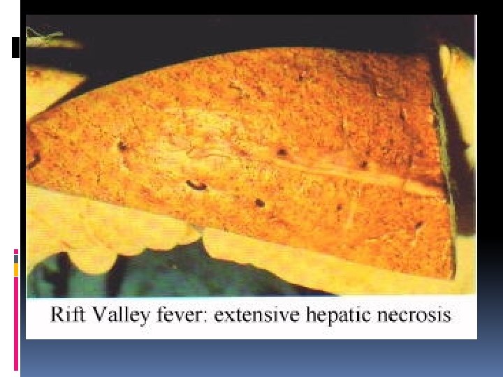
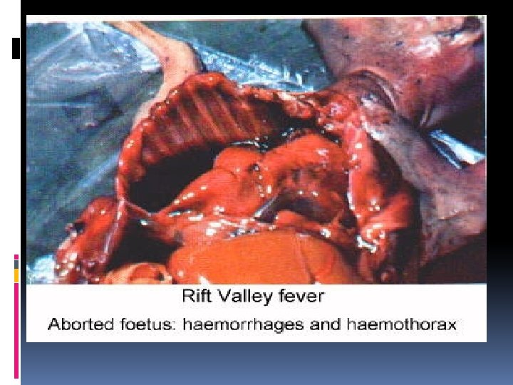
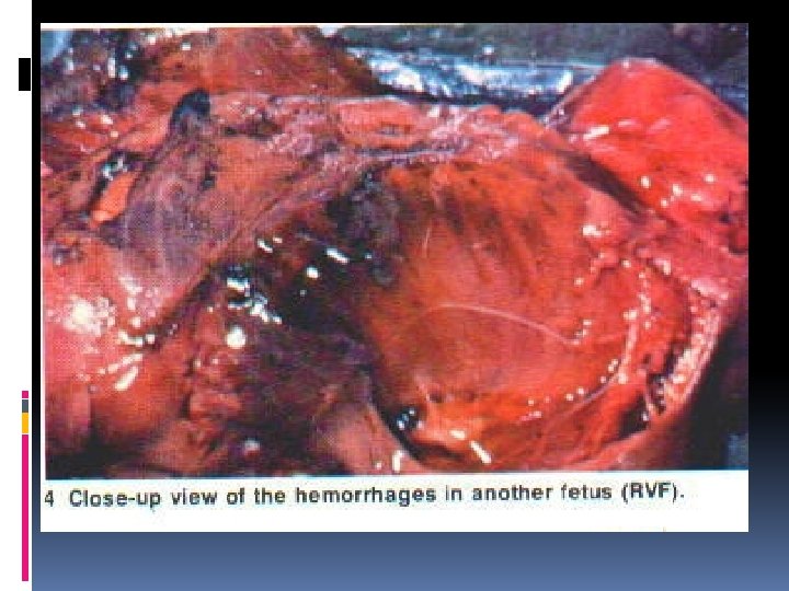
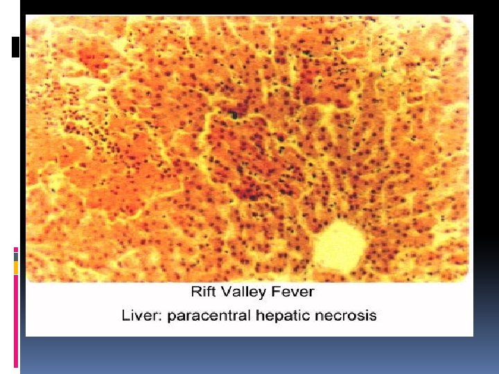
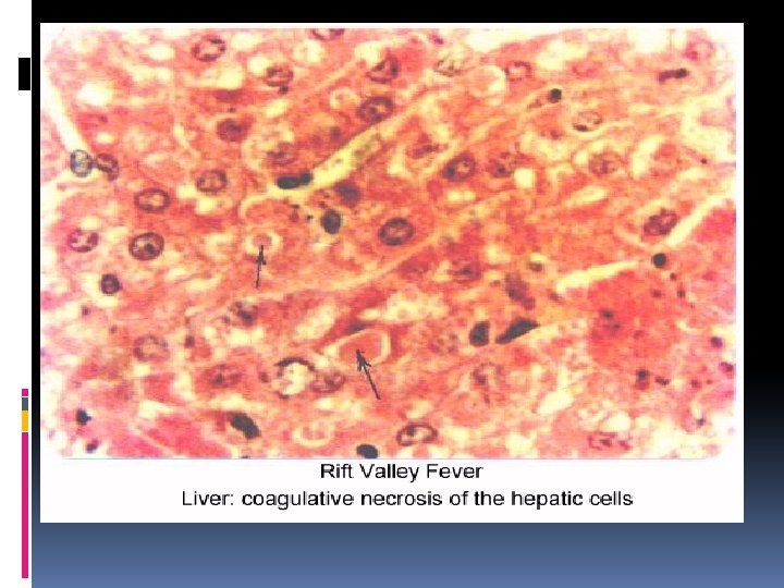
5 -RIFT VALLY FEVER R. V. F (I/N) DEF. Cause R. O. I Lesions (i) MACRO (ii) MICRO Viral disease of sheep & cattle and may human ch’ch’: (a) Hepatitis. (b) High mortality esp. calves&lambs. (c)Abortion in (d)High mortalities in neonates. (e)Zoonotic importance as it transmissible to man leading to death. (Bunyaviridae) R. N. A Viruses Arthropod (mosquito) PATHOGENESIS : Lesions: Bite …………………. viremia ………………liver, spleen, L. N & other organ (a)Viremia(cutanous H. & Pt. hemorrhage in serous membranes with hemorrhagic enteritis) death within 24 hrs. (b)Hepatomegally, yellow colour, with Subcapsular hemorrhage& small white focal necrotic areas. (c)Hemorrhagic and edematous cholangitis. (d)Gall bladder enlarged and edematous. (e)Spleen & L. N ……………. Enlarged, hemorrhagic&necrosed. (f)Pregnant cattle aborted with retained placenta (g)Aborted Foetus : i- Cutaneous hemorrhages and petechial ii- hemorrhages in serous membranes with iii- hemorrhagic enteritis. (a) Acidophilic I/N inclusion bodies in hepatocytes & Kupffer cells. (b) Liver showing; Focal diffuse paracentral coagulative necrosis with living neutrophils -In advanced cases necrotic foci were completely necrosed and appeared (washed out ) (c)Wide areas of blood lagoons. (d) Increase apoptotic cells. (e) Cholestasis in liver, Thrombosis&fibrin in sinusoids. (f) Micro Of Foetus As Those Of adults.
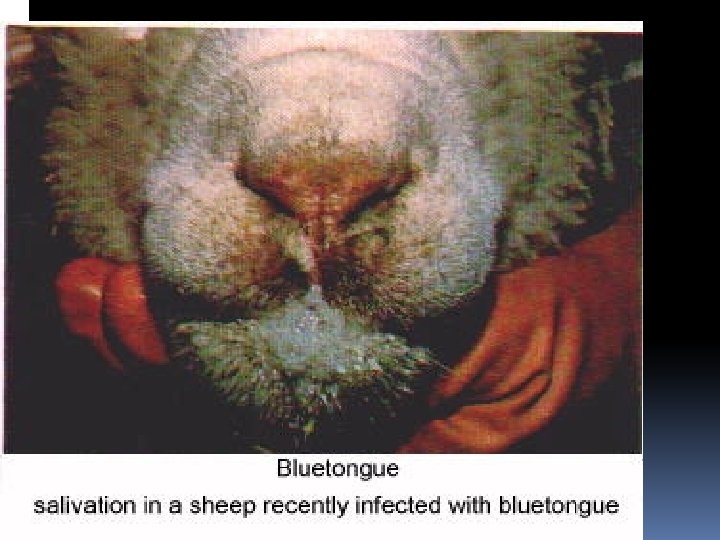
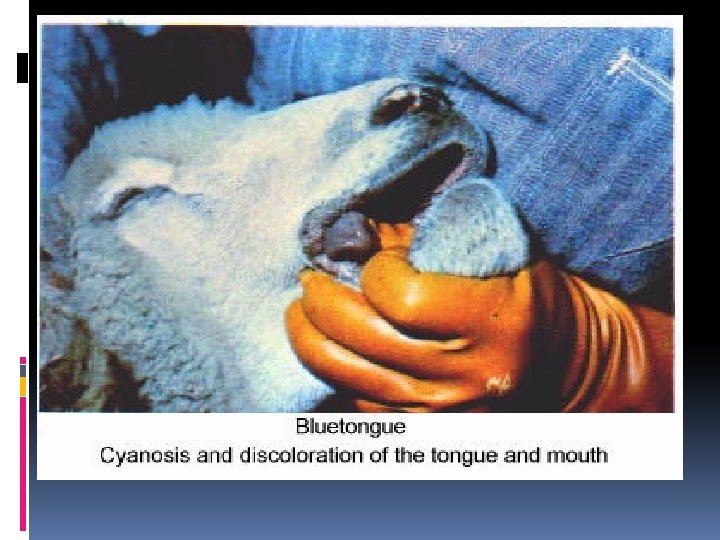
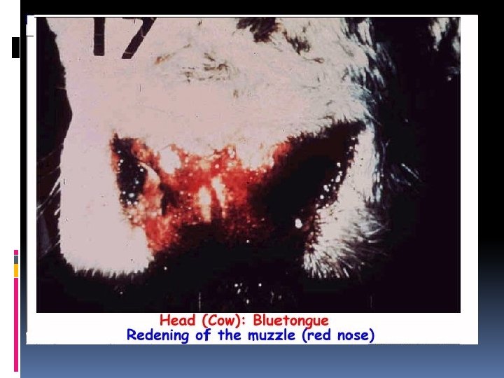
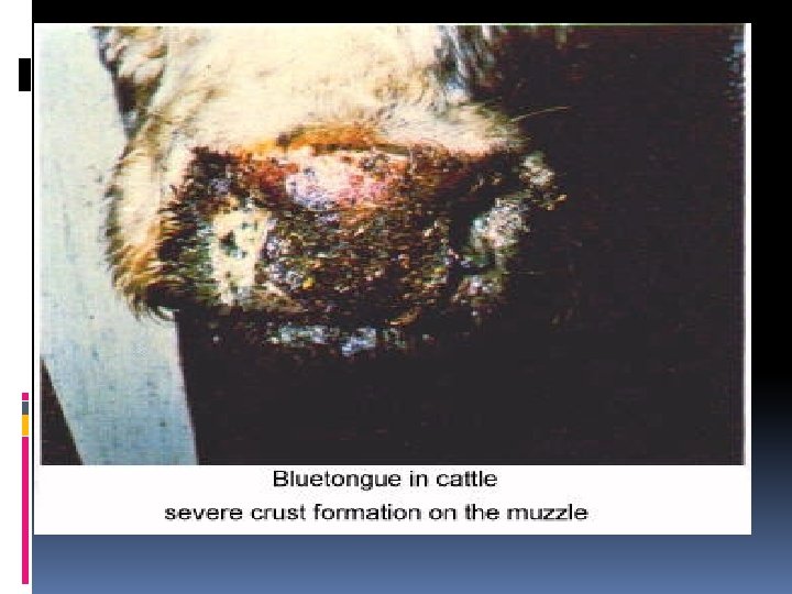
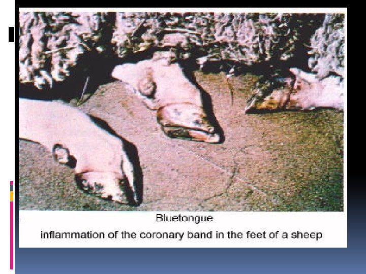
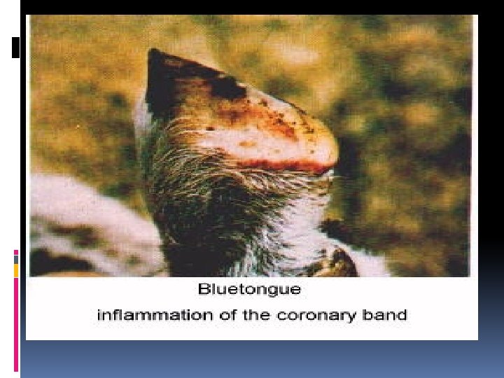
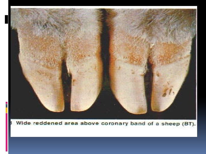
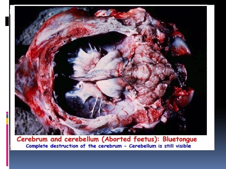
6 - BLUE TONGUE or( SORE MOUTH ) DEF. Cause R. O. I Viral disease of sheep , (rare) in goat & (inapparent) in cattle ch’ch’: Tongue cyanosis & Ulceration. Can cross placenta of pregnant female. (Bunyaviridae) R. N. A Viruses Arthropod (mosquito) PATHOGENESIS : Bite ……viremia ………. endotheliotropic ………. endothelial damage……Vasculitis ……………. vascular permeability (i) Edema&hemorrhage ii) Thrombosis iii) Ischemia & necrosis Lesions: Lesions (i) MACRO (ii) MICRO Per Acute Stage: Pulmonary edema & death due to Asphyxia. Acute stage: (a)Tongue cyanosis due to local v. c. of lingual vein (BT) (b)Ulcer in oral mucosa. ( sore mouth ). (c)Ulcer in omasum, abomasums& intestine. (d)Pt. hemorrhage in abomasum. (e) Pt. hemorrhage in intestine. (f)Congestion of nose ( sore muzzle) (g)Skeletal muscle show edema & hemorrhage. (h)Inflammation of cornet & erosion of interdigital space. (i)Pt. hemorrhage in heart esp. (i)T. adventitia of pulmonary vessels (PATHOGNOMONIC) (ii) T. adventitia of aorta. (iii)subendocardial region. • In advanced stage, dermatitis & loss of wool. • Cross placenta in pregnant females; (i) 50 -55 d ……………Hydran encephaly & Retinal dysplasia. (ii) 75 d ………………. . Porencephaly. (iii) > 100 d ……………. Mild focal Meningeoencephalitis 1. 2. Necrosis of epithelium with lymphocytic infiltration. Vasculitis, thrombosis, venous congestion, edema, and hemorrhages in underlying C. T
VIRAL DISEASE OF CANINES
DEF. 1 - Canine distemper(C. . D) Canine measles Hard pad disease ( I/N + I/C ) Pantropic viral disease of dogs Ch’Ch’ multisystem Affections (Resp. , Nervous, Occular, Skin&Digestive) Cause R. N. A virus ( Paramyxoviridae) R. O. I Inhalation PATHOGEN Inhalation -----Regional L. N (multiplication & replication)--------Thoracic duct-----Circulation-ESIS ------------Target organs 5 Forms Lesions (i) Nervous form : (a) Gliosis with I/N + I/C inclusion bodies. (b) non supp. meningioencephalitis (c) Spongiosis&demylination. (d) Perivascular cuffing. (ii) Occular form : (a)I/N + I/C inclusion bodies. (b) Keratitis (c) Optic neuritis. (iii) Skin form : (a) I/N + I/C inclusion bodies. (b) Hyperkeratosis in foot pad ( HARD PAD DISEASE ). (iv) Respiratory form : (a)I/N + I/C inclusion bodies. (b)Cat. Or purulent rhinopharyngitis. (c)Giant I. S pn. Ch’ch’ hyperplasia of pneumocyte type II with syncytia formation. (v) Alimentry form : (a) Eosinophilic I/N + I/C inclusion bodies. (b) Gastro enteritis. (e) Retinitis
2 - Infectious canine hepatitis I. C. H ( I/N ) DEF. Viral disease of dogs ch’ch’ : Hepatitis Cause DNA virus ( Adenoviridae ) R. O. I Ingestion PATHOG ENESIS Ingestion …………………Blood ( viremia )………………. Liver Lesions MACRO : (i) Liver appear yellow & enlarged. (ii) Edematous cholangitis. (iii) Gall bladder enlarged & hemorrhagic. (iv) Hemorrhage in serous membranes. (v) CORNEAL OPACITY ( In Late stage ) MICRO : (i) I/N inclusion body in hepatocytes & kupffer cells. (ii) Hepatitis (iii) Cholangitis. (iii) Centrolobular necrosis
Canine distemper (intracytoplasmic inclusions) quit previous
3 - Canine parvo virus ( I/N ) DEF. Viral disease of canine Ch’Ch’ : Bloody diarrhea. Cause DNA virus ( Adenoviridae ) R. O. I Ingestion PATHOG Ingestion---Virus replicate in oropharynx------Blood ENESIS i) Bone marrow-----Neutropenia. ii) Lymphopiotic tissues—Lymphopenia. iii) crypt cells of Jejunum&ileum------Necrosis. MACRO : (i) Hemorrhagic gastroenteritis (ii) Myocarditis in puppies associated with edema MICRO : i) I/N inclusion in epithelial cells. ii) Necrosis in gastric and intestinal mucosa. Lesions iii) Lamina propria& submucosa revealed; a. Congested blood vessels. b. Inflammatory cells. c. Large amount of RBCs.
VIRAL DISEASE OF EQUINES
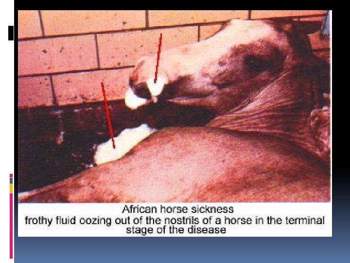
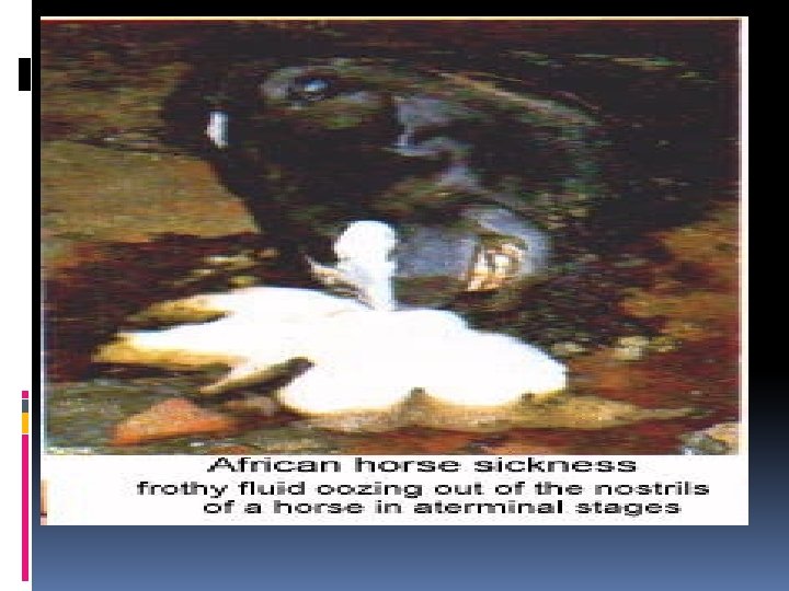
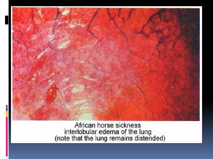
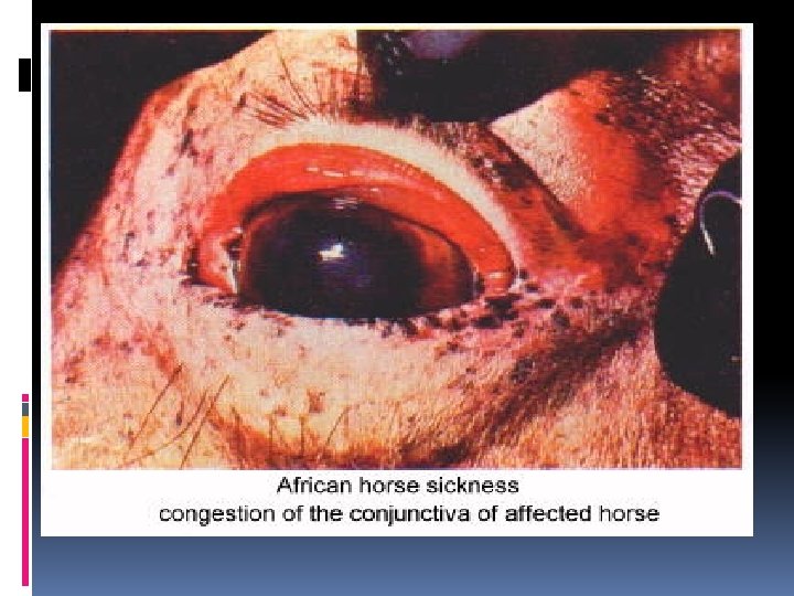
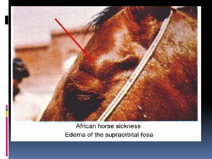
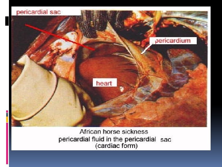
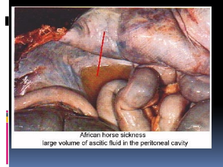
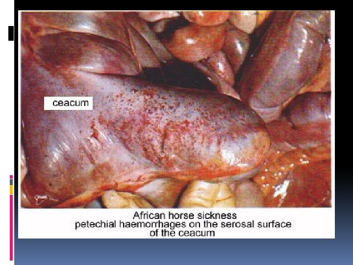
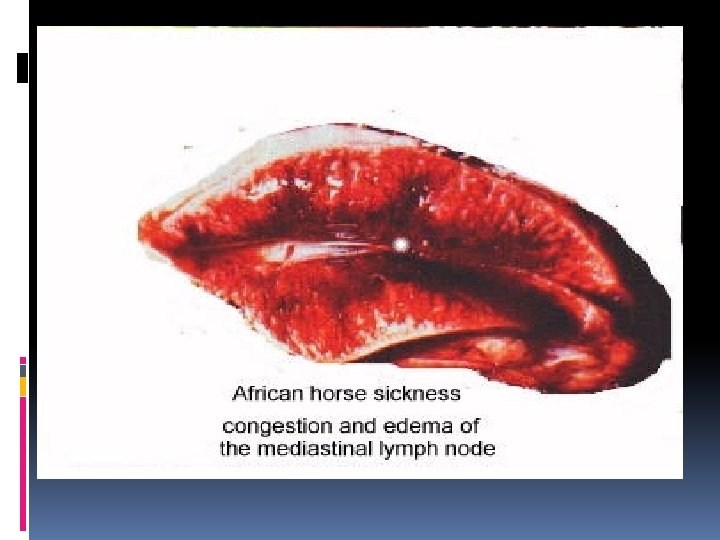
1 - EQUINE ENCEPHALITIS DEF. Cause Viral disease of equine ch’ch ’nervous signs AND brain lesions : Encephalitis & Myelitis Viral disease of equine ch’ch’ : Pulmonary edema & Cardiac edema Associated with i) Frothy exudates from the nostrils ii) Swelling of supra orbital fossa. Horse ……Highly suseptiple. Mule……LESS suseptiple Donky & Zebra………………. . Resistant animals (Togaviridae, 3 strains: Eastern, Western&Venzulian) R. N. A Virus (Reoviridae) R. O. I Mosquito Bite PATHOG Bite------- Viremia -------Nervous ENESIS system Macro: Lesions 2 - AFRICAN HORSE SICKNESS A. H. S NOT CHARACTERESTIC Micro : (i) Encephalitis&Myelitis restricted in grey matter. (ii) Perivascular cuffing. (iii) malacia with Proliferation of microglia cells. Bite-------Viremia--------multiply in endothelium of blood vessels………Vasculitis--------Bl. Permeability------------------Edema & Hemorrhage 4 FORMS (i) Acute Pulmonary Form ( DUNKOP ) : (a) Frothy exudates from the nostrils. (b) Pulmonary edema. (c) Hydrothorax. (d) Pneumonia. (ii) Sub acute cardiac form ( DIKOP ) : (a)Swelling of supra orbital fossa. (b) Hydropericardium & fluid may reach to 2 litres. (c)Epicardium & Sub endocardial hemorrhage. (d) Pericarditis. (e) HEMORRHAGIC GASTRO ENTERITIS (iii) Mixed form : DUNKOP + DIKOP (iv) Mild form : Affect resistant animals ( Donky & Zebra ).
Perivascular lymphocytic cuff Viral encephalitis quit previous
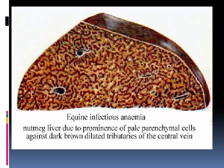
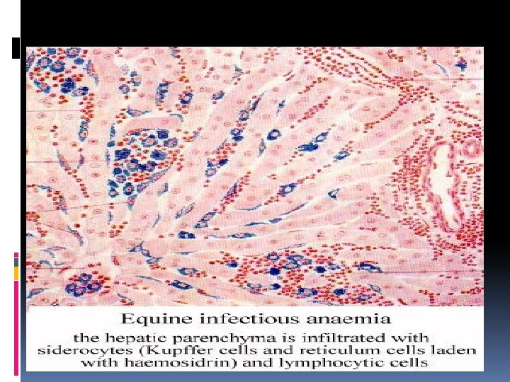
1. EQUINE INFECTIOUS ANEMIA DEF. Cause R. O. I Viral disease of equine Ch’Ch’ : Hemorrhage, Anemia, and Icterus. (Retroviridae) Arthropodes PATH Bite------Blood (viremia)----OGEN Attack RBCs ………………Hemorrhages ESIS in all body. 1. Equine viral arteritis Viral disease of equine Ch’Ch’ : i) Edema & abortion in mares ii) Death in foals. RNA virus ( Arterioviridae) Inhalation------Virus multiply in alveolar macrophages------Lymph node-----Blood (viremia)----Endothelium of small bl. vs------Edema & hemorrhage (vasculitis). i) Pale mucous membranes or may be i) Edema in eye lids & Conjunctivitis. ii) Ascitis, hydrothorax, & hydropericardium. Macro icteric ii) Edema in subcutaneous tissues. iii) Edema in colon & caecum. iii) Enlargement of spleen and lymph iv) Edema in hind limb. nodes. v) Edema in scrotum & prepuse. i) Perivascular lymphocytic infiltration i) Vasculitis Ch’Ch’: in a- Thrombosis b- Fibrinoid necrosis c- Perivascular lymphocytic infiltration Micro most organs. ii) Macrophages infiltration in liver ii) Edema. with iii) Hemorrhages. hemosidrin in kupffer cells.
VIRAL DISEASE WITH NERVOUS MANIFESTATIONS
DEF. Cause R. O. I PATHOGE NESIS Clinical signs MACRO MICRO Rabies Disease Of Hydrophopi Disease Of Photophopia (I/C) Viral encephalitis of all mammals including man Ch’Ch’ nervous signs. R. N. A virus (neurotropic) (Rhabdoviridae). (i) Bite of carnivora through infected saliva. (ii) Wound contamination. * Vampire bats act as reservoir for virus. Incubation Period---------From few days to several months Accord. To site of bite. 1 - Virus replicate at the site of infection in myocytes-------enter neuromuscular junction. 2 -virus transport via sensory nerves by centripedal force to CNS where it replicate. 3 -virus Travel via cranial nerves by centrifugal force to target organs (Salivary Gl& Muscles). 2 Form: (i) Furious form : (a) Restless. (b) irritable. (c) Response to auditory & visual stimulation (d) Excitable (e) Photophopic. (f) Hydrophopic (g) Eat unusuall foreign object (ii) Dump OR Paralytic form : • Dropping of saliva with mouth froth due to parotid adenitis. ( As animal unable to swallow ) • Dropped jaw due to paralysis in masseter muscles. • Change in sound tone and hydrophobia due to paralysis in laryngeal muscles. • Choking sound. (e) Generalized paralysis. NOT CHARACTERSTIC (i) Macerated foot pad. (ii) Foreign bodies in stomach. i) Non Suppurative encephalomyelitis. ii) I/C inclusion bodies (NEGRI BODIES) + clear hallow *Negri bodies demonstrated with (SELLER’S) OR (VAN GIESSON’S) (Present in PURKINGE CELLS in cerebellum in herbivorus animals & in HIPPOCAMPUS in canines). iii) Focal microgliosis in white & grey matter ( BABES NODULES ) iv) Perivascular cuffing. v) Ganglioneuritis in trigeminal ganglia ch’ch; a) Acute degeneration of ganglion cells. B) Microglial nodules.
Intracytoplsmric inclusions (Negri bodies-Rabies) quit previous