CNS pathology Third year medical students lecture 10

 • Occurs in children and")


 • Glial tumors with IDH mutation and no 1 p,")


 • Dense cellularity")


- Slides: 42
CNS pathology Third year medical students lecture 10 CNS tumors/ 2 Dr Heyam Awad FRCPath 2017
Pilocytic astrocytoma • Relatively benign ( WHO grade 1) • Occurs in children and young adults • Mostly: in the cerebellum • Can also involve: third ventricle, optic pathway, spinal cord and rarely cerebral hemispheres
genetics • They have different mutations than diffuse astrocytomas (no IDH mutation, and rarely TP 53 mutation. ) • They have BRAF pathway mutations. . So targeted therapy with BRAF inhibitors can help in treatment, especially in cases were the tumor is not resectable.
morphology • Solid and cystic components • If solid it is usually well defined Microscopically: -bipolar cells with long GFAP positive processes -Rosenthal fibers -eosinophilic granular bodies -microcysts -mitosis and necrosis are rare
Note the well circumscribed lesion in the cerebellum
Pilocytic astro: this example is mainly cystic but has also a solid component
Pilocytic/ microcysts
Pilocytic astrocytoma
Pilocytic astro • Rosenthal fibres: thick , elongated , eosinophilic protein aggregates seen in astrocytic processes. • Note: Rosenthal fibres can also be seen with chronic gliosis.
behavior • Slow growing tumors • Can be treated by resection
oligodendroglioma • 5 -15% of gliomas • 40 -50 years of age • Cerebral hemispheres, mainly in the white matter. • Better prognosis than astrocytoma of the same grade • Well diff (WHO II): 10 -20 years survival; with treatment • Anaplastic (WHO III): 5 -10 years survival; with treatment
genetics • IDH 1 and 2 mutations in 90% of cases. ( mutated tumors have better prognosis than wild type) • Co-deletions of chromosomes 1 p and 19 q in 80% • If the 1 p, 19 q mutation present: highly responsive to chemo and radiotherapy • With the new WHO classification (2016) the 1 p , 19 q co-deletion is essential to diagnose oligodendroglioma.
FYI
New WHO classification (2016) • Glial tumors with IDH mutation and no 1 p, 19 q deletion are called astrocytomas regardless of their morphology. • Glial tumors with IDH mutation and 1 p, 19 q deletion are called oligodendrgliomas regardless of their morphology. • This makes sense because with this definition ALL oligo will have a better prognosis than astro.
morphology • Infiltrative, gelatinous masses • Can have cysts, haemorrhage or calcifications • Micro: sheets of regular cells with spherical nuclei, granular chromatin, clear cytoplasm, rare mitoses • Fried egg appearance of the cells. • Anaplastic: more cellularity, more anaplasia and more mitosis…. Poorer prognosis
oligodendroglioma; / note the white halo around the nuclei giving the fried egg appearance
Oligodendroglioma. .
WHO GRADING of oligodendroglioma • Well differentiated is grade 2 • Anaplastic ( increased cellularity, pleomorphism and mitotic activity) is grade 3 • NOTE: OLIGO CAN PROGRESS TO GLIOBLASTOMA ( grade 4). • NOTE: THERE IS NO WHO GRADE 1 OLIGO
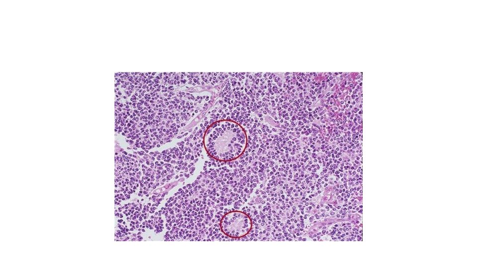
Anaplastic oligo (WHO grade 3) • Dense cellularity
Ependymoma • Arise next to ventricles and central canal of spinal cord. • If they occur in first two decades of life: mostly will arise near the fourth ventricle • In adults : mostly in the spinal cord • Prognosis is better if the tumour is resectable. • Supratentorial and spinal cord tumours are more amenable to complete surgical resection, thus they have a better prognosis than posterior fossa tumors.
morphology • Solid or papillary masses • Regular round nuclei • granular chromatin, • dense fibrillary background ( GFAP POSITIVE). Note that the fibrillary background is seen in all gliomas. • rosette formation around canals and pseudo-rosette around blood vessels • Anaplastic ependymoma : cellular, mitosis, necrosis
Ependymoma/ rosettes note: true rosettes arise around canals
Ependymoma/ pseudorossetes, these arise around blood vessels
Tumors related to the ependymal cells • There are some tumors that occur below the ependymal lining or in association with the choroid plexus. • These tumors include: choroid plexus papilloma, subependymoma and colloid cysts • All the above are benign! And rare. • However because of their location they cause clinical problems including hydrocephalus. • Choroid plexus carcinoma can occur but is a rare tumor.
Neuronal tumors These are rare tumors that have neuronal differentiation. • Central neurocytoma: low grade neoplasm within and adjacent to ventricular system. • ganglioma : glial elements and mature appearing neurones. , usually slow growing but the glial element can progress NOTE: I’M NOT going to ask you about these in the exam. Most likely you will not see a case in your career!
Embryonal neoplasms These are tumors of neuroectodermal origin. • Primitive appearance that resembles the normal progenitor cells that are found in the developing CNS : small round cells, little cytoplasm • Most common type of these tumors: medulloblastoma • Medulloblastoma: 20% of pediatric brain tumors
Medulloblastoma • Occurs predominently in children • Exclusively in the cerebellum • Highly malignant if untreated ( WHO grade 4) • Radiosensitive • Surgery + chemo + radio. . 5 year survival reaches 75%
Histology of medulloblastoma • Highly cellular • Sheets of small blue cells ( small, rounded hyperchromatic nuclei, scanty cytoplasm) • Many mitoses • Homer Wright Rosettes= primitive tumor cells surrounding central neuropil ( pink material formed by neuronal processes
medulloblastoma
Homer Wright Rosettes
genetics • MYC amplification: poor prognosis • WNT signaling pathway mutations: better prognosis • These can help in developing new therapies… because it is better to avoid radiotherapy in young patients
WNT signaling pathway
Beta catenin stain/ if positive then the medulloblastoma has WNT signaling mutation: better prognosis. .
• Activating mutations in beta-catenin in approximately 10% of medulloblastoma represent the WNT subtype. • The identification of nuclear beta-catenin has been demonstrated to be nearly 100% specific and sensitive for the presence of mutation and makes it possible to reliably identify WNT pathway tumors using routine immunohistochemistry.
Treatment of medulloblastma • Medulloblastoma therapy, including craniospinal radiation and multiagent chemotherapy, results in significant long term toxicity for many disease survivors, including neurocognitive impairment, neuropathy, endocrinopathy, impaired bone growth, impaired motor function, hearing loss, and secondary malignancy. • These side effects are closely related to dose of radiation therapy and age at diagnosis, the earlier the age, the worse the neurologic toxicity for the developing brain
• The long-term survival of patients with WNT tumors is excellent, exceeding 90%. • Also, WNT tumors arise exclusively in an older age group of children over the age of three years.
• The rest of the slides in this lecture are FYI. . And to stimulate you to think! • Happy reading !
Palliative care of patients • Palliative care: Aim is to relieve symptoms and side effects caused by cancer or its treatment. Palliative care helps a child with cancer live as comfortably as possible. It also addresses the psychological, social, and spiritual needs of the child and family.
• Palliative care focuses on preventing, managing, and relieving the symptoms of cancer and the side effects of cancer treatment. • It also provides comprehensive support to people living with cancer and their family, friends, and caregivers. • Anyone, regardless of age or type and stage of cancer may receive palliative care before, after, and during treatment. • Talking about palliative care soon after a cancer diagnosis helps patients better understand their prognosis and goals of treatment, clarify their expectations, and maintain their quality of life
The goals of palliative care include: • • • Treating symptoms, including pain, nausea, breathlessness, insomnia, and other physical issues caused by cancer or its treatment Making sure patients and caregivers understand the diagnosis and goals of treatment Providing guidance for making treatment decisions Working with the patient’s other doctors and providing referrals to other health care providers as needed Providing support for the patient’s emotional and social needs, spiritual needs or concerns, and practical needs Providing support for caregivers, and other family members and friends