University of Tehran Faculty of Veterinary Medicine Comparative


 parietal layer b)")

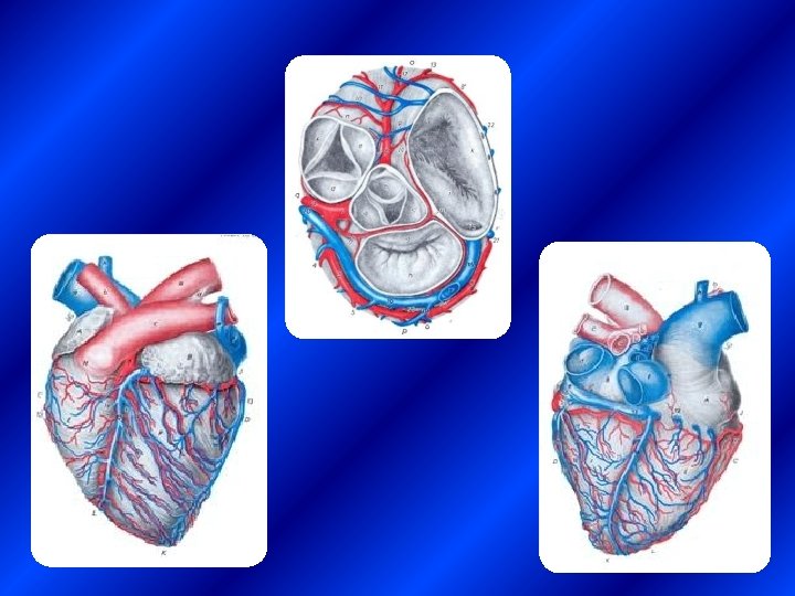
 Cranial Border 2) Caudal Border")
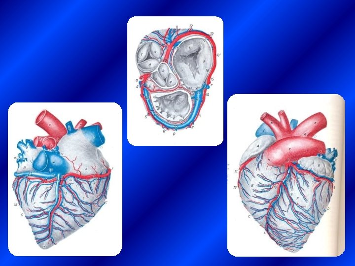
 q Right surface")
 q Subsinusal Interventricular groove(Right surface )")


 2. Papillary muscles 3. Chordae tendineae")
 §")

 2. Trigonal")
")


- Slides: 29
University of Tehran Faculty of Veterinary Medicine Comparative Anatomy of Heart and Vessels By : Dr, N. Goodarzi Ph. D of veterinary anatomy
Topography of the HEART ü obliquely placed in the thorax ü base faces dorsocranially ü apex is directed ventrocaudally.
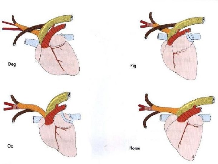
Topography of the HEART Equine 3 -6 Carnivores 3 -7 Ruminants 3 -5
Pericardium has two layers q Fibrous pericardium q Serous pericardium a) parietal layer b) Visceral layer (Epicardium) Pericardial cavity (Liquor pericardii)
Comparative points of pericardium q q Sternopericardial ligament attaches the apex of the heart to the sternum in equine and ruminants. Phrenicopericardial ligament attaches the apex of the heart to the sternum in carnivores
HEART BORDERS 1) Cranial Border 2) Caudal Border
SURFACES OF THE HEART q Left surface (Arterial or Auricular surface) q Right surface (Atrial surface or venous surface)
HEART GROOVES q Paraconal Interventricular groove(Left surface ) q Subsinusal Interventricular groove(Right surface ) q Coronary groove
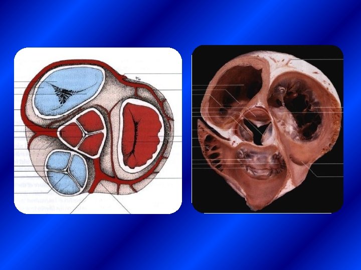
LAYERS OF THE HEART q Epicardium q Myocardium § Pectinate Muscle q Endocardium § Inter atrial Septum § Inter ventricular Septum § Valves (Atrioventricular and semilunar V. )
Atria and Ventricles
Right Atrium 1. Right auricle 2. Sinus venarum cavarum 3. Caudal vena cava 4. Cranial vena cava 5. Intervenous tubercle 6. Terminal crest 7. Coronary sinus 8. Fossa ovalis 9. Limbus fossae ovalis
Left Atrium 1. Left auricle 2. Left atrium 3. 5 -8 pulmonary veins 4. Venarum minimarum
Right Ventricle
Right Ventricle 1. Conus arteriosus 2. Pulmonary trunk 3. Tricuspid valve 4. Papillary muscles 5. Chordae tendineae 6. Trabecula carnea 7. Trabecula septomarginalis (Moderate band)
Left Ventricle 1. Mitral Valve (Bicuspid v. ) 2. Papillary muscles 3. Chordae tendineae 4. Trabecula carnea 5. Trabecula septomarginalis
Valvulae and Cusps of the heart q Right atrioventricular valve (Tricuspid v. ) § Parietal cusp § Septal cusp § Angular cusp § In carnivores : septal and parietal cusp q Left atrioventricular valve (Bicuspid v. ) § Septal cusp § Parietal cusp q Pulmonary valve : right , left and intermediate cusp q Aortic valve : right , left and septal cusp In dog ventral cusp
Ligamentum arteriosum
Skeleton of the HEART 1. Fibrous ring (aroundatrioventricular valves and semilunar valvulae) 2. Trigonal fibrous Ø Ruminant : Os. Cordis Ø Dog : fibrocartilage Ø Cat : irreg. dens. c. t. Ø Equ : hyaline cartilage
Coronary Vesseles q Coronary artery q In dog and ruminant: ( Left coronary type) Right Coronary artery : circumflex a. Left Coronary artery : circumflex a. , paraconal a. , subsinusal a. q In horse : (Right coronary type) Right Coronary artery : circumflex a. , subsinusal a. Left Coronary artery : circumflex a. , paraconal a.
Aorta
Aorta q Ascending Aorta q Aortic arch Brachoicephalicus Trunk q Thoracic Aorta Ø Ø Dorsal intercostal a. Costoabdominals a. Broncho-esophageal a. Cranial phrenic a. ( in horse) q Abdominal Aorta
q. Abdominal Aorta Ø Ø Ø Lumbale a. Caudal phrenic a. Cranial abdominal a. Coeliac a. Cranial Mesentric a. Renal a. Suprarenal a. Testicular a. , Ovarian a. Caudal Mesentric a. External Iliac a. Internal Iliac a. , Median Sacral a.
Brachoicephalicus Trunk q Left and Right subclavian a. Ø cosrocervical trunk dosrsal scapular supreme intercostal Ø Ø Ø Deep cervical Vertebral Axillary Superficial cervical Internal thoracic q Left and Right common carotid
Brachoicephalicus Trunk