HEMODYNAMIC DISORDERS THROMBOSIS AND SHOCK Edema Fluid Balance

HEMODYNAMIC DISORDERS, THROMBOSIS AND SHOCK

Edema

Fluid Balance Across Capillary Walls Factors Involved Interstitial Fluid Photo: Kumar, Cotran, Robbins Basic pathology, 7 th ed. , Saunders, Philadelphia, 2003.

Edema - Pathogenesis

Edema
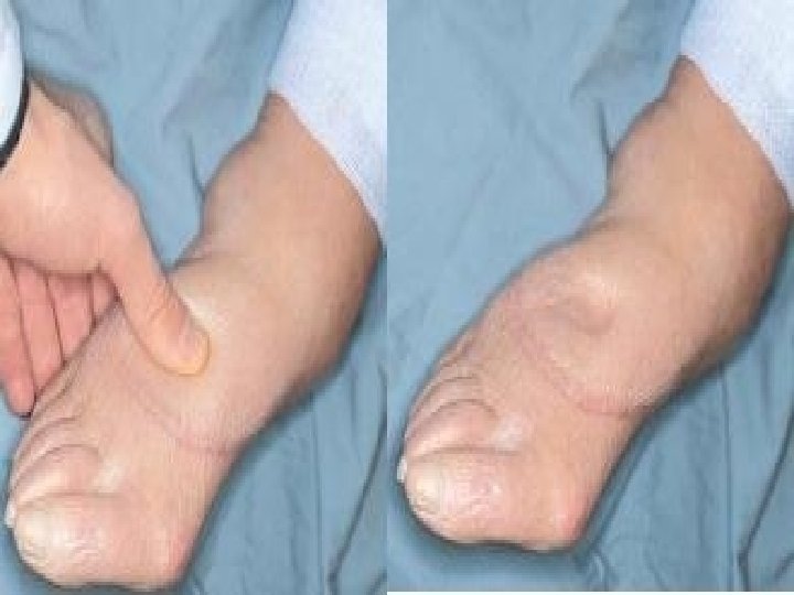

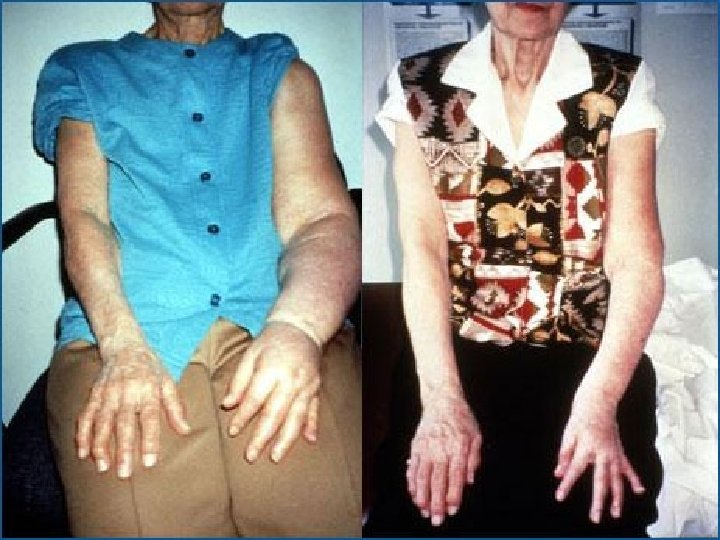
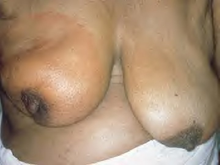
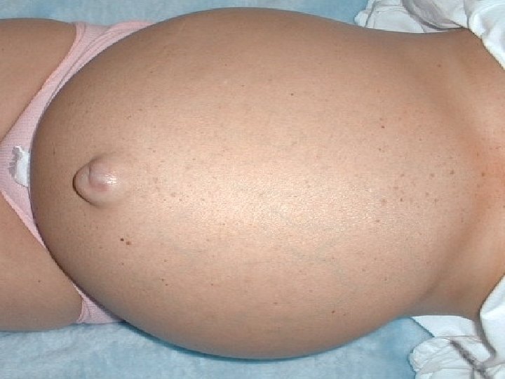
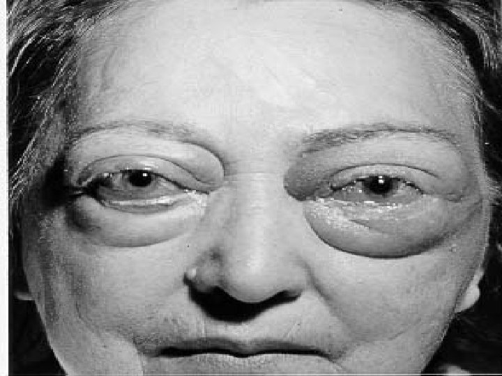
Edema: Lymphatic Obstruction

Pulmonary Edema

Fluid in Trachea/Bronchi

Abdominal Ascites

Normal Brain

Edematous Brain

Hyperemia and Congestion

Congestion and Hyperemia

CONGESTION AND HYPEREMIA

Congested Lungs

Acute Pulmonary Congestion

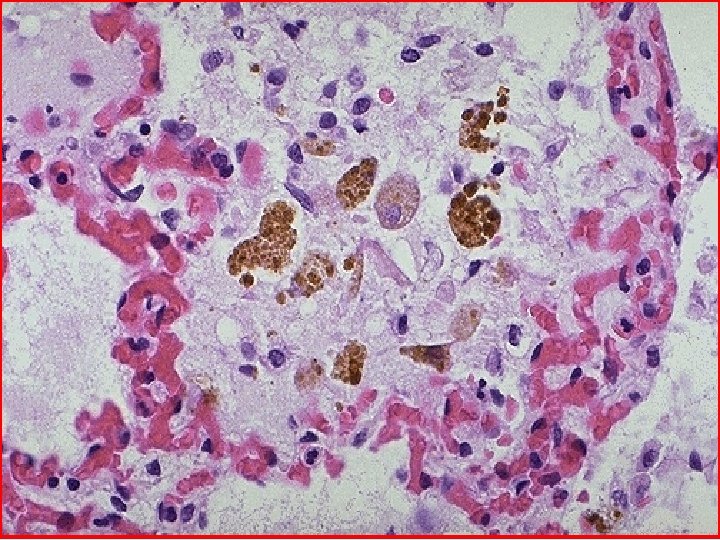
“Heart Failure Cells” in Alveoli Chronic Pulmonary Congestion

Congested and Enlarged Spleen

Nutmeg liver

Microscopically, the nutmeg pattern results from congestion around the central veins, as seen here. This is usually due to a "right sided" heart failure .
 = Nutmeg Liver Right Heart Failure Photo: Stevens A, Lowe J.")
Congested Liver (Passive) = Nutmeg Liver Right Heart Failure Photo: Stevens A, Lowe J. Slide atlas of pathology. Mosby, London, 1995. ; Kumar, Cotran, Robbins Basic pathology, 7 th ed. , Saunders, Philadelphia, 2003.

Hemorrhage
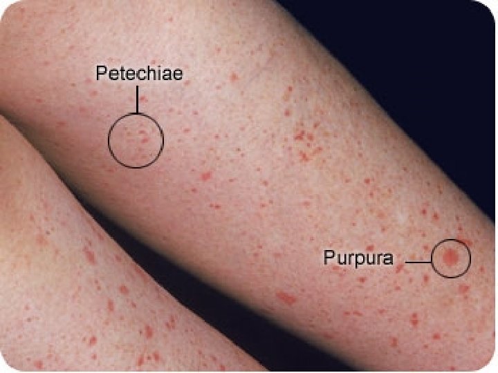
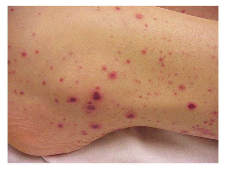

• Here are petechial hemorrhages seen on the epicardium of the heart. Petechiae (pinpoint hemorrhages) represent bleeding from small vessels and are classically found when a coagulopathy is due to a low platelet count. They can also appear following sudden hypoxia.

• The blotchy areas of hemorrhage in the skin are called ecchymoses (singular ecchymosis), or also as areas of purpura. Ecchymoses are larger than petechiae. They can appear with coagulation disorders.
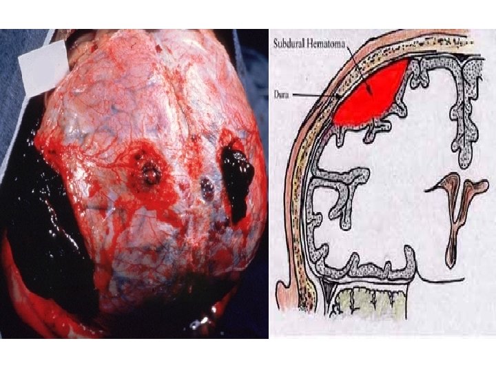

Intracerebral Hemorrhage Photo: Kumar, Cotran, Robbins Basic pathology, 7 th ed. , Saunders, Philadelphia, 2003.

Intracerebral Hemorrhage

Pericardial Hemorrhage

Hemostasis and Thrombosis
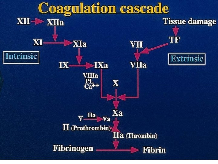

THROMBOSIS -Virchow triad

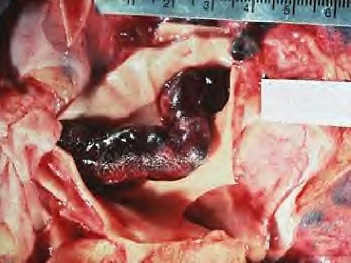
Thrombus - Morphology Arterial Venous Ø Arise in arteries • Ø Grow in retrograde fashion (towards the heart- away from the direction of flow) • Ø Forms at site of Endothelial injury (AS), turbulence (aneurysms) Ø Pale/ white Ø Lines of Zahn Ø Firmly adherent to vessel wall Ø From emboli Cause infarctions (lower extremities – 75%, Brain, Kidney, spleen) Arise in deep veins and superficial veins (popleteal Femoral Iliac), Antigrade (towards the heartdirection of flow) • At site of stasis (lower extremities) • • • Red / dark No lines of Zahn Loosely attached (easily embolize) Emboli cause Pulmonary embolism ( silent in 50% of pts. ) •

• These are "lines of Zahn" which are the alternating pale pink bands of platelets with fibrin and red bands of RBC's forming a true thrombus.

Venous Thrombi: Clinical

Thrombotic Vegetations Mitral Valve Photo: Stevens A, Lowe J. Slide atlas of pathology. Mosby, London, 1995.

Mural Thrombus

Abdominal Aortic Aneurysm Thrombus
")
Deep Vein Thrombosis (DVT)

Plaque with Recent Thrombus

Thrombosis Outcomes Photo: Kumar, Cotran, Robbins Basic pathology, 7 th ed. , Saunders, Philadelphia, 2003.

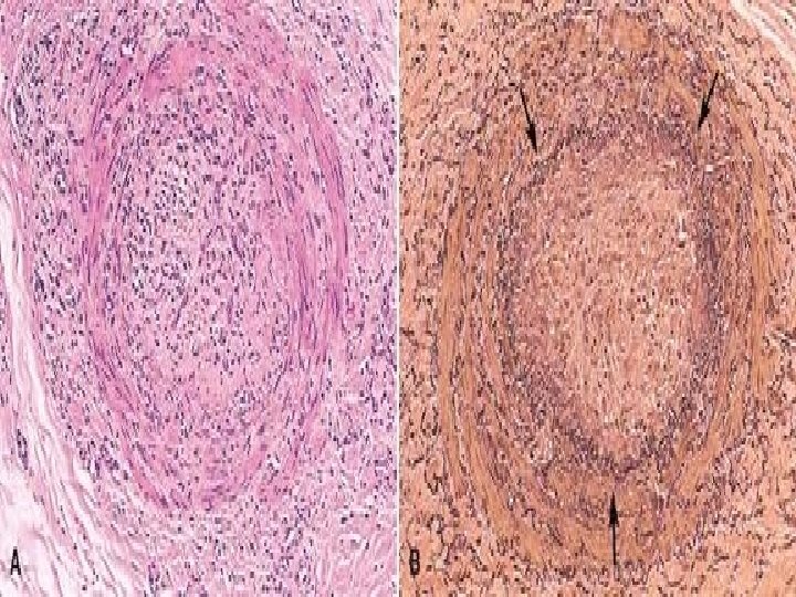
Early Organizing Thrombus

Embolism
 Thromboembolism of Pulmonary Artery Photo: Kumar, Cotran, Robbins Basic pathology, 7 th")
Embolization (Embolus) Thromboembolism of Pulmonary Artery Photo: Kumar, Cotran, Robbins Basic pathology, 7 th ed. , Saunders, Philadelphia, 2003; . Stevens A, Lowe J. Slide atlas of pathology. Mosby, London, 1995.

Right Ventricle Embolus from Leg Vein

Pulmonary Embolus

Bone Marrow Embolus In Pulmonary Vessel Photo: Kumar, Cotran, Robbins Basic pathology, 7 th ed. , Saunders, Philadelphia, 2003.

Infarction
 Lung (Left); Spleen (Right) Photo: Kumar, Cotran, Robbins Basic pathology, 7 th")
Infarction (Infarct) Lung (Left); Spleen (Right) Photo: Kumar, Cotran, Robbins Basic pathology, 7 th ed. , Saunders, Philadelphia, 2003.

Pulmonary Infarction

Small Intestine Infarction
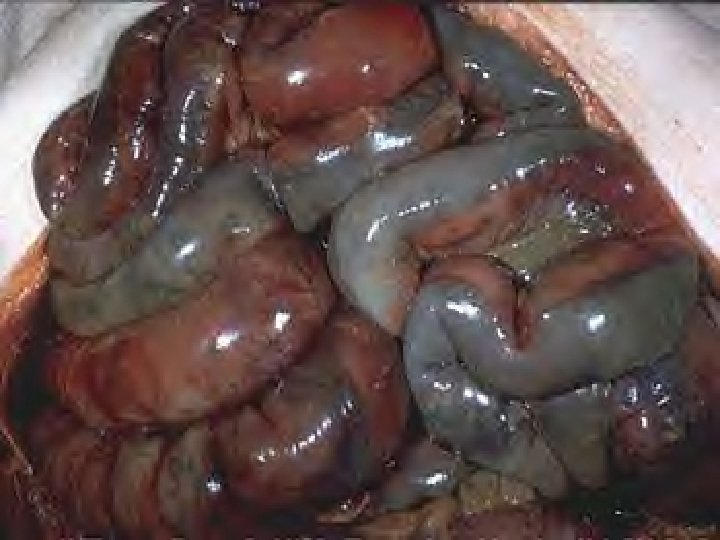
 Photo: Kumar, Cotran, Robbins Basic pathology, 7")
Kidney Infarction Replaced by Fibrotic Scar (Left) Photo: Kumar, Cotran, Robbins Basic pathology, 7 th ed. , Saunders, Philadelphia, 2003.
 of Spleen")
Pale Infarct (Wedge) of Spleen
- Slides: 63