Diseases of Penis PENIS Congenital anomalies Inflammations Tumors


 - Types - Congenital Acquired - Primary , Secondary")


 Involve anterior urethra , �cause muco-purulent discharge + dysuria")
 causes small transient painless")
 – �(H ducreyi ) – Single , multiple ulcers")
 : �Ulcers of irregular margins with")
 � produces irritable vescicles, � soon")
 cause benign warts on the penis")


 Benign �i. Condyloma accuminatum � ii. Bowen’s disease �iii. Erythroplasia of")


 Sq. cell carcinoma - 93 – 95 % Ii )")


 Carcinoma �Varient of sq. cell ca of penis �characterized by mainly")


- Slides: 50
Diseases of Penis
PENIS � Congenital anomalies � Inflammations � Tumors
Congenital lesions
Congenital anomalies � � � Complete absence Hypoplasia Hypertrophic / hyperplasic Epispadies ( Urethral opening on Ventral aspect of shaft of penis ) Hypospadies ( Urethral opening on Dorsal aspect of shaft of penis )
Congenital anomalies Phimosis - “inability to retract the foreskin on the glans penis”. Causes – i) congenital ii) Acquired - infections, scarring,
Congenital anomalies Paraphimosis : “Inability of retracted foreskin to return to normal position” �When phimotic prepuce is forcefully retracted over the glans, marked constriction and subsequent swelling may block the replacement of the prepuce , creating Paraphimosis. �It is painful and may cause urinary retention
Phimosis & Paraphimosis :
Inflammatory lesions
Inflammatory lesions �Acute ◦ Non-specific ◦ Specific �Chronic ◦ Non-specific ◦ Specific
Balanoposthitis � Non-specific infection of the glans and prepuce caused by a wide variety of organisms. � More common agents: Candida albicans, anaerobic bacteria, Gardnerella, and pyogenic bacteria. � Most cases occur as a consequence of poor local hygiene in uncircumcised males, with accumulation of desquamated epithelial cells, sweat, and debris, termed smegma, acting as local irritant. � Persistence of such infections leads to inflammatory scarring and, as mentioned earlier, is a common cause of phimosis.
INFLAMMATORY DISEASES: Acute inflammations �Trauma , Surgery , Infections like - Viral , Bacterial , parasitic , fungal ( e. g. - Syphilis , Gonorrhea, Trichomonas vaginalis , Candida etc ), ischemia �C/F: pain, fever, inguinal lymphadenopathy, and pelvic discomfort associated with discharge from the penis. �On examination –the penis is tender and reddish with seropurulent discharge. �On examination with Gram’s stain or Silver stain or Dark ground illumination the Causative organism is found
Chronic inflammations : �Non-specific – Herpes simples, HPV , others �Specific STD’s : Syphilis , Gonorrhea, LGV, Granuloma inguinale, Soft Sore, HIV, Trichomonas Vaginalis , Candida albicans etc
SYPHILIS ( T pallidum ) - Types - Congenital Acquired - Primary , Secondary , Tertiary stages � Penis lesions -(i) Chancre –(Hard Sore ) occur at penis, scrotum , consists of Single, firm, � non- tender, raised, red lesions , with exudates teeming with spirochetes. �(ii) Condyloma latum – broad based , elevated plaques , (iii) Gumma - Nodular lesions found in testis, , heart, liver, brain etc.
Syphilitic chancre �Photo with hard sore, on the glans penis. �Painless ulcer, erythematous lesion, �Yellow exudate.
Syphilitic chancre �Microphotograph showing =small blood vessel, with peri vascular chronic inflammatory cells, mainly lymphocytes, plasma cells, histiocytes, �Loss of elastic tissue is characteristic.
Syphillis �Condyloma lata of penis
GONORRHEA ( N gonorrheae ) Involve anterior urethra , �cause muco-purulent discharge + dysuria �Asst with fever, pelvic pain, �Urethral discharge on Gram stain shows�Gram negative cocci in the Neutrophils, & �In the exudate
LGV ( Chlamydia trachomatis – 1, 2, 3 types ) causes small transient painless ulcer � Vescicles, papule, � Inguinal Bubo
Chancroid �( Soft Sore ) – �(H ducreyi ) – Single , multiple ulcers painful, with ragged and �Undermined edges
Granuloma Inguinale �( Klebsiella granulomatis –Donovan Bodies ) : �Ulcers of irregular margins with necrotic floor, erythema , indurations, �with Inguinal Bubo
Genital Herpes (HSV- type 1. , 2 ) � produces irritable vescicles, � soon rupture to Produce small, tender, ulcers on the ext genitalia
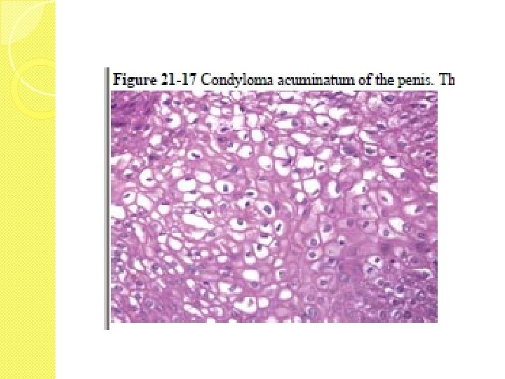
Condyloma accuminatum ( HPV- type 6, 11 ) cause benign warts on the penis � Cause single or multiple exophytic popular or flat warts � Sometimes it may cause giant Condyloma ( Buschke-Lewenstain tumour )
Condyloma accuminatum �Penis showing greywhite papillary mass , arising from coronal sulcus, with red glands
Condyloma acuminatum �Sections shows predominantly papillary lesion, �having thin fibro vascular core and covered by mature stratified squamous epithelium. �Foci of necrosis are seen
Condyloma accuminatum �High power details of the lesion showing broad fronds of mature stratified squamous epithelium, �and parakeratosis, �hyperkeratosis
Other Infections �Molluscum Contagiosum, �Candidiasis , �Trichomonas vaginalis, etc
Tumours
TUMOURS �A ) Benign �i. Condyloma accuminatum � ii. Bowen’s disease �iii. Erythroplasia of Queyrat iv. Others
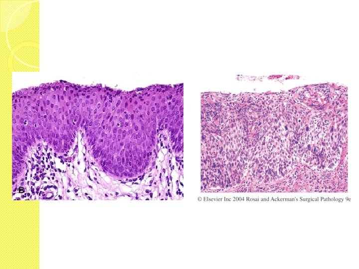
BOWEN’S DISEASE �Common after 35 yrs of age. �Produce single, thick, opaque plaques, �With shallow ulcer , crusting is seen.
BOWEN’S DISEASE �Photo shows scrotum with multiple small, encrusted plaque lesions of 0. 5 – 1 cms in size, �With shallow ulcer crusting is seen. �
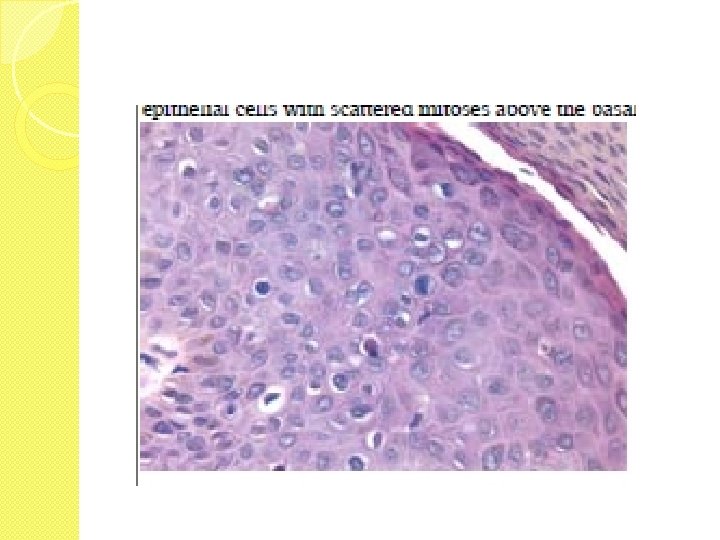
BOWEN’S DISEASE �Micro-shows stratified squamous epithelium, with loss of polarity, �Atypical Sq. cells, in all the layers, �Hyperchromasia �increased mitosis, scanty cytoplasm,
ERYTHROPLASIA OF QUEYRAT �Same as Bowen’s disease �with severe dysplasia changes �at muco-cutaneous regions of the genitals in both sexes. �May progress to carcinoma
MALIGNAT TUMOURS I ) Sq. cell carcinoma - 93 – 95 % Ii ) Verrucus carcinoma - 0 – 2 % Iii ) Basal cell ca 0– 1% Iv ) Malignant melanoma - 0 - 1 % V ) Others - Rare
Epidemiology Increasing age, � Role of smegma , � Poor genital hygiene , � Circumcision Benefit Virus inf : HPV – 16, 18, 31 types , STD’s , Smoking –Polycyclic hydrocarbons ? �
Squamous cell carcinoma �Partial amputated specimen of penis. �There is exophytic growth arising from coronal areas/glands �Greywhite mass, areas of ulceration, hemorrhage �Everted edges
squamous cell carcinoma Cut-sections of the same specimen showing the tumour in the upper areas with corpora , Note the invasive foci Ares of necrosis, hemorrhage
squamous cell carcinoma �Gross of partial amputated penis, �Exophytic growth , with ulceration.
squamous cell carcinoma �Low power view of growth showing tumour composed of sq. cells tumour with �Invasion into deeper tissue
squamous cell carcinoma �High power details showing tumour tissue of Sq. cell type invading the deeper tissue. �Note the anaplasia, mitosis, �Stroma shows chr. inflammatory cells.
squamous cell carcinoma �Micro showing �well diff squamous cells, with formation of epithelial /keratin pearl, �surrounded by immature sq. cells, �Note chr. inflamm cells around the focus
Verrucus carcinoma �Variant of Sq. cell ca, with better prognosis, and slow growing nature. �Specimen shows huge exophytic growth, �everted edges, focal necrosis, hemorrhage
Verrucus carcinoma �Basically Sq. cell ca �Note the broad fronds of invading process. �Mainly consists of mature sq. cells, with areas of kertinization, �There is chr. inflamm cells around the margins
Verrucus carcinoma �Note the broad fronds on the invading tumour cells, �with keratinization.
Basiloid carcinoma �Varient of cancer charactirized by presence of mainly basal cells, �showing invasion, large hyperchromatic nuceli, �scanty cytoplasm.
spindle cell (sarcomatoid) Carcinoma �Varient of sq. cell ca of penis �characterized by mainly spindle cells, with interlacing arrangement �and mimicking sarcoma
Transitional cell carcinoma of penile urethra �Section shows �tumour composed of mainly transitional epithelial cells, �with central core of fibro-vascular areas, covered by abnormal transitional epithelium �Invasion , necrosis seen
Malignant melanoma �Excised specimen of dark pigmented mass of 1 – 2 cms in size, �mainly arising from scrotum, �or skin of penis.