Urinary Bladder Congenital Anomalies trauma cystitis tumors urethral

Urinary Bladder: Congenital Anomalies trauma, cystitis, tumors, urethral trauma Dr. Amit Gupta Associate Professor Dept. of Surgery
 organ that")
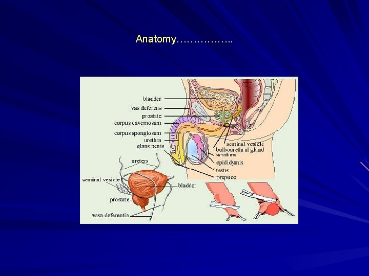
Anatomy The urinary bladder is a hollow, muscular, and distensible (or elastic) organ that sits on the pelvic floor. It is the organ that collects urine excreted by the kidneys prior to disposal by urination. Urine enters the bladder via the ureters and exits via the urethra. In males, the base of the bladder lies between the rectum and the pubic symphysis. It is superior to the prostate, and separated from the rectum by the rectovesical pouch. In females, the bladder sits inferior to the uterus and anterior to the vagina. It is separated from the uterus by the vesicouterine pouch.

Embryology The urogenital sinus is formed by the division of the cloaca by the uro-rectal septum The uro-genital sinus may be divided into three component parts. cranial portion which is continuous with the allantois and forms the bladder proper. The pelvic part of the sinus forms the prostatic urethra and epithelium as well as the membranous urethra and bulbo urethral glands in the male and the membranous urethra and part of the vagina in females. the caudal portion forms the penile urethra in males and the vestibule in females.

Embryology……. . The bladder forms from the cranial end of the urogenital sinus. However, the trigone portion is formed by the caudal ends of the mesonephric ducts. As the bladder expands, the mesonephric ducts begin to become incorporated into the wall of the bladder dragging the ureters along with them. Further growth causes the ureters to eventually have their own opening into the bladder. The bladder is initially continuous with the allantois. Over time, the allantois degenerates to form a cord-like structure, the urachus. The urachus goes from the umbilicus to the apex of the bladder and forms the median umbilical ligament which can be seen in adults. The medial umbilical ligaments may also be seen in adults on both sides of the median umbilical ligament, these are the vestigial remnants of the umbilical arteries.

Congenital Anomalies A major consideration with congenital abnormalities is that they tend to be multiple. All of these anomalies are infrequent or rare, and each condition occurs in both males and females Diagnosed in infancy or childhood Most are discovered in the evaluation of a urinary tract infection or, in the case of urachal anomalies, periumbilical drainage or redness.

Classification Urinary bladder anomalies are– – – – bladder diverticula bladder ears congenital hypoplasia of the bladder megacystis bladder agenesis duplication anomalies of the bladder septa Urachal anomalies are urachal sinus urachal cyst urachal diverticulum patent urachus

Pathophysiology The embryologic cause is unknown. Bladder development occurs during the fifth to seventh week of gestational development. - Development depends upon many factors mesenchymal differentiation mesenchymal growth urine production Bladder cycling, the process of sequential expansion and contraction, is important in the anatomic and physiologic development of the normal bladder.

Bladder Diverticulum Bladder diverticula are herniations of the bladder mucosa through bladder wall musculature (detrusor muscle). • Diverticular size can vary • Diverticula can be wide or narrow mouthed. (The size of diverticular openings has functional implications because narrowmouthed diverticula often empty poorly). • Stasis of urine within diverticula can also lead to stone formation or epithelial dysplasia. • may cause ureteral obstruction, bladder outlet obstruction or vesicoureteral reflux

Bladder Diverticulum…………… MC Site lateral and superior to the ureteral orifices. [dome of the bladder, in disorders as bladder outlet obstruction (ie, posterior urethral valves) or Eagle Barrett syndrome (prune belly syndrome)]. Congenital or acquired. Congenital deficiency or weakness in the Waldeyer fascial sheath has been implicated as a cause. Solitary located at the junction of the bladder trigone and detrusor Congenital diverticula are usually removed surgically

Bladder Diverticula

Bladder ears lateral protrusions of the bladder through the internal inguinal ring and into the inguinal canal. Bladder ears are often observed during voiding cystourethrography (VCUG) or intravenous pyelography (IVP), when the bladder is filled to capacity. Bladder ears have also been seen on CT body imaging. No treatment is necessary Knowledge of this entity is important to surgeons during inguinal herniorrhaphy because occasional reports have been made of partial or near total cystectomy performed under the mistaken notion that this was a large hernia sac.

Bladder agenesis • Rare • generally incompatible with life. • Ureters may enter into the urethra, vagina, Gartner duct cyst (female), prostatic urethra, rectum, or the patent urachus. • Associated hydroureteronephrosis and renal dysplasia (variable) are present. • Other associated anomalies include neurologic, orthopedic, hindgut, and other urogenital anomalies, such as renal agenesis and absence of the prostate, vagina, seminal vesicles, epididymis, or penis This condition requires urinary diversion and subsequent reconstruction

Megacystitis is an enlarged bladder, believed to be secondary to overfilling of the fetal bladder during development Common presentationshydronephrosis. Febrile urinary tract infection high-grade vesicoureteral reflux Megacystitis can be observed in other conditions, such as posterior urethral valves, Ehlers-Danlos syndrome, urethral diverticulum, microcolon hypoperistalsis syndrome, sacral meningomyelocele, sacrococcygeal teratoma, and pelvic neuroblastoma. Clean intermittent catheterization
 v Partial (the bladder joins")
Bladder duplication v Rare v Complete (2 urethras exist) v Partial (the bladder joins distally into one common urethra) v The 2 halves of the bladder are on either side of the midline, with the corresponding ipsilateral ureter draining each bladder half. v Associated anomalies include duplication of the penis, vagina, uterus, lumbar vertebrae, and hindgut. fistulas may be present between the rectum, vagina, and urethra.

Duplication of Ur. bladder

Urachal cyst is a fluid-filled structure occurring in between the two obliterated ends of the urachus. Most occur in the distal third of the urachus. More commonly, the urachal cyst is detected in early childhood or adolescence. Symptoms -infection , inflammation, suprapubic mass, fever, pain, and bladder or irritative voiding symptoms. – (Staphylococcus aureus is the most common bacterial organism). – Death from intra-abdominal rupture has been reported.

Patent urachus Communication from the umbilicus to the bladder. continuous or intermittent drainage from the umbilicus. The tract may become inflamed, resulting in tenderness, periumbilical swelling, and serosanguinous or purulent discharge. May be associated with bladder outlet obstruction, such as posterior urethral valves. Correction of the obstruction may result in the spontaneous resolution of the patent urachus.

Patent Urachus

Patent Urachus

Resected specimen of Urachus

Intra-operative photograph

Urachal sinus derived from a persistently patent urachus Children may present with periumbilical tenderness wet umbilicus granulation tissue at the level of the umbilicus In many instances, these children have undergone multiple sliver nitrate cauterizations under the mistaken notion that this is simply granulation tissue after severance of the remnant umbilical cord. These can be observed in the first 4 -8 weeks of life.

Ectopia Vesicae open to the outside and turned inside-out, so that its inside is visible at birth, protruding from the lower abdomen Symptomsq Exposed posterior bladder wall q Short lower abdominal wall q Inguinal hernia q Separated pubic rami q Epispadias q Incomplete fusion of genital tubercles

q Opening in abdominal wall q Urine excretion through opening in abdominal wall q Incontinence q Dilated ureters q Open pubic arch q Wide-set ischial bones q Constriction between ureter and bladder q Ureteral reflux

Exostrophy of Urinary bladder


Cystitis

Cystitis In women is common due to a number of reasons : - short urethra - pregnancy - decreased estrogen production during menopause. In men: mainly due to persistent bacterial infection of the prostate. In both sexes: common risk factors are : - presence of bladder stone - urethral stricture - catheterization of the urinary tract - diabetes mellitus

Pathogenesis of cystitis Due to frequent irritation of the mucosal surfaces of the urethra and the bladder. Infection results when bacteria ascends to the urinary bladder. These bacteria are residents or transient members of the pereneal flora, and are derived from the large intestine flora. Toxins produced by uropathogens. Condition that create access to bladder are: - Sexual intercourse due to short urethral distance.

Pathogenesis of cystitis Uncomplicated UTI usually occurred in non pregnant , young sexually active female without any structural or neurological abnormality Risk factors : - Catheterization of the urinary bladder , instrumentation - structural abnormalities - obstruction Hematogenous through blood stream ( less common) from other sites of infection
 cause of cystitis. Other Enterobacteria")
Etiologic agents E. coli is the most common (90%) cause of cystitis. Other Enterobacteria include ( Klebsiella pnumoniae, Proteus spp. ) Other gram negative rods eg. P. aeroginosa. Gram positive bacteria : Enterococcus fecalis, group B Strept. and Staphylococcus saprophyticus { honeymoon cystitis}. Candida species Venereal diseases ( gonorrhea, Chlamydia) may present with cystitis. Schistosoma hematobium in endemic areas.

Pathogens involved Uncomplicated UTI Complicated UTI E. coli 64% Enterobacteriaceae 16% Enterococcus spp 20% Pseudomona spp <1% S. aureus <1% Special cases (S. epidermidis) S. saprophyticus Yeasts (catheter related result) Viruses (Adeno, Varicella) Chlamydia trachomatis E. coli % is Enterobacteriaceae not Pseudomonas spp possibl e to Acinetobacter spp (judge often multiresistant strains)
 – Frequency")
Clinical presentation Symptoms usually of acute onset – Dysuria ( painful urination) – Frequency ( frequent voiding) – Urgency ( an imperative call for toilet) – Haematuria ( blood in urine) in 50% of cases. – Usually no fever.
 Candida spp. T. vaginalis Cystitis (80%) E. coli, S. saprophyticus Proteus spp.")
Vaginitis (5%) Candida spp. T. vaginalis Cystitis (80%) E. coli, S. saprophyticus Proteus spp. Klebsiella spp. Dysuria and frequency Non-infectious (<1%) Hypoestrogenism Functional obstruction Mechanical obstruction Chemicals Urethritis (1015%) C. trachomatis, N. gonorrhoeae H. simplex Other bacteria?

How to differentiate between cystitis and urethritis ? • Cystitis is of more acute onset • More sever symptoms • Pain, tenderness on the supra-pubic area. • Presence of bacteria in urine ( bacteriuria) • Urine cloudy, malodorous and may be bloody
 Non-infectious cystitis such as: 1. Traumatic cystitis in")
Differential diagnosis ( types of cystitis) Non-infectious cystitis such as: 1. Traumatic cystitis in women 2. Interstitial cystitis ( unknown cause, may be due to autoimmune attack of the bladder) 3. Eosinophilic cystitis due to S. hematobium 4. Hemorrahagic cystitis due to radiotherapy or chemotherapy.

Laboratory diagnosis of cystitis 1. Specimen collection 2. Microscopic examination 3. Chemical screening tests


Recurrent cystitis 3 or more episodes of cystitis /year Requires further investigations such as Intravenous Urogram ( IVU) or ultrasound to detect obstruction or congenital deformity Cystoscopy required in some cases

Treatment of cystitis • Empiric treatment commonly used depending on the knowledge of common organism and sensitivity pattern • Treatment best guided by susceptibility pattern of the causative bacteria • Common agents: Ampicillin, Cephradine, Ciprofloxacin, Norfloxacin, Gentamicin or TRMSMX.

• Duration of treatment: 3 days for uncomplicated cystitis • 10 -14 days for complicated and recurrent cystitis • Prophylaxis required for recurrent cases by Nitrofurantoin or TRM-SMX • Prevention : drinking plenty of water and prophylactic antibiotic

Bladder cancer

Aetiology • Smoking – 4 x increased risk – Causes 50% of cases • Occupational – rubber/dye industry – Napthylamine, benzidine • Schistosomiasis, chronic infection • Medications – cyclofosfamid, fenacetin

Histology • 90 -95% transitional-cell carcinoma • 3% squamos-cell carcinoma • 2% adenocarcinoma • <1% small-cell carcinoma • 99% primary tumors

Entities • 75 -85% superficial bladder cancer p. Ta, p. Tis, p. T 1 • 10 -15% muscle-invasive bladder cancer p. T 2, p. T 3, p. T 4 • 5% metastatic bladder cancer N+, M+

Presentation • Classically painless frank haematuria, sometimes intermittent • Frequent urination, urgency • symptoms with involvement of neighbouring organs, lymphoedema, pelvic pain

Examination History Physical examination Urine examination / urinalysis, cultivation, cytology – can be only 60% sensitive/ Ultrasound

Examination • Cystoscopy is mandatory • Biopsy or TURBT • Bimanual pelvic examination /before and after TURBT • Chest X-ray • IVU – no routinely, (5% chance upper tract involvement)

Bladder cancer: Stage and Prognosis
 • Precursor infiltrating tumors • Primary or secondary • Subjectively")
Carcinoma in situ (CIS) • Precursor infiltrating tumors • Primary or secondary • Subjectively – frequent urination, urgency, cystalgia • Objectively – no pathologies • Laboratory – Microhematuria – Cytology positive (PAP IV-V)

CIS treatment • Primary CIS: BCG • Secondary CIS: TURB + BCG • Recurrent CIS /after therapy/: cystectomy

Treatment • Superficial Bladder Cancer p. Ta, p. T 1, Tis • Invasive bladder cancer p. T 2 -p. T 4

Superficial Bladder Cancer p. Ta, p. T 1, Tis Standard of care=intravesical therapy transurethral resection bladder tumors /TURBT/ Relapse rate: 70% adjuvant therapy

TURBT

TURBT

TURBT

Superficial Bladder Cancer Adjuvant Therapy • Reduces relapse rate by 30 -80% – Mitomycin C – in patient with intermediate-risk BT – BCG – in patient with CIS, high risk BT

Invasive bladder cancer • Standard of care = Radical cystectomy with pelvic lymphadenectomy • Only about 50% of patients with high-grade invasive disease are cured

Radical cystectomy

Radical cystectomy

Chemotherapy for bladder cancer • Bladder cancer is a chemosensitive disease • Active single agents RR – Cisplatin – Carboplatin – Gemcitabine – Ifosfamide 30% 20 -30% 20%
 – 13% reduction in")
Neoadjuvant chemotherapy • Meta-analysis of ten randomised trials (2688 patients) – 13% reduction in risk of death – 5% absolute benefit at 5 years – OS increased from 45% to 50% – ABC Meta-analysis Collaboration. Lancet 2003; 361: 1927

Bladder-sparing therapy for invasive bladder cancer • High probability of subsequent distant metastasis after cystectomy or radiotherapy alone (50% within 2 years) • Radiotherapy in comparison with cystectomy has inferior results (local control 40%) • muscle-invasive bladder cancer is often a systemic disease combined modality therapy
 Cystoscopy after 1")
Bladder-sparing protocol Transurethral resection Induction Therapy: Radiation + chemotherapy (cisplatin, paclitaxel) Cystoscopy after 1 month no tumor Consolidation: RT + CT tumor cystectomy

Urinary diversion • Diversion of urinary pathway from its natural path • Types: – Temporary – Permanent

A nephrostomy is a surgical procedure by which a tube, stent, or catheter is inserted through the skin into the kidney
")
Cutaneous Ureterostomy… One kidney drainage, with short-live prognosis Complications (infection, stone, stenosis)

Permanent urinary diversion • Uretero – sigmoidostomy • Ileal conduit • Colon conduit • Ileocaecaecal segment
 A 12 cm loop of ileum led")
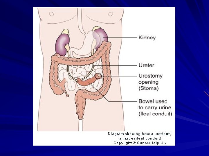
Cutaneous urinary diversions Ileal conduit (ileal loop) A 12 cm loop of ileum led out through abdominal wall Stents used The space at cystectomy site drained by a drainage system After surgery a skin barrier and a transparent disposable urinary drainage bag Constantly drains

Complications of ileal conduit • • • Wound infection Wound dehiscence Urinary leakage Ureteral obstruction Small bowel obstruction Ileus Stomal gangrene Narrowing of the stoma Pyelonephritis Renal calculi

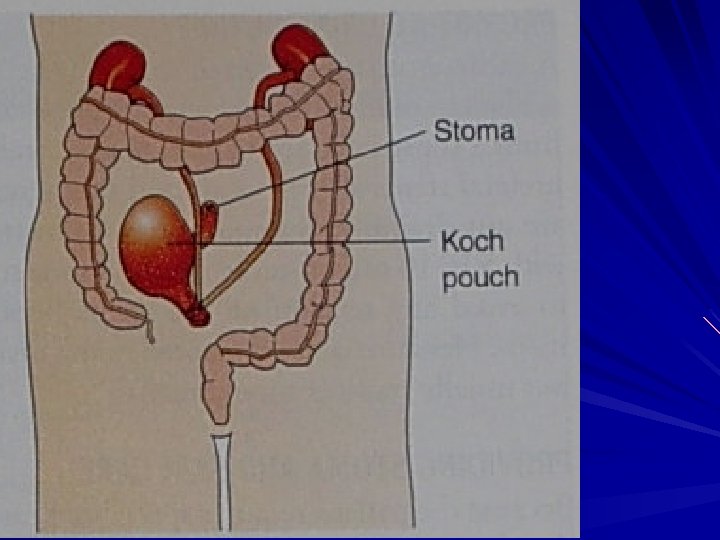
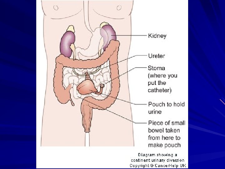
Continent Urinary Diversions • Continent Ileal Urinary Reservoir • • • Indiana Pouch Most common continent urinary diversion Periodically catheterized Koch Pouch Ureterosigmoidostomy Voiding occurs from rectum

Ureterosigmoideostomy

Uretero- sigmoidostomy • Complications: – Reflux of urine – Hyperchloraemic acidosis (ammonium chloride reabsorption, bicarbonates secretion) – Renal infection – Stricture formation


Potential complications • Peritonitis due to disruption of anastomosis • Stoma ischaemia and necrosis due to compromised blood supply to stoma • Stoma retraction and separation of mucocutaneous border due to tension or trauma

Bladder Reconstruction

INJURIES OF THE URINARY BLADDER • Bladder injuries occur most often from external force and are often associated with pelvic fractures. • Iatrogenic injury may result from gynecologic and other extensive pelvic procedures as well as from hernia repairs and transurethral operations.

Classification • closed and open • • isolated and combined intraperitoneal, retroperitoneal and mixed.

Clinical Findings • There is usually a history of lower abdominal trauma. • Blunt injury is the usual cause • Patients ordinarily are unable to urinate, but when spontaneous voiding occurs, gross hematuria is usually present • Most patients complain of pelvic or lower abdominal pain.

Clinical Findings • Heavy bleeding associated with pelvic fracture may result in hemorrhagic shock, usually from venous disruption of pelvic vessels. • An acute abdomen indicates intraperitoneal bladder rupture. • A palpable mass in the lower abdomen usually represents a large pelvic hematoma. • On rectal examination, landmarks may be indistinct because of a large pelvic hematoma.

Clinical Findings • Laboratory Findings: • Catheterization usually is required in patients with pelvic trauma but not if bloody urethral discharge is noted. • When catheterization is done, gross or, less commonly, microscopic hematuria is usually present. • Urine taken from the bladder at the initial catheterization should be cultured to determine whether infection is present.

X-Ray Findings • A plain abdominal film generally demonstrates pelvic fractures. There may be haziness over the lower abdomen from blood and urine extravasation. • An intravenous urogram should be obtained to establish whether kidney and ureteral injuries are present.

X-Ray Findings • Bladder disruption is shown on cystography. • Retrogradual cystography help to differentiate penetrating and unpenetrating, intraperitoneal and retroperitoneal ruptures of the bladder, locate urinary flow and approximate site of rupture. • The sign of retroperitoneal rupture is accumulation of X-ray contrast matter in perivesical fat tissue. • With intraperitoneal extravasation, free contrast medium is visualized in the abdomen, highlighting bowel loops.

X-Ray Findings

Treatment • A. Emergency Measures: Shock and hemorrhage should be treated. • B. Surgical Measures

Treatment In a case of retroperitoneal complete rupture of the bladder it is exposed by suprapubic extraperitoneal access carefully inspected and is juncture by two-row catgut junctures. Drainage by means of epicystostomy is necessary. Operation finish with drainage of perivesical and pelvic tissue. In order to prevent formation of urinary flow, in all cases of retroperitoneal rupture of urinary bladder, perivesical space is drainage through obturatorial foramen or ischiorectal space.

Treatment Intraperitoneal bladder ruptures should be repaired via a transperitoneal approach after careful transvesical inspection and closure of any other perforations. The peritoneum must be closed carefully over the area of injury. The bladder is then closed in separate layers by absorbable suture. All extravasated fluid from the peritoneal cavity should be removed before closure. At the time of closure, care should be taken that the suprapubic cystostomy is in the extraperitoneal position.

Urethral Injuries • Urethral injuries are uncommon and occur most often in men, usually associated with pelvic fractures or straddle-type falls. • Various parts of the urethra may be lacerated, transected, or contused. • Management varies according to the level of injury. • The urethra can be separated into 2 broad anatomic divisions: the posterior urethra, consisting of the prostatic and membranous portions, and the anterior urethra, consisting of the bulbous and pendulous portions

Clinical Findings Symptoms: • lower abdominal pain and inability to urinate. A history of crushing injury to the pelvis is usually obtained. Signs: • Blood at the urethral meatus is the single most important sign of urethral injury (Urethroragia). • Suprapubic tenderness and the presence of pelvic fracture are noted on physical examination. • A large developing pelvic hematoma may be palpated. • Perineal or suprapubic contusions are often noted. • Rectal examination may reveal a large pelvic hematoma with the prostate displaced superiorly.

Clinical Findings • Laboratory Findings: • Anemia due to hemorrhage may be noted. • Urine usually cannot be obtained initially, since the patient should not void and catheterization should not be attempted.

X-Ray Findings Fractures of the bony pelvis are usually present. A urethrogram (using 20 -30 ml of watersoluble contrast material) shows the site of extravasation. Ordinarily, there is free extravasation of contrast material into the perivesical space. Incomplete prostatomembranous disruption is seen as minor extravasation, with a portion of contrast material passing into the prostatic urethra and bladder.

X-Ray Findings

Treatment • Conservative therapy is effective for patients with recent nonpenetrating damage of urethra: rest, cool compresses, and antibiotics. • Within 7 -8 days after trauma thermal procedures and resorption agents are prescribed. • In case of ischuria instead of a high cystotomy it is possible to perform troacar epicystostomy. • Shock and hemorrhage should be treated.

Treatment Surgical Measures: • Urethral catheterization should be avoided. • suprapubic cystostomy to provide urinary drainage. • The bladder should be opened and carefully inspected for lacerations. If a laceration is present, the bladder should be closed with absorbable suture material and a cystostomy tube inserted for urinary drainage. • The suprapubic cystostomy is maintained in place for about 3 months. This allows resolution of the pelvic hematoma, and the prostate and bladder will slowly return to their anatomic positions.

Film Suprapubic Cystostomy

Treatment Urethral reconstruction – • Reconstruction of the urethra after prostatic disruption can be undertaken within 3 months. • Before reconstruction, a combined cystogram and urethrogram should be done to determine the exact length of the resulting urethral stricture. • The preferred approach is a single-stage reconstruction of the urethral rupture defect with direct excision of the strictured area and anastomosis of the bulbous urethra directly to the apex of the prostate. • • • A 16 F silicone urethral catheter should be left in place along with a suprapubic cystostomy. Catheters are removed within a month, and the patient is then able to void

Re-do end to end

Complications • Stricture, impotence, and incontinence are complications of prostato membranous disruption. • Stricture following primary repair and anastomosis occurs in about onehalf of cases. If the preferred suprapubic cystostomy approach with delayed repair is used, the incidence of stricture can be reduced to about 5%. • The incidence of impotence after primary repair is 30 -80% (mean, about 50%). • Incontinence in primary reanastomosis is noted in one-third of patients.
- Slides: 102