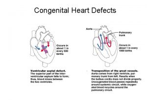
DISEASES OF THE CARDIOVASCULAR SYSTEM CONGENITAL DEFECTS CONGENITAL


- Slides: 25

DISEASES OF THE CARDIOVASCULAR SYSTEM: CONGENITAL DEFECTS

CONGENITAL DEFECTS: PATENT DUCTUS ARTERIOSUS CHIHUAHUAS, MALTESE, POODLE, POMERANIAN, SHELTIE PUPPIES COMMONLY AFFECTED

CONGENITAL DEFECTS: PATENT DUCTUS ARTERIOSUS Normally, the ductus arteriosus carries blood from the pulmonary artery to the aorta during fetal development. It bypasses the lungs of the fetus.

CONGENITAL DEFECTS: PATENT DUCTUS ARTERIOSUS The duct should close in the first 1224 hours after birth. If it does not, the blood begins to shunt from the aorta into the pulmonary artery and hyperperfuse the lungs. The left side of the heart will have an increase in blood return and become volume overloaded. THIS IS CALLED A LEFT-TO-RIGHT SHUNT

CONGENITAL DEFECTS: PATENT DUCTUS ARTERIOSUS (PDA)

CONGENITAL DEFECTS: PATENT DUCTUS ARTERIOSUS � CLINICAL SIGNS: � A loud murmur best � Sometimes called a heard over the left base “machinery” murmur or a continuous murmur � If the shunt is small some animals may be asymptomatic � In large shunts the animal will develop left-sided heart failure Pulmonary edema Cough Exercise intolerance Tachypnea Weight loss � ECG: wide range of arrhythmias including APCs and VPCs � Echocardiography (ultrasound) � Radiographs: left atrial and ventricular enlargement

PATENT DUCTUS ARTERIOSUS: TREATMENT EXCELLENT PROGNOSIS WITH SURGICAL CORRECTION: LIGATION OF THE DUCTUS ARTERIOSUS

PATENT DUCTUS ARTERIOSUS: TREATMENT � CLIENT INFO: � 64% OF ANIMALS WILL DIE WITHIN 1 YEAR IF NOT TREATED SURGICALLY � Dogs with this condition should not be used for breeding

CONGENITAL DEFECTS: ATRIAL AND VENTRICULAR SEPTAL DEFECTS Atrial Septal Defect During fetal life, the foramen ovale is an openingi n the interatrial septum, allowing shunting of blood from the right atrium to the left atrium in order to bypass the nonfunctioning fetal lungs. It should close at birth. If it doesn’t, after birth, the blood will shunt from left to right resulting in overload of the right side of the heart.

CONGENITAL DEFECTS: ATRIAL AND VENTRICULAR SEPTAL DEFECTS � CLINICAL SIGNS: ATRIAL SEPTAL DEFECTS � Result in overload of the right side of the heart → dilation and hypertrophy of the right-sided chambers � Systolic murmur � Right-sided heart failure � Radiographs: right ventricular enlargement � Echo: right ventricular dilatation

CONGENITAL DEFECTS: ATRIAL AND VENTRICULAR SEPTAL DEFECTS Blood is shunted from the oxygen-rich left ventricle into the right ventricle. The blood goes through pulmonary circulation and right back into the left atrium and ventricle resulting in volume overload of the left side of the heart. The right ventricle may dilate as well.

CONGENITAL DEFECTS: ATRIAL AND VENTRICULAR SEPTAL DEFECTS � CLINICAL SIGNS: VENTRICULAR SEPTAL DEFECTS: � Animals with small defects may have minimal or no signs � Larger defects may result in acute left-sided heart failure, usually by 8 weeks of age � A harsh holosystolic murmur � CLIENT INFO: � Repair of these defects requires open-heart surgery or cardiopulmonary bypass. These procedures are uncommon in the dog and cat � Most of these animals will eventually experience development of congestive heart failure

CONGENITAL DEFECTS: PULMONIC STENOSIS Chihuahuas, English Bulldogs, are commonly affected. CAUSE: polygenic inheritance

PULMONIC STENOSIS In pulmonic stenosis, the right ventricular outflow tract is narrowed, either at the valve itself, just below it, or just after it.

PULMONIC STENOSIS The most common form of pulmonic stenosis involves a deformed pulmonary valve such that the valve leaflets are too thick, the opening is too narrow, or the valve cusps are fused. The heart must pump extra hard to get blood through This unusually narrow, stiff valve. The right ventricle becomes thickened from all this extra work. The right atrium May become dilated and hypertrophied.

CONGENITAL DEFECTS: PULMONIC STENOSIS NORMAL CANINE CHEST RADS THIS DOG HAS PULMONIC STENOSIS – THE HEART LOOKS “PREGNANT” IN THE FRONT DUE TO RIGHT VENTRICULAR ENLARGEMENT

CONGENITAL DEFECTS: PULMONIC STENOSIS � CLINICAL SIGNS: � Syncope � Tiring on exercise � Right-sided congested heart failure � Left basilar (base) murmur � Right ventricular enlargement � Radiographs: right ventricular enlargement, dilation of the pulmonary artery, pulmonary underperfusion � Echo: right ventricular hypertrophy and enlargement, dilation of the main pulmonary artery

PULMONIC STENOSIS: TREATMENT A special balloon is inserted into the valve where it is inflated and the obstruction is broken down. Unfortunately, medical management is not very beneficial in these cases. Beta-blockers may be used to relax the heart muscle and possibly dilate the stenosis.

CONGENITAL DEFECTS: SUBAORTIC STENOSIS Newfoundland, Boxer, Golden Retriever, and Bull Terrier are most commonly affected LESION DEVELOPS IN THE FIRST 4 -8 WEEKS OF LIFE

CONGENITAL DEFECTS: SUBAORTIC STENOSIS There is a scar-like narrowing just below the aortic valve. The heart must pump extra hard to get blood through the narrowed area. The blood is pushed through in a turbulent fashion creating a heart murmur.

CONGENITAL DEFECTS: SUBAORTIC STENOSIS THE HARD WORK RESULTS IN LEFT VENTRICULAR HYPERTROPHY, LEFT ATRIAL ENLARGEMENT, AORTIC DILATION

CONGENITAL DEFECTS: SUBAORTIC STENOSIS: � CLINICAL SIGNS: � Fatigue � Exercise intolerance (low cardiac output) � Syncope � Systolic murmur at the left heart base � ECG: evidence of left ventricular enlargement - ↑ QRS height � Echo: left ventricular hypertrophy, subvalvular fibrous ring, aortic dilation

CONGENITIAL DEFECTS: SUBAORTIC STENOSIS TREATMENT Balloon catheter dilation – has been done with variable and temporary results Medical management: THE GOAL IS TO SLOW THE HEART RATE AND DECREASE CONTRACTILITY; PROPRANOLOL (BETA-BLOCKER WILL DO THIS)

CONGENITAL DEFECTS: SUBAORTIC STENOSIS � CLIENT INFO: � Should not be used for breeding � Acute, left-sided congestive heart failure is possible � Sudden death is not uncommon

DCM HCM PDA Aortic stenosis Pulmonic stenosis • 1 – dogs • Enlarged Heart bronchile constriction • Dilated Flappy muscle • Nutritional: no taurin in cats • 1 – Cats • Aorta – • Saddle thrombus pulmonary a – • Rarely in dogs lungs back L side (hereditary) • Noncompliant heart muscle • Stenotic aortic valve causes LV hypertrophy • High pressure in aortic valve can lead to aortic dilatation • Stenotic pulmonic valve • Pregnant heart • L sided heart failure (HF) • LV hypertrophy • RV hypertrophy • R sided HF • Increased HR • Cough • Increased HR • Weakness in hindlimbs, acute pain, rear cold feet • Pulmonary edema • Sudden death if aorta ruptures • Digoxin: increased contractibility • Beta blocker: Slow HR • Diuretic • Blood thinner • Treat surgically or die • No breeding • Balloon valvuloplasty
 Cardiovascular system diseases and disorders chapter 8
 Venous blood
 What makes up the circulatory system
 Rat cardiovascular system simulation
 Totally tubular dude
 Crash course cardiovascular system
 Cengage learning heart diagram
 Chapter 11 the cardiovascular system figure 11-3
 Chapter 11 the cardiovascular system figure 11-3
 Chapter 11 the cardiovascular system
 Lesson 11 cardiovascular system
 Agranulocytes
 Chapter 11 the cardiovascular system
 Introduction to cardiovascular system
 Ptca
Ptca Anatomy and physiology unit 7 cardiovascular system
 Chapter 13 cardiovascular system
 Chapter 11 the cardiovascular system figure 11-2
 The cardiovascular system includes the
 Cardiovascular system
 Venule
 Chapter 6 musculoskeletal system
 Chapter 17 reproductive system diseases and disorders
 Chapter 15 nervous system diseases and disorders
 10 diseases of lymphatic system
 Respiratory system
