COMMON MISCELLANEOUS DISEASES ENCOUNTERED IN POULTRY of Dr

 High Altitude Disease, Heart Failure")


, reduce oxygen-carrying capacity of")
 may result in flock outbreaks")


 are unsatisfactory for meat-type chickens, and growth must")


. �Bright lighting.")


 is a widely prevalent")
")


")
")


, which can then")


 infection is a contagious disease in domestic ducks,")


 � day old")


")


- Slides: 157
COMMON MISCELLANEOUS DISEASES ENCOUNTERED IN POULTRY of. Dr. Ahmed Ibrahim Dept of Poultry diseases
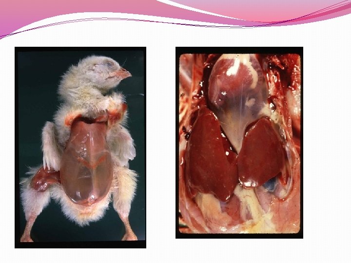
Ascites Syndrome in Poultry (Pulmonary hypertension syndrome, Water belly) High Altitude Disease, Heart Failure Syndrome
Definition �Ascites is an accumulation of non inflammatory transudate in one or more of the peritoneal cavities or potential spaces. �The fluid, which accumulates most frequently in the two ventral hepatic, peritoneal, or pericardial spaces,
�Pulmonary hypertension occurs frequently in chickens secondary to high altitude-associated hypoxia with resultant �may contain yellow protein clots �Ascites may result from increased vascular hydraulic pressure, vascular damage,
The most common cause of ascites �The genetics of meat birds has changed dramatically in the last ten years. �Today's broilers grow much faster, eating less feed. The growth of the heart and lungs has not increased in size proportional to the increase in body weight and breast meat yield. �The rapid growth of the bird means more oxygen demand, requiring more work out of the heart and lungs. .
�Anything that limits oxygen uptake from the lungs is going to cause the heart to work harder. Diseases of the lungs and poor ventilation may be involved.
�Excess levels of sodium in the water or salt in feed leads to increased blood pressure in the lungs �Levels of sodium over 400 ppm could cause problems in broilers.
�High altitudes have long been known to cause heart failure and ascites. �Chilling is a common cause in small flocks. It causes an increased blood flow through the lungs.
�it is usually caused by primary or spontaneous pulmonary hypertension because of insufficient capacity of the pulmonary capillaries. � Cold stress, even briefly, during the first 3 wk of life is known to markedly increase predisposition to ascites syndrome.
� liver damage may be caused by aflatoxin or by toxins from plants such as Crotalaria. �In broiler chickens, obstructive cholangiohepatitis (caused by Clostridium perfringens infection) is the most common cause of the liver damage, which results in ascites. �In both meat-type ducks and breeders, amyloidosis of the liver may also cause ascites.
� However, rapid growth rate is a known predisposing factor, and sometimes the largest broilers are affected, with occurrence in males more frequent than in females.
Pathogenesis and Epidemiology: �Pulmonary hypertension syndrome is caused by increased pressure in the pulmonary arteries when the heart tries to pump more blood through the lungs to meet the body’s oxygen requirement.
�The resultant volume and pressure overload on the right ventricle cause dilatation and hypertrophy of the right ventricular wall, valvular insufficiency, RVF, and ascites. �particularly if increased sodium or lung pathology (eg, aspergillosis). Bacterial or viral affection.
Predisposing factors �Predisposing factors that increase oxygen demand �(eg, cold), reduce oxygen-carrying capacity of the blood (eg, acidosis, carbon monoxide), � increase blood volume (eg, sodium), or interfere with blood flow through the lung (eg, lung pathology that narrows or occludes capillaries,
�increased RBC rigidity, or polycythemia with increased blood viscosity) may result in flock outbreaks of pulmonary hypertension syndrome with or without ascites. �Immediate causes for hypoxaemia are the inadequate ventilation, the high ammonia or dust levels in premises, low temperatures, stimulating the metabolic processes
incidence �The incidence of pulmonary hypertension syndrome is >2% in some broiler and many roaster flocks �occasionally 15%– 20% in other roaster flocks.
�Right ventricular hypertrophy is the response to an increased workload and eventually leads to RVF if the volume or pressure load persists. �Hypertrophy and dilatation of the right ventricular wall is pulmonary hypertension directly related to
Signs �Poor development. �Progressive weakness and abdominal distension. �Recumbency. �Dyspnoea. �Sudden deaths in rapidly developing birds. �Possibly cyanosis.
�The ascites increases the respiratory rate Affected broilers frequently die on their backs. �Not all broilers that die from pulmonary hypertension syndrome have ascites. �Death may occur suddenly before signs are seen.
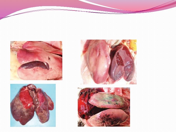
Post-mortem lesions �Thickening of right-side myocardium. �Dilation of the ventricle. �Thickening of atrioventricular valve. �General venous congestion. �Severe muscle congestion.
�Lungs and intestines congested. �Liver enlargement. �Ascites. �Pericardial effusion. �Microscopic - cartilage nodules increased in lung.
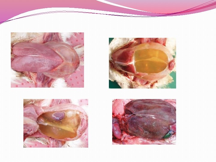
�Hydro pericardium is mild to marked, and occasionally there is pericarditis with adhesions, usually from secondary infections. ight ventricular dilatation and mild to marked hypertrophy of the right ventricular wall may be noted.
�Hydropericardium is mild to marked, and occasionally there is pericarditis with adhesions, usually from secondary infections. �Right ventricular dilatation and mild to marked hypertrophy of the right ventricular wall may be noted.
�The right atrium and vena cava are markedly dilated in most cases. �Occasionally, there is thinning of the left ventricle. The lungs are extremely congested and edematous. �The intestine may or may not be empty.
Diagnosis: �Broilers that die from ascites or suddenly as the result of RVF or pulmonary hypertension can be identified by the enlarged heart. �Enlarged, thickened right ventricle; or fluid in the body cavities and heart sac.
�If the wall of the right ventricle is enlarged or thickened, the broiler has probably died from pulmonary hypertension syndrome, even if there is no fluid in the body or heart sac.
Control: �Reducing the birds’ metabolic oxygen requirement by �slowing growth or reducing feed density or availability can prevent ascites caused by pulmonary hypertension syndrome.
�Environmental temperature, humidity, and air movement should be controlled to prevent excessive loss of body heat, particularly in the early neonatal period. �Ascites caused by other factors (eg, sodium, lung damage, liver damage, etc) can be prevented by avoiding the etiologic agents involved.
�Altitudes >3, 000 ft (900 m) are unsatisfactory for meat-type chickens, and growth must be slowed to prevent mortality. �More care to prevent chilling is also necessary at higher altitudes.
�Research has demonstrated that broilers can be genetically selected for both resistance and susceptibility to pulmonary hypertension syndrome and associated ascites.
CANNIBALISM IN POULTRY �It’s a bad vices in Chickens, turkeys, pheasants and quail will literally pick each other to death at times. �This problem can be very expensive for the producer and can make life for the flock very uncomfortable. �Once cannibalism starts, it readily becomes a habit that must be stopped.
�cannibalism includes feather pulling, toe pecking and head, wing, and tail picking. feather pulling �Prevention is much easier for man and bird than is treatment.
Cause �It is usually impossible to pinpoint any one reason for the start of this behavioral problem in birds. �There are many management conditions that are known to be involved with or related to an outbreak. Some of these are:
�Overcrowding �Insufficient feeder, waterer or nesting space. �Flock nervousness or overexcitement (may be breed related). �Dietary absences or deficiencies. �Incorrect lighting (usually too much light). �Lame birds left in the flock.
�Stresses due to moving birds or making other necessary management changes. �Prolapse of another egg laying female. �Females laying on the floor rather than in a nest or cage. �Timid birds in the flock that are not getting enough feed or water. �Keeping different ages or colors together. Any off- colored chicks in a flock do not have a ghost of chance.
�Extremely high environmental temperatures. �Abrasions or tears that may be the result of an accident or mating. �feed, water, perch or nesting space. �Strain differences in propensity to feather peck and display cannibalism
�Insufficient access to resources, including. �Dietary absences or deficiencies (example: salt deficiency). �Bright lighting. �Injured birds left in the flock. �Large group size.
Prevention �Outbreaks can occur in the best-managed flock. �problems are less likely to arise if preventative measures are in place.
�The first step in a cannibalism control program is to select a genetic stock that is not prone to cannibalism and then �give the birds the best care possible, including adequate feed, carefully managed lighting, safe housing and environmental enrichment.
�Providing a complete ration to meet the nutrient needs for age and type of flock is also very important. Cannibalism has been linked to deficiencies in protein, sodium and phosphorus. �Protein requirements change as chicks grow and should be adjusted based on a recommended feeding schedule.
�Adequate feeder space for all birds to eat simultaneously helps prevent underweight birds that are frequently victims of cannibalism. �A combination of good management, correct lighting and beak trimming will prevent the problem.
�Beak trimming can be used to control the malady even when management is not good. However, trimming alone does not correct poor management and can serve to temporarily "cover-up" management problems that may result in poor performance from the flock, so good management is essential.
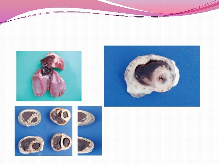
Visceral Gout, Nephrosis, Baby Chick Nephropathy �All poultry species are susceptible �Visceral gout is the deposition of white urates, which are normally excreted as a white cap on well formed faeces, in various tissues. �Urates are also often deposited in joints and in the kidney.
�This condition can occur as an individual problem at any age. �Outbreaks are seen in young chicks in the first week of life (baby chick nephropathy) or in flocks suffering kidney damage, or reduced water intake
�The kidney damage can arise from infection with certain strains of Infectious Bronchtiis, IBD virus, exposure to some mycotoxins or inadequate water intake. � Possible causes for this could be obstruction of ureters, renal damage or dehydration.
�A number of aetiological factors are related to this condition: �protein excess, calcium excess (3% or more), sodium bicarbonate toxicity, mycotoxins (ochratoxin etc. ), vitamin A deficiency and nephrotropic strains bronchitis virus. of the infectious
�Most commonly, visceral gout following dehydration is observed in newly hatched chickens after overheating or a more prolonged stay in the hatchery. �Visceral gout outbreaks are related to vitamin A deficiency, treatment with bicarbonate, mycotoxicoses etc. sodium
�Urolithiasis is an aetiologically unknown state, occurring primarily in cage layer hens, characterized by obstruction of one or both ureters with urates, atrophy of one or more renal lobes and a various degree of renal and visceral gout �The lower phosphorus levels (under 0. 6%) are probably helping the manifestation of the disease.
�A number of aetiological factors are related to this condition: � protein excess, calcium excess (3% or more), sodium bicarbonate toxicity, mycotoxins (ochratoxin etc. ), vitamin A deficiency and nephrotropic strains of the infectious bronchitis virus. The lower phosphorus levels (under 0. 6%) are probably helping the manifestation of the disease
Signs �Depression. �Low feed intake and growth. �There are no specific clinical signs except for the depression and the weight loss. �The death rate could increase and persist around 2 -4% monthly during the productive period. �The total mortality is heavily affected flocks could reach 50%.
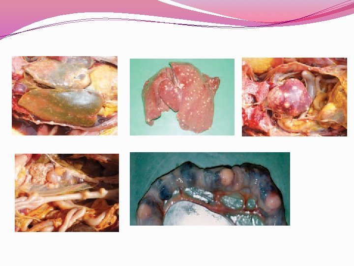
Post-mortem lesions �Chalky white deposits on pericardium, liver, air sacs, peritoneum. �Similar deposits may be present in joints and are usually present in the kidney.
Diagnosis �Lesions.
Treatment �This is based on correcting any management errors and encouraging water intake. �Avoid any intentional or unintentional restriction in water intake. � Sodium bicarbonate at 1 g/litre water is mildly diuretic, however it could be counter-productive if water intake is in any way restricted.
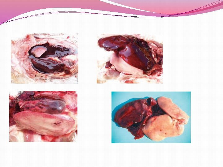
Fatty Liver Haemorrhagic Syndrome �The fatty liver haemorrhagic syndrome (FLHS) is a widely prevalent sporadic disease mainly among commercial layers. �The FLHS outbreaks are often associated with hot weather and a period of extensive egg-laying. �
�The hens in the flock are overweight (on the average by 20% or more) and a sudden drop in egg production is observed. �The birds are discovered suddenly dead, with pale head skin. �In the abdomen, large blood clots are detected.
�Subcapsular parenchymal haematomas are possible. It is assumed that high energy forages and the restricted locomotion are prerequisites for fattening of the liver.
�Other possible contributing factors are the deficiency of lipotropic agents, necessary for fat mobilization by the liver, aflatoxins, genetic factors etc. �Frequently, FLHS and cage layer fatigue are diagnosed at a time.
Signs �Overweight typically by 25%. �Sudden death. �Sudden drop in egg production. �Some birds with pale comb and wattles.
Post-mortem lesions �Obesity. �Headparts pale. �Liver yellow, greasy and soft with numerous haemorrhages. �Death by internal exsanguination after rupture of haematocyst.
Diagnosis �Lesions, history. Treatment �Reduce energy intake, supplement with choline, vitamin E, B 12 and inositol. �Clinically healthy birds in the flock could also exhibit liver haematomas, dark red (fresh) or green to brown (old). Considerable amounts of fat are detected in the abdominal cavity.
Prevention �Feed to avoid obesity, avoid mycotoxins and stress. �The only successful approach for prevention is the Omphalitis reduction of obesity in layers. �The use of lipotropic agents such as vitamin E, vitamin B 12 and choline chloride gives conflicting results. �The avoidance of heat stress and moulded forages could be also helpful.
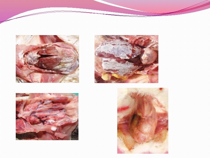
Omphalitis. Mushy Chick Disease Yolk Sack infection �Omphalitis is a common cause of death in chicks during the first week of life and most common with artificially hatched chicks. �It is a bacterial infection of the yolk sac. Various bacteria may be involved in yolk sack infection, E. coli, Staphylococci, Clostridia, fecalis and Pseudomonas. Proteus,
CAUSES �Omphalitis may be caused by a bacterium that enters through the porous egg shell. � incubation conditions are ideal for breeding bacteria as well as incubating eggs. �As the chick is hatching its exposed umbilicus (navel) can easily be infected by bacteria.
�More susceptible are those newly hatched chicks resting on dirty eggs, un-sanitized incubators, and also with assisted hatching. �A chick removed from the incubator prior to complete healing of the navel is also giving a chance of infection in the brooder.
�Transmission: Omphalitis occurs during the first few days of life, so it cannot be considered transmissable from bird to bird. �It is transmitted from unsanitary equipment in the hatchery to newly hatched birds having unhealed navels.
�Symptoms �The chicks become lethargic and die quickly either during incubation or after hatching, with most dying within 48 hours from the infection.
�Symptoms may VARY depending on bacteria and can include �poorly healed, open navels or enlarged navels, �subcutaneous edema (large fluid volume in the abdomen), bluish color of the abdominal muscles and often �unabsorbed yolk materials that could even smell putrid.
�Often yolks are ruptured and peritonitis (inflammation of the inner wall of the abdomen) is common. �Chicks have little interest in food and water and are attracted to heat. � Mortality often begins at hatching and can continue for 10/14 more days.
subcutaneous edema (large fluid volume in the abdomen)
unhealed navel after yolk sack rupture below
unhealed OPEN Navel
Treatments �There is no specific treatment for omphalitis. Most affected birds die. �Prevention is the best method.
�Watch brooder temperatures as Chilling & overheating may increase losses at this stage. Isolate chick from others to prevent spread of bacteria and avoid pecking injuries. �Keep the brooder as clean and fresh as possible. Keep in mind that it may be wiser to humanely destroy the more severely affected chicks.
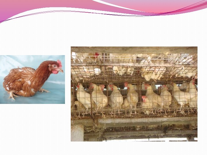
CAGE LAYER FATIGUE �cage layer fatigue syndrome in birds is characterized by an inability to stand on their feet and fragile bones. �It is mainly observed in young layer hens reared in batteries in the period of maximum egg-laying. �Affected birds lie down and stopped eating. Egg shells become thin.
Causes �Reduced intake of calcium, phosphorus and vitamin D 3. �Dietary deficiency of the calcium and phosphorus. �reduced activity within the cage is a predisposing factor.
�High density cages, poor feeder space and fewer feeding periods can generate competition for calcium intake contribute to CLF in dominated birds. and
�Poor absorption of calcium and phosphorus through the gut. �Poor feeding system, generating separation of fine particle limestone in the feeders. �The condition is rarely seen in floor-housed birds, suggesting that
�Signs �High-producing laying hens maintained in cages sometimes show paralysis during and just after the period of peak egg production due to a fracture of the vertebrae that subsequently affects the spinal cord. Birds become lame and are reluctant to stand in the cage.
�Affected birds are invariably found on their sides in the back of the cage. �At the time of initial paralysis, birds appear healthy and often have a shelled egg in the oviduct and an active ovary. �Death occurs from starvation or dehydration because the birds cannot reach feed or water. Egg shells become thin.
Treatment and Management � by giving pullets a high calcium diet (minimum 4. 0% Ca) for at least 7 days prior to first oviposition. �Diets must provide adequate quantities of calcium and phosphorus deficiencies. However, to prevent
�Some cases can be treated with the removal of the scab and the application of Vetericyn 23 times a day until healed. � More advanced cases may need to be surgically treated and some cases may require a course of antibiotics
�A readily assimilable calcium and/or calcium phosphate supplement is effective if started very soon after paralysis due to calcium deficiency develops. � Adding a vitamin/electrolyte supplement to drinking water is recommended in any age bird suffering from this condition.
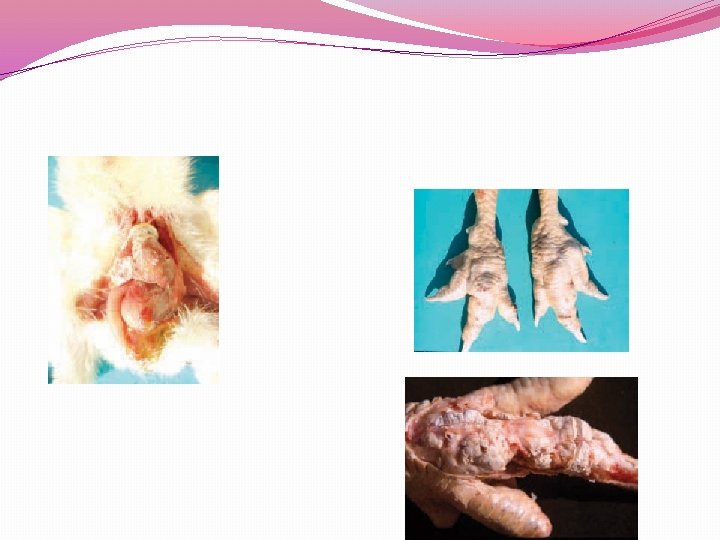
BUMBLEFOOT �Bumblefoot is also known as “plantar pododermatitis” and �is an infection found on the bottom of the feet of chickens and other poultry, which is typically identified by swelling and a dark scab, and/or limping in more advanced cases. Left untreated, it can be fatal.
�Causes: �Bumblefoot can be caused by a cut, scrape or injury to the foot pad, commonly occurring from a splintered roost or repetitive, heavy landings from management. heights or poor litter
�The compromised skin allows an entry point for bacteria (eg: staphylococcus), which can then lead to a pus-filled abscess. � A less common cause of bumblefoot is a vitamin A deficiency.
�Detection: �Regular inspection of your birds’ feet is recommended. �The most common symptoms of bumblefoot include limping or lameness. �Inspection of the foot pad may reveal redness, swelling and either a callous-looking lesion or a black scab.
�Prevention: Provide your chickens with a good, balanced diet, (e. g. : layer pellets for egg-producing hens) proper roosts that are splinter-free and less than 18” in height and properly maintained litter conditions.
Treatment: The affected foot should be cleaned thoroughly with a Betadine solution. �Mild cases can take a "wait and see" approach, but they tend to get worse.
�Some cases can be treated with the removal of the scab and the application of Vetericyn 23 times a day until healed. � More advanced cases may need to be surgically treated and some cases may require a course of antibiotics.
Avian tuberculosis is a chronic infectious disease characterized by the formation of granulomatous lesions in viscera, a progressive weight loss and death. It is usually encountered sporadically in birds reared in small yards, zoos and is a problem among caged exotic birds.
Aetiological agent �The aetiological agent is Mycobacterium avium, a very resilient and acid-resistant microorganism. �It is resistant to temperature changes, drying, p. H changes, to many disinfectants and survives in the soil for years. �Along the small intestine, single or multiple subserously prominating tubercles are detected.
�Causes: �Bumblefoot can be caused by a cut, scrape or injury to the foot pad, commonly occurring from a splintered roost or repetitive, heavy landings from heights or poor litter management.
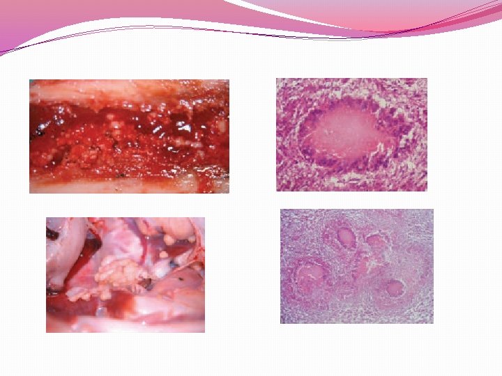
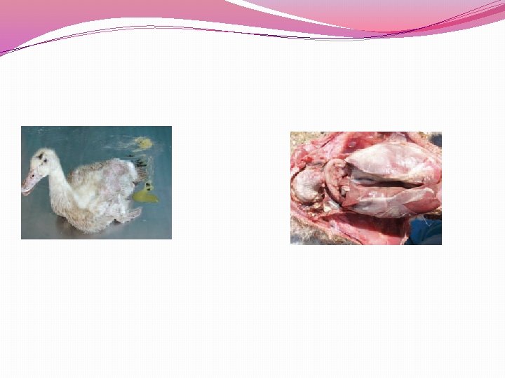
RIEMERELLA ANATIPESTIFER INFECTIONS �Riemerella anatipestifer (RA) infection is a contagious disease in domestic ducks, turkeys and other fowl. It is encountered as acute or chronic septicaemia and is characterized with serous fibrinous polyserosites. The respiratory tract could be also affected. The ducklings at the age of 18 weeks are especially susceptible. Clinically, sneezing, cough, trembling of the head and neck, ataxia and greenish diarrhoea could be present.
�The most characteristic gross lesion is the deposit of fibrinous exudate on the pericardium, the liver capsule or air sacs. The chronic lesions affect the skin and the joints. Although a tentative diagnosis could be made on the basis of observed clinical symptoms and lesions, it is confirmed upon the isolation and identification of RA. The RA infection should be distinguished from septicaemiae due to P. multocida, E. coli, Salmonella etc. The treatment with antibiotics (Flumequine) and sulfonamides IJrimetoprim, Sulfadiazine) has a varying success.
Erysipelas in Poultry �Erysipelas is a bacterial disease caused by infection with Erysipelothrix rhusiopathiae. �The disease is most often seen as septicemia,
Etiology: �E rhusiopathiae is a facultative, anaerobic bacterium. � E rhusiopathiae stains gram-positive but tends to decolorize, particularly in older cultures. � The organism is small, non-acid fast, nonmotile, does not form spores, and produces no known toxins. �There is no flagellum, but a capsule has been demonstrated.
�E rhusiopathiae has three colony types and grows readily on ordinary culture media containing the blood or sera of various animals. � Growth is enhanced by reducing the oxygen content or increasing the carbon dioxide level to 5%– 10%. �Optimal incubation temperature is 35°– 37°C, and the optimal p. H range is 7. 4– 7. 8
�The organism is not readily destroyed by the usual laboratory disinfectants �It may survive in litter or soil for various lengths of time. �It is inactivated by a 1: 1, 000 concentration of bichloride of mercury, 0. 5% sodium hydroxide solution, 3. 5% liquid cresol, 5% solution of phenol, quaternary ammonium, chlorine, or 0. 5% formalin as long as it is not in organic matter.
�Although 26 different serotypes of E rhusiopathiae were described based on an agar gel diffusion test � E rhusiopathiae serotypes 1, 2, and 5 have been most frequently isolated from poultry.
�Erysipelas occurs sporadically in poultry of all ages. �It is ubiquitous in nature and found where nitrogenous substances decompose. �Turkeys are susceptible regardless of sex or age, although under field conditions it is more common in older birds. �The incidence in males is reported to be higher
� Infection results from entrance of the organisms through �breaks in the skin, through the mucous membranes such as during artificial insemination, �by ingestion of contaminated foodstuffs (particularly cannibalism of infected carcasses), and possibly by mechanical transmission via biting insects. �The poultry red mite can harbor the organism and may serve as a mechanical vector. � Fighting and cannibalism increase losses.
�The organism is shed in feces from infected animals and contaminates the soil, in which it may survive for long periods depending on temperature and p. H. �Poultry, as well as other animals, may be carriers and shed the organism without showing clinical signs of disease. �Carriers can shed from feces, urine, saliva, and nasal secretions. � Transmission into poultry houses via rodents can occur.
�In nonvaccinated flocks, morbidity and mortality rates may reach 40%– 50%, but mortality is usually <15%. �In vaccinated flocks, some birds may be depressed for a short period and recover. Mortality in vaccinated and nonvaccinated poultry is influenced by the virulence of the organism.
Clinical Findings: �Erysipelas is primarily an acute infection that results in sudden death. �In an affected flock, a few birds may be depressed but easily aroused; within 24 hr, a few birds will be dead. � Just before death, some birds may be very droopy, with an unsteady gait.
�Chronic clinical disease in a flock is not usual but does occur; birds may have cutaneous lesions and swollen hocks. �Turkeys with vegetative endocarditis usually do not have clinical signs and may die suddenly. �Erysipelas should be suspected in flocks that have been artificially inseminated 4– 5 days before an episode of death without clinical signs.
�Clinical signs in chickens include general weakness, depression, diarrhea, and sudden death. � Most sick birds die. �In laying hens, egg production may drop markedly. �Decreased egg production and conjunctival edema can be seen in organic, cage-free flocks.
Lesions: �At necropsy, a generalized darkening of the skin or various sized areas of diffuse darkening is common. �The liver and spleen are usually enlarged and friable and may be mottled. �Other gross lesions such as peritonitis, pericarditis, petechiation of the heart, catarrhal exudate in the GI tract, and degeneration of fat associated with the thigh and heart may be noted. �Vascular damage and fibrin thrombi are common findings on microscopic examination.
Diagnosis: �A presumptive diagnosis can be based on an impression smear of the liver or spleen or on a smear of cardiac blood or bone marrow that shows gram-positive, slender, pleomorphic rods. � Bone marrow is the tissue of choice in partially decomposed specimens. �Isolation and identification of E rhusiopathiae is necessary for definitive diagnosis
�. Identification can be made by fluorescent antibody staining and PCR can distinguish E rhusiopathiae from E tonsillarum �Infections with Escherichia coli or Pasteurella multocida, as well as salmonellosis and peracute Newcastle disease, may be confused with the septicemic form of erysipelas. Urticaria and endocarditis may be caused by other miscellaneous bacterial or fungal pathogens
�The antibiotic of choice is a rapid-acting penicillin such as potassium or sodium penicillin. As soon as a presumptive diagnosis is made, penicillin should be administered IM at 22, 000 U/kg body wt
What is a disease Any condition that results in deviation from normal function
How do diseases occur? AGENT HOST ENVIRONMENT
ETIOLOGY Infectious Agents �Bacteria �Viruses � Parasites � Fungi Non-infectious agents �Chemical � Physical �Lack or excess of certain vitamins and minerals �Toxins
General Signs of Disease �Poor appetite �Huddling �Depression �Runting/stunting �poor uniformity � Ruffled feathers �Coughing, sneezing, �oculo-nasal discharge, �difficult breathing �Bloody or wet litter � Increased mortality
VIRAL DISEASES
FOWL POX � Viral disease of domestic fowl � development of nodular proliferative Skin lesions on the featherless parts of the body. � fibrino necrotic and proliferative lesions in the mucous membranes
ETIOLOGY �Family: Pox viridae �Genus: avipoxvirus �Double stranded DNA
CLINICAL SIGNS �Appearance of nodular lesions �combs �wattles �eyelids �other unfeathered areas of the body.
PREVENTION AND CONTROL �Prophylactic vaccination �Fowl pox vaccine at 4 -6 weeks of age �Second dose at 12 -14 weeks of age.
RANIKHET DISEASE �New castle disease �Viral disease of domestic fowl is characterised by � respiratory signs �often associated with nervous and digestive disorders �high mortality.
ETIOLOGY �Paramyxoviridae �Paromyxovirus-1
CLINICAL SIGNS opisthotonus �Listlessness �Increased respiration �Weakness �Edema around the eyes �Torticolis �Paralysis of legs
Prevention and control �Prophylactic vaccination �Lentogenic strain (F or B 1) � day old chicks � intranasal � intraocular route � drinking water. �Mesogenic strain (R 2 B) � 6 -8 weeks of age � intramuscular �Subcutaneous route.
BACTERIAL DISEASES
INFECTIOUS CORYZA �FOWL CORYZA �Highly contagious �acute disease of upper respiratory tract of chickens, �turns into a chronic respiratory disease.
ETIOLOGY �Haemophilus paragallinarum �Small cocoid or gram negative rod �Non motile �Exhibits bipolar staining
CLINICAL SIGNS �Serous to mucoid nasal discharges with foul smelling �Facial edema �Conjunctivitis �Swollen wattles �Diarrhoea �Reduced feed and water consumption.
Treatment and control �Gentamicin �Penicillin �streptomycin �delivered in feed or drinking water. �Proper Disinfection
BACILLARY WHITE DIARRHOEA �PULLORUM DISEASE �Fatal septicemia of young chicks. �Salmonella. pullorum
CLINICAL SIGNS �Somnolecence �Weakness �Loss of appetite �Chalky white diarrhoea �Stained greenish brown(sometimes) in and around vent
TREATMENT �Enrofloxacin �Parenteral injections �Oral liquids �Supportive therapy
BUMBLE FOOT PODODERMATITIS Injury to the lower surface of the foot and subsequent infection with Staphylococcus bacteria
Common causes of injury: • Rough perches • Splinters • Wire floors • Poor litter or bedding �quality
CLINICAL SIGNS �Lameness �Swelling of the foot pad �Hard, pus‐filled abscess on foot pad
TREATMENT �Soak foot in warm water and Epsom salts. �disinfect with alcohol. � If skin is open, drain pus from abscess. �Flush abscess cavity with hydrogen peroxide to cleanout pus and debris. �Pack the cavity with antibiotic ointment. �wrap the footwith gauze and elastic bandage. �Repeat daily until foot heals.
PREVENTION AND CONTROL �Provide good quality litter or bedding. �Keep bedding clean, dry, and deep. � Keep perches less than 18 inches from the floor to prevent foot damage due to impact from jumping. �Remove potential sources of injury such as sharp objects and/or surfaces.
DEFECIENCY DISEASES
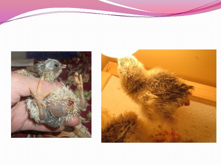
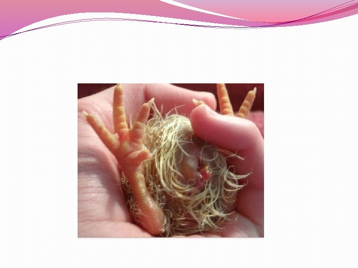
CURLED TOE PARALYSIS �Defeciency of Riboflavin �Poor growth �Weakness �Emaciation and diarrhoea �unable to walk as their toes are turned inwards �Drooping of wings
TREATMENT �Riboflavin @3. 6 mg/kg of feed in chicks �Riboflavin @ 1. 8 mg/kg of feed in growers �Riboflavin @ 2. 2 mg/kg of feed in layers
THANKYOU