Chapter 4 The Tissue Level of Organization Copyright


- Slides: 81

Chapter 4 The Tissue Level of Organization Copyright © John Wiley & Sons, Inc. All rights reserved.

Tissues q Tissues are a group of cells with a common embryonic origin that function together to carry out specialized activities. § They include various types, ranging from hard (bone) to semisolid (fat) to liquid (blood). Copyright © John Wiley & Sons, Inc. All rights reserved.

Tissues q Histology is the study of the microscopic anatomy of cells and tissues. q Of the 10 trillion cells in our body, no single cell type can said to be “typical”. A trained histologist can recognize over 200 distinct human cell types under the microscope and is able to distinguish a cell from pancreatic tissue as opposed to a cell from the skin. • Each cell type has features particular to its function. Copyright © John Wiley & Sons, Inc. All rights reserved.

Intercellular Junctions q Tissues are formed by grouping cells together using a variety of Intercellular Junctions. § Intercellular Junctions connect adjacent cells mechanically at the cell membranes or through cytoskeletal elements within and between cells. Copyright © John Wiley & Sons, Inc. All rights reserved.

Intercellular Junctions q Tight Junctions are found where a leakproof seal is needed between cells. § They keep materials from leaking out of organs like the stomach and bladder. Copyright © John Wiley & Sons, Inc. All rights reserved.

Intercellular Junctions q Adherens Junctions make an adhesion belt (like the belt on your pants) that keeps tissues from separating as they stretch and contract like in the intestines. q Cadherin is a glycoprotein that forms the belt-like “plaque”. Copyright © John Wiley & Sons, Inc. All rights reserved.

Intercellular Junctions q Desmosomes act as “spot welds” to prevent separation under tension. They also use cadherin glycoprotein (plus intermediate filaments) to hook into the cytoplasm. q Common in epidermis and cardiac muscle. Copyright © John Wiley & Sons, Inc. All rights reserved.

Intercellular Junctions q Hemidesmosomes are half-welds that join cells to the basement membrane. Copyright © John Wiley & Sons, Inc. All rights reserved.

q q Intercellular Junctions Gap Junctions have pores (connexons) that allow small substances like ions to pass between cells. If one of the cells gets sick or dies, these seal like a hatch to prevent damage to other cells. Allow nerve and muscle Copyright © John Wiley & Sons, Inc. All rights reserved.

The 4 Basic Tissues q Connective tissues (C. T. ) protect, support, and bind organs. § Fat is a type of C. T. that stores energy. § Red blood cells, white blood cells, and platelets are all C. T. Copyright © John Wiley & Sons, Inc. All rights reserved.

The 4 Basic Tissues q Muscular tissues generate the physical force needed to make body structures move. They also generate heat used by the body. q Nervous tissues detect changes in the body and respond by generating nerve impulses. Copyright © John Wiley & Sons, Inc. All rights reserved.

The 4 Basic Tissues q Of all the cells in the body, they combine to make only 4 basic tissue types: § Epithelial tissues § Connective tissues § Muscular tissues § Nervous tissues Copyright © John Wiley & Sons, Inc. All rights reserved.

The 4 Basic Tissues q Epithelial tissues cover body surfaces and form glands and line hollow organs, body cavities, and ducts. Copyright © John Wiley & Sons, Inc. All rights reserved.

Epithelium q Epithelium is used to line surfaces and form protective barriers. Epithelium is also good at secreting things like mucous, hormones, and other substances. q All epithelia have a free apical surface and an attached basal surface. Copyright © John Wiley & Sons, Inc. All rights reserved.

Epithelium q The basal layer of the epithelium secretes a basal lamina; the underlying C. T. secretes a reticular lamina. § Together the basal lamina and the reticular lamina form a noncellular basement membrane on which the epithelium sits. Copyright © John Wiley & Sons, Inc. All rights reserved.

Epithelium q Epithelia are named according to the shape of their cells, and the thickness or arrangement of their layers (of cells). Copyright © John Wiley & Sons, Inc. All rights reserved.

Epithelium Copyright © John Wiley & Sons, Inc. All rights reserved.

Epithelium q Naming epithelia according to shape Flat, wide “paving stone” cells Cells as tall as they are wide Cells taller than they are wide Copyright © John Wiley & Sons, Inc. All rights reserved.

Epithelium q Naming epithelia according to arrangement One layer. All cells in contact with basement membrane Appears to have layers, but in reality all cells go from the apex to the base Two or more layers. Only basal layer in contact with basement membrane Copyright © John Wiley & Sons, Inc. All rights reserved.

Epithelium q Simple Squamous Epithelium is composed of a single layer of flat cells found: § In the air sacs of lungs § In the lining of blood vessels, the heart, and lymphatic vessels § In all capillaries § As the major part of a serous membrane simple squamous pseudostratified squamous simple cuboidal pseudostratified cuboidal simple columnar pseudostratified columnar transitional Copyright © John Wiley & Sons, Inc. All rights reserved.

Epithelium q Simple Cuboidal Epithelium is composed of a single layer of cube shaped cells. § It is often found lining the tubules of the kidneys and many other glands. simple squamous pseudostratified squamous simple cuboidal pseudostratified cuboidal simple columnar pseudostratified columnar transitional Copyright © John Wiley & Sons, Inc. All rights reserved.

Epithelium q Simple Columnar Epithelium forms a single layer of column-like cells, ± cilia, ± microvilli, ± mucous (goblet cells). Found in entire gastrointestinal tract § Goblet cells are simple columnar cells that have differentiated to acquire the ability to secrete mucous. simple squamous pseudostratified squamous simple cuboidal pseudostratified cuboidal simple columnar pseudostratified columnar transitional Copyright © John Wiley & Sons, Inc. All rights reserved.

q Epithelium Pseudostratified Columnar Epithelium appears to have layers, due to nuclei which are at various depths. In reality, all cells are attached to the basement membrane in a single layer, but some do not extend to the apical surface. . Found in respiratory tract § Ciliated tissue has goblet cells that secrete mucous. simple squamous pseudostratified squamous simple cuboidal pseudostratified cuboidal simple columnar pseudostratified columnar transitional Copyright © John Wiley & Sons, Inc. All rights reserved.

Epithelium q Stratified Squamous Epithelium has an apical surface that is made up of squamous (flat) cells. § The other layers have different shapes, but the name is based on the apical layer. § The many layers are ideal for protection against strong friction forces. simple squamous pseudostratified squamous simple cuboidal pseudostratified cuboidal simple columnar pseudostratified columnar transitional Copyright © John Wiley & Sons, Inc. All rights reserved.

Epithelium q Stratified Cuboidal Epithelium has an apical surface made up of two or more layers of cube-shaped cells. § Locations include the sweat glands and part of the male urethra q Stratified Columnar Epithelium is very rare § Lines esophageal glands and part of anal mucous membrane simple squamous pseudostratified squamous simple cuboidal pseudostratified cuboidal simple columnar pseudostratified columnar Copyright © Johntransitional Wiley & Sons, Inc. All rights reserved.

Epithelium q The cells of Transitional Epithelium change shape depending on the state of stretch in the tissue. § The apical “dome cells” of the top layer (seen here in relaxation) are an identifiable feature and signify an empty bladder. § In a full bladder, the cells are flattened. simple squamous pseudostratified squamous simple cuboidal pseudostratified cuboidal simple columnar pseudostratified columnar transitional Copyright © John Wiley & Sons, Inc. All rights reserved.

Epithelium q Although epithelia are found throughout the body, certain ones are associated with specific body locations. § Stratified squamous epithelium is a prominent feature of the outer layers of the skin. Copyright © John Wiley & Sons, Inc. All rights reserved.

Epithelium § Simple squamous makes up epithelial membranes and lines the blood vessels. § Columnar is common in the digestive tract. § Pseudostratified ciliated columnar is characteristic of the upper respiratory tract. § Transitional is found in the bladder. § Cuboidal lines ducts and sweat glands. Copyright © John Wiley & Sons, Inc. All rights reserved.

q Tissues of the body develop from three primary germ layers: Endoderm, Mesoderm, and Ectoderm § Epithelial tissues from all three germ layers § C. T. and muscle are derived from mesoderm. § Nervous tissue develops from ectoderm. Copyright © John Wiley & Sons, Inc. All rights reserved.

Covering and Lining Epithelium q Endothelium is a specialized simple squamous epithelium that lines the entire circulatory system from the heart to the smallest capillary – it is extremely important in reducing turbulence of flow of blood. q Mesothelium is found in serous membranes such as the pericardium, pleura, and peritoneum. § Unlike other epithelial tissue, both are derived from embryonic mesoderm (the middle layer of the 3 primary germ layers of the embryo). Copyright © John Wiley & Sons, Inc. All rights reserved.

Connective Tissue q Connective Tissues are the most abundant and widely distributed tissues in the body – they are also the most heterogeneous of the tissue groups. § They perform numerous functions: • Bind tissues together • Support and strengthen tissue • Protect and insulate internal organs • Compartmentalize and transport • Energy reserves and immune responses Copyright © John Wiley & Sons, Inc. All rights reserved.

Connective Tissues q Collagen is the main protein of C. T. and the most abundant protein in the body, making up about 25% of total protein content. q Connective tissue is usually highly vascular and supplied with many nerves. § The exception is cartilage and tendon - both have little or no blood supply and no nerves. Copyright © John Wiley & Sons, Inc. All rights reserved.

Connective Tissues q Although they are a varied group, all C. T. share a common “theme”: § Sparse cells § Surrounded by an extracellular matrix q The extracellular matrix is a non-cellular material located between and around the cells. § It consists of protein fibers and ground substance (the ground substance may be fluid, semifluid, gelatinous, or calcified. ) Copyright © John Wiley & Sons, Inc. All rights reserved.

Cells Of Connective Tissues q Common C. T. cells § Fibroblasts are the most numerous cell of connective tissues. These cells secrete protein fibers (collagen, elastin, & reticular fibers) and a “ground substance” which varies from one C. T. to another. Copyright © John Wiley & Sons, Inc. All rights reserved.

Cells of Connective Tissues q Of the other common C. T. cells: § Chondrocytes make the various cartilaginous C. T. § Adipocytes store triglycerides. § Osteocytes make up bone. § White blood cells provide immune response. Copyright © John Wiley & Sons, Inc. All rights reserved.

Connective Tissues q There are 5 types of white blood cells (WBCs): § Macrophages are the “big eaters” that swallow and destroy invaders or debris. They can be fixed or wandering. § Neutrophils are also macrophages (“small eaters”) that are numerous in the blood. § Mast cells and Eosinophils play an important role in inflammation. § Lymphocytes secrete antibody proteins and attack invaders. Copyright © John Wiley & Sons, Inc. All rights reserved.

Connective Tissues q C. T. cells secrete 3 common fibers: § Collagen fibers • Strong and tension resistant • Usually found in parallel bundles • Found in most connective tissues but very abundant in dense regular, bone, and cartilage § Elastic fibers • Can be stretched and then returned to original shape • Found in skin, blood vessel walls, lung tissue, and bone coverings Copyright © John Wiley & Sons, Inc. All rights reserved.

Connective Tissues q Reticular fibers § Provide support and strength § Create a “net-like” framework that is abundant in reticular tissue § Thinner than collagen fibers Copyright © John Wiley & Sons, Inc. All rights reserved.

Connective Tissue Classification Outline q Embryonic connective tissue § Mesenchyme § Mucous connective tissue q Mature connective tissue § Loose connective tissue § Dense connective tissue § Cartilage § Bone § Liquid Copyright © John Wiley & Sons, Inc. All rights reserved.

Embryonic Connective Tissues q There are 2 Embryonic Connective Tissues: § Mesenchyme gives rise to all other connective tissues. § Mucous C. T. is a gelatinous substance within the umbilical cord and is a rich source of stem cells. Copyright © John Wiley & Sons, Inc. All rights reserved.

Mature Connective Tissues q Loose Connective Tissues § Areolar Connective Tissue is the most widely distributed in the body. It contains several types of cells and all three fiber types. • It is used to attach skin and underlying tissues, and as a packing between glands, muscles, and nerves. § Adipose § Reticular Copyright © John Wiley & Sons, Inc. All rights reserved.

Mature Connective Tissues q Loose Connective Tissues § Loose areolar § Adipose tissue is located in the subcutaneous layer deep to the skin and around organs and joints. • It reduces heat loss and serves as padding and as an energy source. § Reticular Copyright © John Wiley & Sons, Inc. All rights reserved.

Mature Connective Tissues q Loose Connective Tissues § Loose areolar § Adipose § Reticular connective tissue is a network of interlacing reticular fibers and cells. • It forms a scaffolding used by cells of lymphoid tissues such as the spleen and lymph nodes. Copyright © John Wiley & Sons, Inc. All rights reserved.

Mature Connective Tissues q Dense Connective Tissues § Dense Irregular Connective Tissue consists predominantly of fibroblasts and collagen fibers randomly arranged. • It provides strength when forces are pulling from many different directions. Occur in sheets around organs and in the dermis § Dense regular § Elastic Copyright © John Wiley & Sons, Inc. All rights reserved.

Mature Connective Tissues q Dense Connective Tissues § Dense Irregular § Dense regular Connective Tissue comprise tendons, ligaments, and other strong attachments where the need for strength along one axis is mandatory (a muscle pulling on a bone). § Elastic Copyright © John Wiley & Sons, Inc. All rights reserved.

Mature Connective Tissues q Dense Connective Tissues § Dense Irregular § Dense regular § Elastic Connective Tissue consists predominantly of fibroblasts and freely branching elastic fibers. • It allows stretching of certain tissues like the elastic arteries (the aorta). Copyright © John Wiley & Sons, Inc. All rights reserved.

Mature Connective Tissues q Cartilage is a tissue with poor blood supply that grows slowly. When injured or inflamed, repair is slow. § Hyaline cartilage is the most abundant type of cartilage; it covers the ends of long bones and parts of the ribs, nose, trachea, bronchi, and larynx. • It provides a smooth surface for joint movement. § Fibrocartilage § Elastic cartilage Copyright © John Wiley & Sons, Inc. All rights reserved.

Mature Connective Tissues q Cartilage § Hyaline cartilage § Fibrocartilage, with its thick bundles of collagen fibers, is a very strong, tough cartilage. • Fibrocartilage discs in the intervertebral spaces and the knee joints support the huge loads up and down the long axis of the body. § Elastic cartilage Copyright © John Wiley & Sons, Inc. All rights reserved.

Mature Connective Tissues q Cartilage § Hyaline cartilage § Fibrocartilage § Elastic cartilage consists of chondrocytes located in a threadlike network of elastic fibers. • It makes up the malleable part of the external ear and the epiglottis. Copyright © John Wiley & Sons, Inc. All rights reserved.

Mature Connective Tissues q Cartilage § Two types of growth • Interstitial – from within • Appositional – growth from outer surface Copyright © John Wiley & Sons, Inc. All rights reserved.

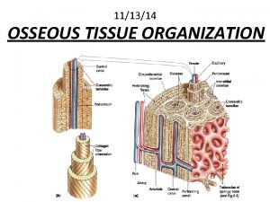
Mature Connective Tissues q Bone is a connective tissue with a calcified intracellular matrix. In the right circumstances, the chondrocytes of cartilage are capable of turning into the osteocytes that make up bone tissue. Copyright © John Wiley & Sons, Inc. All rights reserved.

Mature Connective Tissues q Blood and lymph are atypical liquid connective tissues. As we have seen, blood has many cells. It also has fibers (such as fibrin that makes blood clot). Copyright © John Wiley & Sons, Inc. All rights reserved.

Summary of Mature Connective Tissues

Muscle and Nerve Tissues q Muscles and nerve tissues are the last of the 4 basic tissue types. q Neurons and muscle fibers are considered excitable cells because they exhibit electrical excitability, the ability to respond to certain stimuli by producing electrical signals such as action potentials. § Action potentials can propagate (travel) along the plasma membrane of a neuron or muscle fiber due to the presence of specific voltage-gated ion channels. Copyright © John Wiley & Sons, Inc. All rights reserved.

Muscle Tissue q Skeletal Muscle § Under voluntary control § Attached to bones via tendons § Functions: Provide motion, heat production, protection, posture § Multi-nucleated § Contains long, cylindrical fibers that are striated • Striations – alternating light and dark bands (stripes) Copyright © John Wiley & Sons, Inc. All rights reserved.

Skeletal muscle fiber (cell) Nucleus Striations LM 400 x Longitudinal section of skeletal muscle tissue Skeletal muscle fiber Copyright © John Wiley & Sons, Inc. All rights reserved.

Muscle Tissue q Cardiac Muscle § Under involuntary control § Found in the heart wall § Function: Pump blood to the body § Usually one nucleus per cell § Has branched, striated fibers with intercalated discs • Intercalated discs – help hold the tissue together under a lot of stress and allow for quick electrical signals Copyright © John Wiley & Sons, Inc. All rights reserved.

Nucleus Cardiac muscle fiber (cell) Intercalated disc Heart Striations LM 500 x Longitudinal section of cardiac muscle tissue Cardiac muscle fibers Copyright © John Wiley & Sons, Inc. All rights reserved.

Muscle Tissue q Smooth Muscle § Under involuntary control § Found in the walls of hollow structures like blood vessels and intestines § Function: Motion § One nucleus per cell § No visible striations § Cells are tapered at each end Copyright © John Wiley & Sons, Inc. All rights reserved.

Smooth muscle fiber (cell) Nucleus of smooth muscle fiber Artery LM 500 x Longitudinal section of smooth muscle tissue Smooth muscle fiber Copyright © John Wiley & Sons, Inc. All rights reserved.

Nervous Tissue q Consists of Neurons (nerve cells) and neuroglia (support cells) q Neurons transmit nerve impulses throughout the body. We will talk about this in much more detail later in the year. Copyright © John Wiley & Sons, Inc. All rights reserved.

Dendrite Nucleus of neuroglial cell Nucleus in cell body Spinal cord Axon LM 400 x Neuron of spinal cord Copyright © John Wiley & Sons, Inc. All rights reserved.

Muscle and Nerve Tissues Copyright © John Wiley & Sons, Inc. All rights reserved.

Epithelial Membranes q Combining two tissues creates an organ. However, most of the organs and all of the organs systems studied this year contain all 4 basic types of tissues. § Epithelial membranes are the simplest organs in the body, constructed of only epithelium and a little bit of connective tissue. Copyright © John Wiley & Sons, Inc. All rights reserved.

Epithelial Membranes q Epithelial membranes = epithelium + connective tissue § Mucous membranes § Serous membranes § Cutaneous membrane = skin • Skin is not a simple organ Copyright © John Wiley & Sons, Inc. All rights reserved.

Epithelial Membranes q Mucous membranes line “interior” body surfaces open to the outside: § Digestive tract § Respiratory tract § Reproductive tract q Serous membranes line some internal surfaces: § Parietal layer next to body wall § Serous fluid between layers § Visceral layer next to organ Copyright © John Wiley & Sons, Inc. All rights reserved.

Synovial Membranes q Synovial membranes enclose certain joints and are made of connective tissue only. Copyright © John Wiley & Sons, Inc. All rights reserved.

Glands q Epithelial glands are another example of simple organs § Glands that secrete their contents directly into the blood are called endocrine glands. § Glands that secrete their contents into a lumen or duct are called exocrine glands. Copyright © John Wiley & Sons, Inc. All rights reserved.

Exocrine Glands q Exocrine glands secrete substances through ducts to the surface of the skin or into the lumen of a hollow organ. § Secretions of the exocrine gland include mucus, sweat, oil, earwax, saliva, and digestive enzymes. Copyright © John Wiley & Sons, Inc. All rights reserved.

Exocrine Glands q The two criteria for categorizing multicellular glands according to structure: § Whether the ducts are branched or unbranched… • In a simple gland the duct does not branch. • In a compound gland the duct branches. § … and the shape of the secretory portion of the gland • Tubular glands have tubular secretory parts. • Acinar glands have rounded secretory parts. • Tubuloacinar glands have features of both. Copyright © John Wiley & Sons, Inc. All rights reserved.

Exocrine Glands unbranched duct (simple) branched duct (compound) Copyright © John Wiley & Sons, Inc. All rights reserved.

Exocrine Glands tubular shape in secretory portion Copyright © John Wiley & Sons, Inc. All rights reserved.

Exocrine Glands acinar or alveolar (grape-like) shape in secretory portion Copyright © John Wiley & Sons, Inc. All rights reserved.

Exocrine Glands q The criteria for categorizing multicellular glands according to function is based on the manner in which the gland secretes its product from inside the cell to the outside environment. § Merocrine § Apocrine § Holocrine Copyright © John Wiley & Sons, Inc. All rights reserved.

Exocrine Glands q Merocrine secretion is the most common manner of secretion. § The gland releases its product by exocytosis and no part of the gland is lost or damaged. Copyright © John Wiley & Sons, Inc. All rights reserved.

Exocrine Glands q Apocrine glands “bud” their secretions off through the plasma membrane, producing membrane-bound vesicles in the lumen of the gland. § The end of the cell breaks off by “decapitation”, leaving a milky, viscous odorless fluid. § This type of sweat only develops a strong odor when it comes into contact with bacteria on the skin surface. Copyright © John Wiley & Sons, Inc. All rights reserved.

Exocrine Glands q Holocrine secretions are produced by rupture of the plasma membrane, releasing the entire cellular contents into the lumen and killing the cell (cells are replaced by rapid division of stem cells. ) § The sebaceous gland is an example of a holocrine gland, because its secretion (sebum) is released with remnants of dead cells. Copyright © John Wiley & Sons, Inc. All rights reserved.

Tissue Repair q A convenient way to refer to certain cells when discussing a tissue is Parenchyma or Stroma. § The parenchymal cells of an organ consist of that tissue which conducts the specific function of the organ. Cells of the stroma are everything else— connective tissue, blood vessels, nerves. q For example: The parenchyma of the heart is cardiac muscle cells. The nerves, intrinsic blood vessels, and connective tissue of the heart comprise the stroma. Copyright © John Wiley & Sons, Inc. All rights reserved.

Tissue Repair q Parenchyma is interesting. Because organ-specific function usually centers on parenchymal cells (“how’s your heart working? ”), histological and physiological descriptions of the tissues of an organ often emphasize parenchyma. q Unfortunately, stroma is commonly ignored as just boring background tissue. No organ, however, can function without the mechanical and nutritional support provided by the stroma. Copyright © John Wiley & Sons, Inc. All rights reserved.

Tissue Repair q When tissue damage is extensive, return to homeostasis depends on active repair of both parenchymal cells and stroma. § Fibroblasts divide rapidly. § New collagen fibers are manufactured. § New blood capillaries supply materials for healing. q All of these processes create an actively growing connective tissue called granulation tissue. Copyright © John Wiley & Sons, Inc. All rights reserved.

Aging and Tissues q Tissue heals faster in young adults. q Surgery of a fetus normally leaves no scars. q Young tissues have a better nutritional state, blood supply, and higher metabolic rate. q Extracellular components also change with age. q Changes in the body’s use of glucose, collagen, and elastic fibers contribute to the aging process. Copyright © John Wiley & Sons, Inc. All rights reserved.
 Chapter 4 the tissue level of organization
 Chapter 4 the tissue level of organization
 Chapter 4 the tissue level of organization
 Porifera and cnidaria
 Jaringan epitel dapat ditemukan di
 Chapter 3 the cellular level of organization
 The chemical level of organization
 The chemical level of organization chapter 2
 Process organization in computer organization
 Block essay
 Food pyramid biology
 Hát kết hợp bộ gõ cơ thể
 Ng-html
 Bổ thể
 Tỉ lệ cơ thể trẻ em
 Chó sói
 Glasgow thang điểm
 Hát lên người ơi alleluia
 Các môn thể thao bắt đầu bằng tiếng bóng
 Thế nào là hệ số cao nhất
 Các châu lục và đại dương trên thế giới
 Cong thức tính động năng
 Trời xanh đây là của chúng ta thể thơ
 Mật thư anh em như thể tay chân
 Phép trừ bù
 Phản ứng thế ankan
 Các châu lục và đại dương trên thế giới
 Thơ thất ngôn tứ tuyệt đường luật
 Quá trình desamine hóa có thể tạo ra
 Một số thể thơ truyền thống
 Cái miệng bé xinh thế chỉ nói điều hay thôi
 Vẽ hình chiếu vuông góc của vật thể sau
 Thế nào là sự mỏi cơ
 đặc điểm cơ thể của người tối cổ
 V. c c
 Vẽ hình chiếu đứng bằng cạnh của vật thể
 Tia chieu sa te
 Thẻ vin
 đại từ thay thế
 điện thế nghỉ
 Tư thế ngồi viết
 Diễn thế sinh thái là
 Dạng đột biến một nhiễm là
 Số nguyên là gì
 Tư thế ngồi viết
 Lời thề hippocrates
 Thiếu nhi thế giới liên hoan
 ưu thế lai là gì
 Sự nuôi và dạy con của hổ
 Sự nuôi và dạy con của hươu
 Hệ hô hấp
 Từ ngữ thể hiện lòng nhân hậu
 Thế nào là mạng điện lắp đặt kiểu nổi
 Organization level
 Organization-level diagnostic model
 Middle level management examples
 What is the least complex level of organization
 Ecology levels of organization
 Level of organization organ system
 Levels of mdm
 Smallest to largest level of organization
 Cellular level of organisation
 Example of management information system
 Three level cache organization
 What level of organization
 Lower level management
 Levels of ecological organization
 Porifera
 Molecular level vs cellular level
 Isis level 1 vs level 2
 Dr shaffi
 Isis level 1 vs level 2
 Significance level and confidence level
 Confidence level and significance level
 Data flow diagram level 0 1 2 examples
 Security level 0
 Level questions
 Thread level parallelism in computer architecture
 Low-level thinking in high-level shading languages
 What is poly foundation programme
 Chapter 14 bleeding shock and soft tissue injuries
 Muscles and muscle tissue chapter 9

