Topical Agents Definition Topical Place the use of


 is a silver- white metal as protective agents")


 • Pronounced – (an-jee-oh-kar-dee-OG-rah-fee) – (CAR-dee-ak kath-eh-ter-ih-ZAY-shun) • Defined – Specialized")
 • Defined – Series of X-ray films allowing visualization")
 – (SER-eh-bral an-jee-OG-rah-fee) • Defined – Injection")
 • Defined – X-ray visualization of internal")
 • Defined – X-ray visualization of arteries following the")
 • Defined – Process of taking X-rays of the")
 • Pronounced – (BAH-ree-um EN-eh-mah) • Defined – Infusion of a")
 • Pronounced – (BAH-ree-um SWALL-oh) • Defined – Oral")
 • Defined – Bronchial examination via X-ray following the")
 • Pronounced – (koh-lan-jee-OG-rah-fee) – (in-trah-VEE-nus) • Defined – Visualizing and outlining")
 • Pronounced – (koh-lan-jee-OG-rah-fee) – (per-kyoo-TAY-nee-us trans-heh-PAT-ik) • Defined – Examination")
 • Pronounced – (koh-lan-jee-oh-pan-kree-ah-TOG-rah-fee) – (en-doh-SKOP-ic RET-roh-grayd) • Defined – Procedure")
 • Pronounced – (koh-lee-sis-TOG-rah-fee) • Defined – Visualization of the gallbladder through")
 • Defined – Diagnostic technique combining the techniques of")
 • Pronounced – (kom-PEW-ted AK-see-al toh-MOG-rah-fee) • Defined –")

 • Defined – X-ray visualization of the")
 • Pronounced – (DIJ-ih-tal sub-TRAK-shun an-jee-OG-rah-fee) • Defined – X-ray")
 • Defined – Diagnostic procedure for studying the structure")
 • Defined – Radiological technique used to examine the")
 • Defined – X-ray assessment of uterus and fallopian")
 • Defined – X-ray assessment of lymphatic system following")
 • Pronounced – (mag-NET-ik REZ-oh-nans IM-ij-ing) • Defined – Noninvasive")
 • Defined – Process of taking X-rays of the")
 • Defined – Introduction of contrast medium into the")
 (IVP) • Pronounced – (pye-eh-LOG-rah-fee) – (in-trah-VEE-nus) • Defined – Radiographic procedure")
 • Defined – Delivery of ionizing radiation")

 • Defined – Examination that")
 • Pronounced – (SCAN-ing) • Defined – Scanning is")
 • Defined – Image of the area being studied")


 • Pronounced – (single FOH-ton ee-MISH-un kom-PEW-ted toh-MOG-rah-fee) •")
 • Defined – Oral administration")
 • Defined – X-ray technique used to construct a")
 • Pronounced – (ull-trah-son-OG-rah-fee) • Defined – Procedure in which sound waves")


 • Defined – Technique used to prepare an X-ray")
 • Defined – Use of high-energy electromagnetic waves, passing")

 • X-rays pass through the posterior")
- Slides: 69
Topical Agents
Definition • Topical : Place the use of these drugs or compounds on the surface • Ex: Antiseptic • Systemic: The drugs are absorbed into circulatory system and distributed to various organs or tissues.
Types of inorganic topical agents • The compounds used topically will be divided into broad categories based on their usual action or uses. • There are TWO types: • 1 - Protective agents. • 2 - Antimicrobial agents.
Protective agents • Are substances which may be applied to skin to protect certain areas from irritation. • Properties of protective agents are insoluble in water ( H 2 O), and chemically inert ( unreactive), in order to prevent interactions between the protective substance and the tissue. • Also, small fine partials(large surface area).
Classification of protective agents • 1 - Dusting powders. • 2 -Suspensions. • 3 - Ointments
Talc • Mg. O. 4 Si. O 2. H 2 O • Hydrous magnesium silicate, very fine white powder, smoothly, greasy feeling to the touch( soapstone). • Talc is characterized as fine powder ( 80/100) mesh particle size, odorless, insoluble in water , dil HCl, bases.
Uses of Talc • 1 - It is useful in lubricating, protective dusting, • Can be prevent any friction. • 2 -Used for wound & surgical incision because can produce sterile abscesses • 3 - Used in medical gloves. ( plastic disposable) • 4 - Used in cosmetic may be perfumed • Medical Talc by mixing boric acid with talc as antimicrobial agents.
Zinc Oxide • The chemical formula for Zinc oxide is Zn. O • Is a very fine, oderless, amorphous , white or fait yellow powdwer, can heated 400 -500 ∙C • Zno is insoluble in water, alcohol, but react with dil HCl • Zn. O + HCl ----- Zn. Cl 2 + H 2 O
Uses of Zinc Oxide • 1 - Is a mild astringent and weak antimicrobial agents. Used as powder, ointments to protect the skin. • 2 - Dusting powder used in the treatment of skin ulceration & other dermatological problems. • Medicated zinc oxide by mixing with boric acid as antimicrobial agents.
Calamine • The formula is Zn. O. x. Fe 2 O 3 • Can be synthesized by mixing Zn. O with ferric oxide. • It is a fine powder, water insoluble, alcohols , adhering to the skin.
Uses of Calamine • Calamine can be used as topical protective agent. • 1 - It is USP product used as dust powder, calamine lotion ( applied to skin )pink color. • Calamine is applied to the skin for its adsorbent, protective properties, used in dermatological problems.
• The calamine lotion contain Zn. O and ferric oxide equally mixed Bentonite magma in the solution of Calcium hydroxide. • Phenolated calamine lotion( USP ) contains 1% liquid which provide a local anesthetic and anti- itching action.
Titanium Dioxide • Ti. O 2 as topical protective agent, it is characterized by off white powder color, tasteless, odorless. A 1 to 10 aqueous suspension of the solution is neutral to litmus paper. • It is insoluble in water , HCl. HNO 3, dil H 2 SO 4. • It is soluble in HF, Conc. H 2 SO 4. Also, in H 2 O 2 • Ti. O 2 + H 2 O 2 ------- • Tritium dioxide Ti. O 3 Titanium trioxide
Uses of Ti. O 2 • Ti. O 2 is a protective agent, topically, used as Sun screen light ( UV light radiation) due to its highly refractive index. • Many pharmaceutical preparation as Ti. O 2 cream, 5, 10, . . 25% ointments. • Other organic sun screen agent p- aminobenzoic acid ( PABA). • Ti. O 2 used in cosmetic and paints.
Aluminum • Aluminum ( Al ) is a silver- white metal as protective agents , highly affinity to O 2 forming Al 2 O 3 as protective layer. • It is insoluble in water, alcohols, and uncreative towards HNO 3, H 2 SO 4 • It react rapidly with dil HCl • HCl + Al ------ Al. Cl 3. 6 H 2 O • Aluminum, paste ( Zn. O with base ) used for prevent irritation.
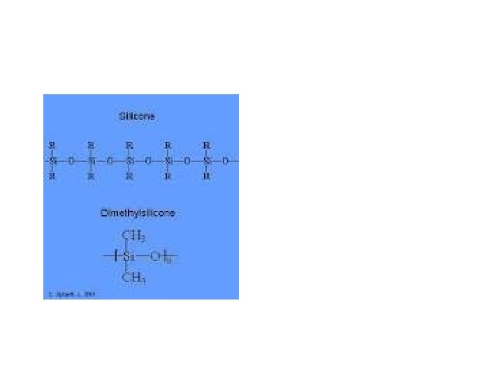
Silicon Polymer • There are inert protective substances occurring in liquid form known as Silicon oils; e. g. dimethyl silicon ( Dimethicon, or Simethicone ) • CH 3 • CH 3 Si-O- ( - Si - O ) ------Si - CH 3 • CH 3 • Very well adhere to skin, but not wound, be avoided contact to the eyes.
Radiology and Diagnostic Imaging
Radiology and Diagnostic Imaging Overview • X-rays – High-energy electromagnetic waves – Travel in straight lines – Shorter wave length than visible light – Able to penetrate solid materials of varying densities – Capable of exposing a photographic plate (Xray film) • Much the same way as a camera exposes film
Radiology and Diagnostic Imaging Overview • X-rays – Used to visualize internal organs and structures of body – Provide valuable means for verifying presence of illness or disease • Radiology – Study of the diagnostic and therapeutic uses of X -rays
PROCEDURES AND TECHNIQUES Radiology and Diagnostic Imaging
Angiocardiography (Cardiac Catheterization) • Pronounced – (an-jee-oh-kar-dee-OG-rah-fee) – (CAR-dee-ak kath-eh-ter-ih-ZAY-shun) • Defined – Specialized diagnostic procedure in which a catheter is introduced into a large vein or artery – Usually of an arm or a leg, and is then threaded through circulatory system to the heart
Angiography • Pronounced – (an-jee-OG-rah-fee) • Defined – Series of X-ray films allowing visualization of internal structures after the introduction of a radiopaque substance
Cerebral Angiography • Pronounced – (seh-REE-bral an-jee-OG-rah-fee) – (SER-eh-bral an-jee-OG-rah-fee) • Defined – Injection of a radiopaque contrast medium into an arterial blood vessel (carotid, femoral, or brachial) to make visualization of the cerebral vascular system via X-ray possible
Renal Angiography • Pronounced – (REE-nal an-jee-OG-rah-fee) • Defined – X-ray visualization of internal anatomy of the renal blood vessels (blood vessels of the kidney) after injection of a contrast medium
Arteriography • Pronounced – (ar-tee-ree-OG-rah-fee) • Defined – X-ray visualization of arteries following the introduction of a radiopaque contrast medium into the bloodstream through a specific vessel by way of a catheter
Arthrography • Pronounced – (ar-THROG-rah-fee) • Defined – Process of taking X-rays of the inside of a joint, after a contrast medium has been injected into the joint • Contrast medium makes the inside of the joint visible
Barium Enema (BE) • Pronounced – (BAH-ree-um EN-eh-mah) • Defined – Infusion of a radiopaque contrast medium, barium sulfate, into the rectum – Contrast medium is retained in lower intestinal tract while X-ray films are obtained of the lower GI tract
Barium Swallow (Upper GI Series) • Pronounced – (BAH-ree-um SWALL-oh) • Defined – Oral administration of a radiopaque contrast medium, barium sulfate, which flows into the esophagus as the person swallows • X-rays are taken as barium sulfate flows into the upper GI tract
Bronchography • Pronounced – (brong-KOG-rah-fee) • Defined – Bronchial examination via X-ray following the coating of the bronchi with a radiopaque substance
Cholangiography (Intravenous) • Pronounced – (koh-lan-jee-OG-rah-fee) – (in-trah-VEE-nus) • Defined – Visualizing and outlining of the major bile ducts following an intravenous injection of a contrast medium
Cholangiography (Percutaneous Transhepatic) • Pronounced – (koh-lan-jee-OG-rah-fee) – (per-kyoo-TAY-nee-us trans-heh-PAT-ik) • Defined – Examination of bile duct structure using a needle to pass directly into an intrahepatic bile duct to inject a contrast medium • Also known as PTC or PTHC
Cholangiopancreatography (Endoscopic Retrograde) • Pronounced – (koh-lan-jee-oh-pan-kree-ah-TOG-rah-fee) – (en-doh-SKOP-ic RET-roh-grayd) • Defined – Procedure that examines the size of and the filling of the pancreatic and biliary ducts through direct radiographic visualization with a fiberoptic endoscope
Cholecystography (Oral) • Pronounced – (koh-lee-sis-TOG-rah-fee) • Defined – Visualization of the gallbladder through X-ray following the oral ingestion of pills containing a radiopaque iodinated dye
Cineradiography • Pronounced – (sin-eh-ray-dee-OG-rah-fee) • Defined – Diagnostic technique combining the techniques of fluoroscopy, radiography, and cinematography by filming the images that develop on a fluorescent screen with a movie camera
Computed Axial Tomography (CT, CAT) • Pronounced – (kom-PEW-ted AK-see-al toh-MOG-rah-fee) • Defined – Painless, noninvasive diagnostic X-ray procedure using ionizing radiation that produces a crosssectional image of the body
Computed Axial Tomography
Voiding Cystourethrography • Pronounced – (VOYD-ing sis-toh-yoo-ree-THROG-rah-fee) • Defined – X-ray visualization of the bladder and urethra during the voiding process, after the bladder has been filled with a contrast material
Digital Subtraction Angiography (DSA) • Pronounced – (DIJ-ih-tal sub-TRAK-shun an-jee-OG-rah-fee) • Defined – X-ray images of blood vessels only, appearing without any background, due to the use of a computerized digital video subtraction process
Echocardiography • Pronounced – (ek-oh-kar-dee-OG-rah-fee) • Defined – Diagnostic procedure for studying the structure and motion of the heart via ultrasound • Useful in evaluating structural and functional changes in a variety of heart disorders
Fluoroscopy • Pronounced – (floor-or-OSS-koh-pee) • Defined – Radiological technique used to examine the function of an organ or a body part using a fluoroscope
Hysterosalpingography • Pronounced – (his-ter-oh-sal-ping-OG-rah-fee) • Defined – X-ray assessment of uterus and fallopian tubes by injecting a contrast material into these structures
Lymphangiography • Pronounced – (lim-fan-jee-OG-rah-fee) • Defined – X-ray assessment of lymphatic system following injection of a contrast medium into lymph vessels in the hand or foot
Magnetic Resonance Imaging (MRI) • Pronounced – (mag-NET-ik REZ-oh-nans IM-ij-ing) • Defined – Noninvasive scanning procedure that provides visualization of fluid, soft tissue, and bony structures without the use of radiation
Mammography • Pronounced – (mam-OG-rah-fee) • Defined – Process of taking X-rays of the soft tissue of the breast to detect various benign and/or malignant growths before they can be felt
Myelography • Pronounced – (my-eh-LOG-rah-fee) • Defined – Introduction of contrast medium into the lumbar subarachnoid space through a lumbar puncture to visualize the spinal cord and vertebral canal through X-ray examination
Pyelography (Intravenous) (IVP) • Pronounced – (pye-eh-LOG-rah-fee) – (in-trah-VEE-nus) • Defined – Radiographic procedure that provides visualization of the entire urinary tract: kidneys, ureters, bladder, and urethra • Also known as intravenous pyelogram or excretory urogram
Radiation Therapy • Pronounced – (ray-dee-AY-shun THAIR-ah-pee) • Defined – Delivery of ionizing radiation to accomplish one or more of the following: • • • Destruction of tumor cells Reduction of tumor size Decrease in pain Relief of obstruction To slow or stop the spread of cancer cells
Radiation Therapy • Radiation therapy – Destroys rapidly multiplying cells regardless of whether they are cancerous – Goal is to reach maximum tumor control with no, or minimal, normal tissue damage – May be delivered by teletherapy (external) – May be delivered by brachytherapy (internal)
Radioactive Iodine Uptake • Pronounced – (ray-dee-oh-AK-tiv EYE-oh-dine UP-tayk) • Defined – Examination that determines the position, size, shape, and physiological function of the thyroid gland through the use of radionuclear scanning • Image of the thyroid is recorded and visualized after a radioactive substance is given
Scanning (Bone, Brain, Liver, Lungs) • Pronounced – (SCAN-ing) • Defined – Scanning is the process of recording emission of radioactive waves, using a gamma camera (scanner) • After an intravenous injection of a radionuclide material into the particular part of the body being studied
Scanning (Bone, Brain, Liver, Lungs) • Defined – Image of the area being studied is displayed by recording concentration or collection of a radioactive substance specifically drawn to that area
Scanning • Bone – Involves intravenous injection of a radionuclide material absorbed by bone tissue • Used to detect spread of cancer to the bones, osteomyelitis, and other destructive changes in the bones
Scanning • Brain – Nuclear scanning of cranial contents two hours after an intravenous injection of radioisotopes • Useful in diagnosing abnormal findings such as an acute cerebral infarction, cerebral neoplasm, cerebral hemorrhage, brain abscess, aneurysms, cerebral thrombosis, hematomas, hydrocephalus, cancer metastasis to the brain, and bleeds
Scanning • Liver – Noninvasive scanning technique that enables the visualization of the shape, size, and consistency of the liver after the IV injection of a radioactive compound • Useful in detecting cysts, abscesses, tumors, granulomas, or diffuse infiltrative processes affecting the liver
Scanning • Lung – Visual imaging of the distribution of ventilation or blood flow in the lungs by scanning the lungs after the patient has been injected with or has inhaled radioactive material
Scanning • Spleen – Noninvasive scanning technique that enables the visualization of the shape, size, and consistency of the spleen after injection of radioactive red blood cells • Useful in detecting damage, tumors, and other problems
Single-Photon Emission Computed Tomography (SPECT) • Pronounced – (single FOH-ton ee-MISH-un kom-PEW-ted toh-MOG-rah-fee) • Defined – Nuclear imaging procedure that shows how blood flows to tissues and organs • Tracking of radioactive material allows physician to see perfusion of blood to tissues and organs
Small Bowel Follow-Through • Pronounced – (Small Bowel Follow-Through) • Defined – Oral administration of a radiopaque contrast medium, barium sulfate, which flows through the GI system • X-ray films are obtained at timed intervals to observe progression of barium through small intestines
Tomography • Pronounced – (toh-MOG-rah-fee) • Defined – X-ray technique used to construct a detailed cross -section, at a predetermined depth, of a tissue structure • Useful in identifying space-occupying lesions in the liver, brain, pancreas, and gallbladder
Ultrasonography (Ultrasound) • Pronounced – (ull-trah-son-OG-rah-fee) • Defined – Procedure in which sound waves are transmitted into body structures as a small transducer is passed over the patient’s skin • Sound waves are reflected back into the transducer and are interpreted by a computer that converts waves to a composite picture form
Ultrasonography • Abdominal ultrasound – Use of reflected sound waves to provide reliable visualization of the liver, gallbladder, bile ducts, pancreas, kidneys, bladder, and ureters
Ultrasonography • Pelvic ultrasound – Noninvasive procedure that uses high-frequency sound waves to examine the abdomen and pelvis – Can be used to locate a pelvic mass, an ectopic pregnancy, or an intrauterine device, and to inspect and assess the uterus, ovaries, and fallopian tubes
Ultrasonography • Renal ultrasound – Noninvasive ultrasound of the kidneys that is useful in distinguishing between fluid-filled cysts and solid masses, detecting renal calculi, identifying obstructions, and evaluating transplanted kidneys • Thyroid Echogram (ultrasound) – Ultrasound examination important in distinguishing solid thyroid nodules from cystic nodules
Venography • Pronounced – (vee-NOG-rah-fee) • Defined – Technique used to prepare an X-ray image of veins – Veins are injected with a radiopaque contrast medium – Phlebography
X-rays • Pronounced – (ECKS-rays) • Defined – Use of high-energy electromagnetic waves, passing through the body onto a photographic film, to produce a picture of the internal structures of the body for diagnosis and therapy
X-rays • Chest X-ray – Visualization of interior of chest – Provides diagnostic information about: • Tumors, inflammation, accumulation of fluid, accumulation of air, bone fractures, diaphragmatic hernia, size of heart, calcification, placement of centrally located intravenous access devices
X-rays • Chest X-ray views – Posteroanterior (PA) • X-rays pass through the posterior (back) to the anterior (front) – Lateral • X-rays pass through the person’s side – Oblique • X-rays are taken from different angles – Decubitus • X-rays are taken with person in recumbent lateral position – aids in localizing fluid