Pregnancy and peripheral neuropathy Mozhdehi panah h Neurology


 is the clinical diagnosis that describes the constellation of symptoms")


")
")


 �True entrapment neuropathy of UNW is uncommon; usually UNW occurs")


, refers to the syndrome of numbness, paresthesia, and pain")


 Nerve � Anatomy � The peroneal nerve is composed of the L")


- Slides: 131
Pregnancy and peripheral neuropathy Mozhdehi panah. h Neurology department
Entrapment neuropathy H. Mozhdehi panah. MD Neurologist QUMS. Neurology department
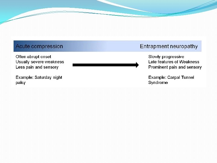
INTRODUCTION �Compression neuropathy describes a heterogeneous group of focal neuropathy syndromes related to peripheral nerve compression resulting in pain, paresthesia, and loss of function of that nerve
� The term entrapment neuropathy, although frequently used interchangeably with compression neuropathy, describes a subset of compression neuropathy related to chronic compression.
Predisposing condition �Acute or chronic compression-related injury may occur in essentially any peripheral nerve, but certain considerations such as anatomic course, superficial location, or adjacent fibrous or osseous structures may predispose certain nerves to intrinsic or extrinsic compression-related injury.
� Mechanical pressure leads to associated ischemia and edema of the compressed nerve. �Acute compression leads to focal demyelination, and if compression is sufficiently prolonged Wallerian-like axonal degeneration will occur.
�The clinical presentations of specific compression or entrapment syndromes depend on factors such as chronicity, location, severity, and mechanism of involvement of a particular nerve.
�In cases of acute or subacute compression-related neuropathy such as Saturday night palsy (radial neuropathy at the spiral groove), treatment may be simply be supportive, and improvement is expected over time without intervention. �In other cases of chronic compression or entrapment neuropathy, such as carpal tunnel syndrome, symptoms may continue to progress despite modification of activities and avoidance of provocative activities. �Such cases often require more aggressive intervention.
Entrapment neuropathy �Most common: -upper limb. CTS. Ulnar entrapment at elbow. Radial entrapment at spiral groove -lower limb. Meralgia paresthetica. Peroneal entrapment at fibular head. Tarsal tunnel syndrome
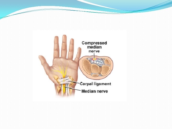
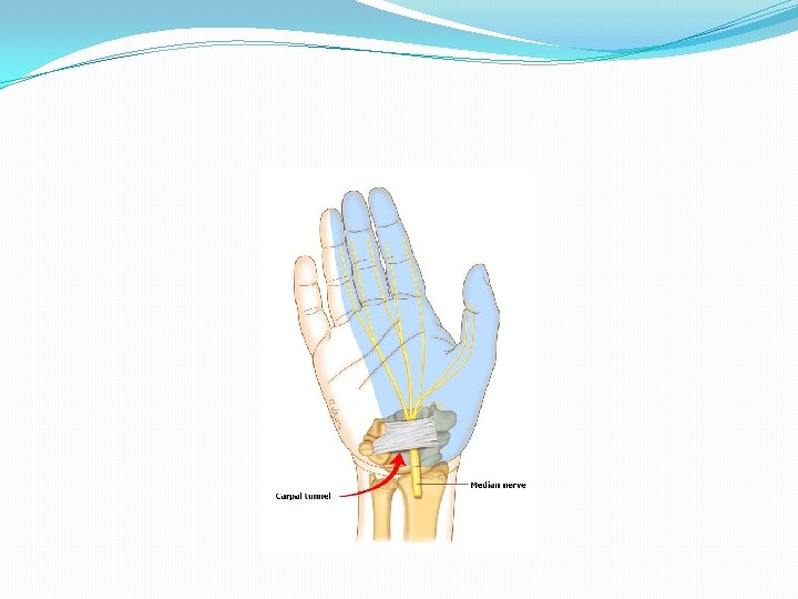
Carpal tunnel syndrome
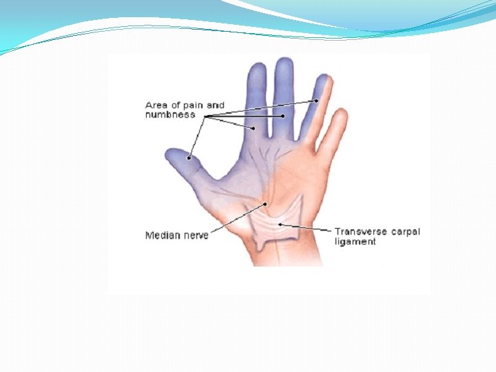
�Carpal tunnel syndrome (CTS) is the clinical diagnosis that describes the constellation of symptoms associated with median nerve entrapment at the wrist. �CTS is attributed to chronic compression of the median nerve within the carpal tunnel. �It is a common clinical problem and the most common entrapment neuropathy, with an estimated prevalence of 2% in men and 3% in women.
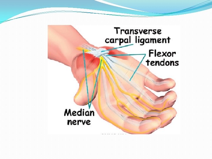
Median Nerve �Anatomy �The median nerve is formed from fibers of the ventral roots of spinal nerves C 5 -T 1. � The fibers of C 5 -C 7 travel within the lateral cord of the brachial plexus to merge with fibers traveling within the medial cord from the ventral roots of spinal nerves C 8 and T 1. �The median nerve supplies no significant motor or sensory branches above the elbow.
�Within the forearm the median nerve provides motor innervation to all of the flexor muscles, excluding flexor carpi ulnaris and the fascicles of flexor digitorum profundus, to the ring and small fingers. �Before passing under the transverse carpal ligament and through the carpal tunnel, the palmar cutaneous nerve branches from the median nerve to supply sensation over thenar eminence �As a consequence the sensation over thenar eminence is always spared in carpal tunnel syndrome.
� Within the hand, the median nerve and its branches supply sensory innervation of the lateral portion of the hand the palmar surface of the thumb, index, middle and lateral half of the ring finger, and motor innervation to abductor pollicis brevis, opponens pollicis, superficial head of the flexor pollicis brevis, and the first and second lumbrical muscles. �The thenar compartment muscles including abductor pollicis brevis, opponens pollicis, and the superficial head of flexor pollicis brevis are innervated by the recurrent motor branch.
�The recurrent motor branch usually branches from the median nerve prior to the carpal tunnel, but travels along with the median nerve under the transcarpal ligament. �Because of this, the recurrent motor branch may rarely be selectively or more severely involved in comparison with the main median nerve during compression at the carpal tunnel, leading to motorpredominant presentation.
�Compression of the median nerve within the carpal tunnel leads to inhibition of axonal transport and reduction of epineurial blood flow, leading to intraneural edema, myelin thinning, and eventual nerve-fiber degeneration and fibrosis.
�Most CTS is idiopathic, with no specific predisposing condition identified. �Systemic predisposing conditions include diabetes, pregnancy, thyroid disorders, chronic kidney disease, acromegaly, and obesity. �Underlying peripheral neuropathymay also contribute to the development of CTS, particularly certain types of neuropathy, including hereditary neuropathy with predisposition to pressure palsies and familial amyloidosis related to transthyretin mutation. � A family history of carpal tunnel may be present in up to 30% cases.
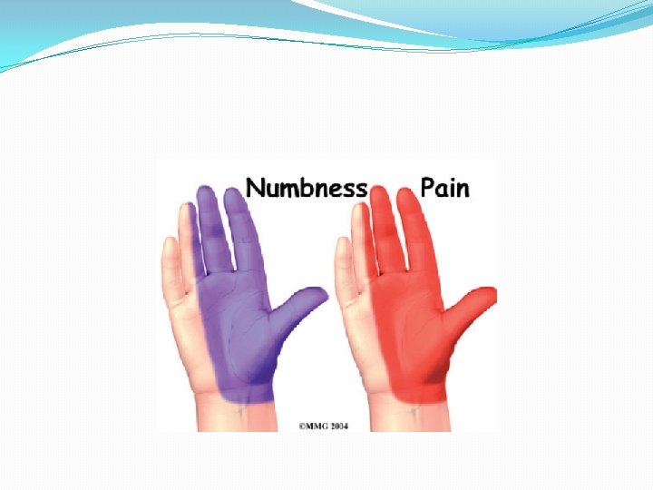
�Patients with CTS usually present with predominantly sensory features of distal median nerve dysfunction including pain, numbness, and tingling affecting the thumb, index, and middle fingers. �These symptoms are typically intermittent early in the course of disease, and nocturnal worsening of symptoms or worsening symptoms with activity of the hands are common. �Classically the sensory examination will demonstrate impairment of the distal medial distribution but spare the palm.
�Classically the sensory examination will demonstrate impairment of the distal medial distribution but spare the palm.
�Frequently, particularly early in the course of disease, the physical examination may be normal. �Around 50%of patients with defined carpal tunnel will have a normal examination, and even with quantitative sensory testing around 20% of patients with defined carpal tunnel syndrome will have normal sensory findings.
�Pain may be a prominent feature in CTS, and it is not uncommon for patients to report symptoms of pain or numbness outside of the median distribution with numbness of the “whole hand, ” and pain radiating proximally to the forearm and elbow. �Infrequently some patients do report associated referred pain to the shoulder.
�Motor findings involving the distal median innervated muscles may be seen in CTS, but significant weakness or atrophy is usually only seen in severe or very chronic cases. �Patients will often complain of weakness or specifically “dropping” objects, which is usually due to sensory and proprioceptive defects rather than weakness; true weakness is not usually an early or prominent feature.
tinel sign
phallen test
manual carpal compression test �Applying pressure over the transverce carpal ligament for 30 seconds causes paresthesia
Hand elevation test �Raising the hand above the head for at least 1 min
Test sensitivity specificity Tinel 50 77 Phalen 68 73 manual carpal compression test 64 83
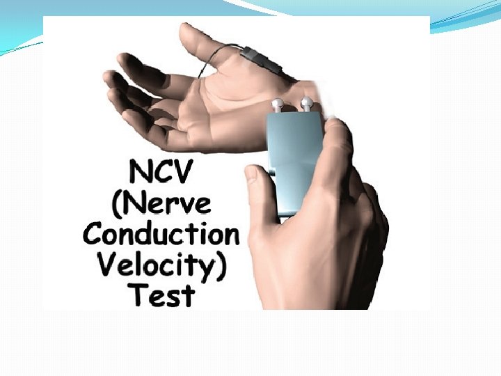
Diagnostic modalities �CTS is usually diagnosed on a clinical basis or in combination with Edx testing. �Edx testing provides additional benefits of defining severity and excluding coexistent or mimicking conditions. �There are numerous Edx techniques that may be used to confirm median neuropathy at the wrist.
�Although dependent on choice of technique, Edx testing has an approximate sensitivity of 85% and specificity of 95% in detecting median neuropathy at the wrist in the setting of defined CTS.
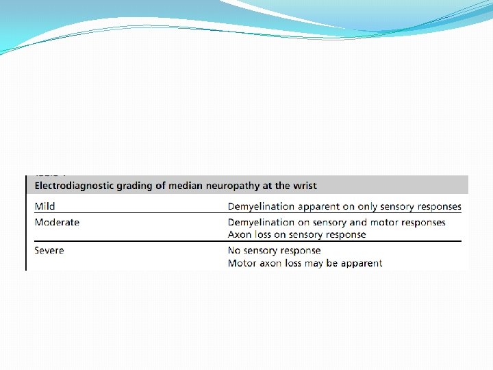
�Edx findings may be used to characterize the severity of median neuropathy at the wrist �Although it may be helpful to use this classification to guide treatment strategies, severity of symptoms often do not correlate closely with Edx findings.
�There is no role for universal laboratory screening for associated systemic disorders in patients diagnosed with CTS, unless other systemic signs are evident by history or examination.
�Ultrasound imaging is a rapidly advancing area of the assessment of focal neuropathies, and there has been progress recently in the diagnosis of CTS. �Although peripheral nerve ultrasound testing is not yet universally available and Edx testingremains the gold standard for compressive neuropathy, ultrasonography does offer several potential advantages including less pain than with Edx tests, structural assessment of the involved nerve and surrounding structures, and cost efficiency. �Other diagnostic imaging modalities such as magnetic resonance imaging (MRI) or plain radiographs may also be helpful in select cases.
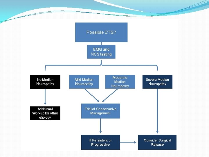
Therapeutic strategy �Up to 20%of patients with CTS will spontaneously improve without any intervention. �To determine the best treatment strategy, factors such as severity and duration of symptoms and the impact on the patient’s function should be considered.
�Although Edx testing will often not correlate with symptom severity, Edx grading can be helpful in guiding treatment strategies. �For most patients with mild or moderate median neuropathy on nerve conduction studies, a trial of conservative treatment should be attempted before aggressive strategies �If significant median nerve injury is evident, early surgical intervention should be considered to prevent further injury
Therapeutic strategy �Modifying of habits �Non pharmacologic treatment �pharmacologic treatment -NSAID, corticosteroids, …. . �Surgery
Non surgical treatment �Despite a lack of data to support or refute whether activity modification will modify the natural history of CTS, avoiding aggravating activities is reasonable and may be recommended.
�Try to reduce all non-essential activities such as shopping, lifting, computer work, typing and writing �Try to keep your wrist straight to avoid pressure on the nerve.
�Increase the grip size of your pen/cutlery by placing foam around the handle
�Wrist splints are an appropriate first-line treatment in all patients, excluding those with severe median neuropathy. �Splints can be effective in up to 60% of patients and do not involve any risk. �Splints should be designed to maintain neutral wrist position and worn at night. �There is no additional benefit derived from customdesigned splints or from daytime wear.
Wrist rest splint
�Elevate your forearm on a pillow at night �Try placing an ice pack over the wrist for 10 minutes or massage the wrist with an ice cube �Alternatively, try contrast baths – dip your hand(s) into warm (not hot) water for 30 seconds, then cold water for 30 seconds. Alternate for 3 -5 minutes.
�Gentle exercise can be helpful if done regularly through the day to help reduce swelling. �Gentle massage of hands and arms may offer some relief too. �Elevation – where possible, try to elevate your wrists/arms on pillows, to help reduce the swelling
Make and release a fist (this can also be done with your hands elevated) 3 -4 times a day
Stretch your wrists backwards and forwards (3 -4 times a day)
Wrist flexor stretch. Hold a gentle stretch for 30 seconds. Perform 3 repetitions, 2 -3 times daily.
Doorway Stretch. Hold onto doorframe as pictured above for 30 seconds. Perform 3 repetitions 2 -3 times daily.
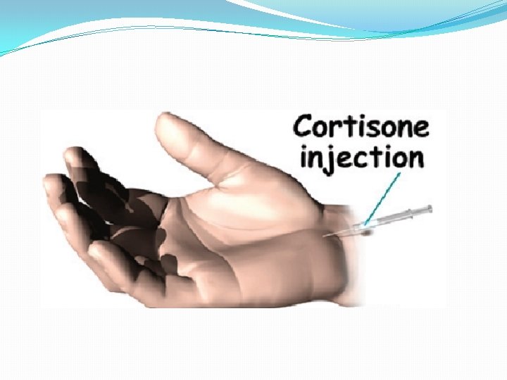
Pharmacologic treatment �NSAIDS �Oral prednisolone �Local injection of prednisolone
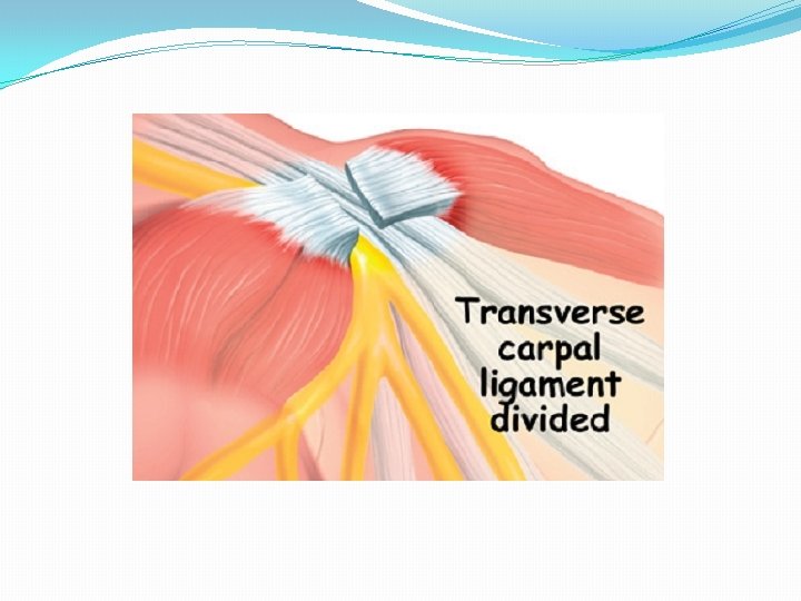
Surgery �Surgery can be considered if conservative measures fail or in severe acute cases with debilitating functional impairment
Other site of median entrapments
� These cases can usually be distinguished by weakness of proximal median innervated muscles and the presence of sensory loss in the palmar aspect of the hand. � Entrapment syndromes of the proximal median nerve have been described that are related to compression by the ligament of Struthers, biceps brachii aponeurosis, pronator teres muscle, and flexor digitorum superficialis muscle. � Pronator syndrome is an ill-defined entrapment syndrome involving compression of the median nerve as it passes between the pronator teres muscle. � Symptoms include aching pain in the forearm associated with sensory and/or motor deficits in the distribution of the proximal median nerve. � Anterior interosseous syndrome describes symptoms of deep aching pain in the forearm followed by weakness in anterior interosseousinnervated
�Anterior interosseous syndrome describes symptoms of deep aching pain in the forearm followed by weakness in anterior interosseous-innervated muscles. �Symptoms of slowly progressive pain localized to only the forearm associated with deficits in only the distribution of the anterior interosseous nerve may suggest chronic compression. �Conversely, an abrupt onset of severe pain or weakness is usually less likely related to anterior interosseous nerve entrapment or compression.
Ulnar Nerve �Anatomy �The ulnar nerve originates from the lower trunk and medial cord of the brachial plexus. �It is formed by sensory and motor axon contributions from the ventral roots of spinal nerves C 8 and T 1. � The ulnar nerve supplies no sensory or motor innervation above the elbow.
�Within the forearm the ulnar nerve innervates the flexor carpi ulnaris and flexor digitorum profundus to digits 4 and 5. �There are 2 sensory branches of the ulnar nerve that occur within the forearm. �The palmar cutaneous nerve, which supplies sensation to the hypothenar region, leaves the ulnar nerve at the mid forearm and passes superficial to the Guyon canal.
� Entering the hand, the ulnar nerve passes through the Guyon canal and splits into 2 terminal branches. �The superficial terminal branch supplies motor innervation to palmaris brevis and sensation to the distal medial palm and the palmar surfaces of the small finger, and the medial half of the ring finger. �The deep terminal branch is a pure motor branch supplying motor innervation to the hypothenar, interossei, the third and fourth lumbrical, and adductor pollicis muscles.
Ulnar neuropathy �Ulnar nerve compression most commonly occurs in the region of the elbow, and ulnar neuropathy at the elbow (UNE) is the second most common entrapment neuropathy. �Because of anatomic factors, UNE may occur in the setting of chronic entrapment or acute compression. �UNE usually occurs at the condylar groove and less frequently within the cubital tunnel.
�Compression at the condylar groove frequently occurs because of the superficial location and minimal protection of the ulnar nerve at this site. �With compression at the cubital tunnel, known as cubital tunnel syndrome, the ulnar nerve is usually compressed 1. 5 to 3 cm distal to the epicondyle. �UNE may occur more acutely in relation to trauma or positioning, and it is the most common focal neuropathy reported during general anesthesia for surgery
� Less commonly, the ulnar nervemay also be compressed at the wrist. � Ulnar neuropathy at the wrist (UNW) is usually classified according to which of the distal branches are involved. � Type I involves compression proximal to the Guyon canal leading to involvement of the superficial sensory, hypothenar motor, and deep motor branches. � Type II involves compression within the canal, with involvement of only the superficial sensory branch. � Type III involves compression distal to the superficial sensory branch, with compression of the hypothenar and the deep motor branch. � Type IV involves compression distal to the superficial sensory and hypothenar motor branches, and involves only the deep motor branch. � Type V involves compression of the deep motor branch just proximal to the branches to the adductor pollicis and first dorsal interosseous muscles.
�Typically UNE is associated with sensory loss and paresthesia, initially intermittent, of the medial hand, dorsal and palmar aspects, and the medial half of the fourth and the fifth digit.
�In mild or early UNE, similar to CTS, the sensory examination may be normal. �Patients may complain of an ache-like pain at the elbow, and will often self-report a Tinel phenomenon of the ulnar nerve at the elbow in the region of the ulnar groove. �If UNE is severe, motor axon loss may occur and lead to weakness of all ulnar innervated muscles usually affecting the distal muscles more severely. �Occasionally some patients with UNE will lack prominent sensory symptoms, and present with significant weakness and atrophy.
�Whether or not sensation of the dorsal medial hand is involved can help localize ulnar neuropathy (always spared in UNW), but some patients with definite UNE will have sparing of sensation of the dorsal hand.
�There are several special tests or signs that have been described for the diagnosis of ulnar neuropathy. �Though useful, these only supplement the detailed examination of individual muscles of the upper limb in the patient with a suspected peripheral nerve injury or entrapment.
�The Wartenberg sign is an abduction of the fifth digit caused by weakness of the third palmar interosseous muscle. �The Froment sign is a classically described sign of weakness of adductor pollicis, flexor pollicis brevis, and first dorsal interosseous muscles leading to functional substitution of flexor pollicis longus. �The ulnar claw hand, or Duchenne sign, is the clawing of the fourth and fifth digits.
Wartenberg sign
Froment sign
Diagnostic modalities �Edx studies are the diagnostic test of choice in ulnar neuropathy. �Most patients with ulnar neuropathy should undergo testing to confirm the presence of ulnar neuropathy, determine the severity, and to exclude other mimicking conditions, in particular disorders such as C 8 radiculopathy or lower trunk/medial cord brachial plexopathy. �The diagnostic sensitivity of Edx in ulnar neuropathy is approximately 80%.
�Similar to the median nerve, ultrasonography may be used to image the ulnar nerve to identify evidence of compression and occasionally identify the structural lesion predisposing to the development of neuropathy. �While Edx continues to be the gold standard of diagnosis for ulnar neuropathy, ultrasonography may occasionally confirm cases with normal Edx results. �Other imaging modalities should also be used in cases with clinically apparent malalignment or a history of traumatic osseous injury (tardy ulnar palsy).
Therapeutic strategy �All patients with ulnar neuropathy should be educated regarding the basic path of the ulnar nerve at the elbow and wrist, and should be instructed on ways to avoid excessive pressure or compression on the ulnar nerve. � Treatment strategies are broadly grouped into either conservative (aimed at reducing mechanical pressure on the nerve) or surgical intervention.
�Conservative treatment measures include life style modification to avoid increased pressure on the ulnar nerve, wearing external padding to reduce pressure at the elbow, and sleeping with straight elbows with or without the assistance of splinting.
�Initial conservative therapeutic measures are appropriate for patients with mild (manual muscle testing of intrinsic hand muscles equal to grade 4 or greater) or no weakness, unless there is evidence for a structural abnormality at the elbow. �Patients with weakness evident on examination, even if mild, should be followed closely for signs of progression.
�Surgical intervention should be considered in patients who experience progression of weakness despite conservative management. � Initial surgical management should be considered for patients with significant motor weakness or patients with persistent or progressive symptoms despite conservative treatment.
�One important exception is the patient who presents with an abrupt onset of UNE associated with findings of prominent ulnar motor conduction block (>50%) on Edx testing, in which case conservative management may be reasonable. �There is a variety of surgical approaches : including medial epicondylectomy, cubital tunnel decompression, and ulnar nerve transposition. � In general, the most minimal surgical approach that will address any structural or anatomic concerns should be used.
Ulnar Neuropathy at Wrist(UNW) �True entrapment neuropathy of UNW is uncommon; usually UNW occurs in the setting of acute or subacute compression. � Typically patients will be able to describe an antecedent trauma preceding the onset of numbness or motor symptoms. � Ulnar neuropathy at the wrist is also known as cyclist’s palsy, owing to the incidence of UNW in cyclists in relation to trauma at the wrist from the handlebars. �Occasionally a structural lesion (ie, ganglion cyst) may predispose an individual to the development of true ulnar nerve entrapment at the wrist and, if no clear history of trauma can be identified, evaluation for a structural lesion should be considered. �Surgery is not indicated in most cases of UNW if there is no clear structural compression of the nerve.
Radial Nerve �Anatomy �The radial nerve originates from the posterior cord of the brachial plexus and is formed from fibers of the ventral roots of spinal nerves C 5 -T 1. �It provides motor innervation of the dorsal arm muscles (triceps brachii and anconeus) and muscles of the extensor compartment of the forearm as well as sensory innervation of the dorsal aspects of the thumb, index, and middle fingers and the dorsal lateral portion of the hand.
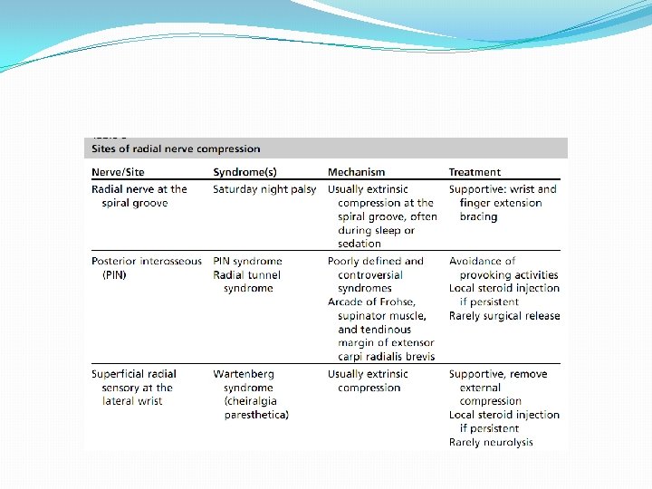
�There are several potential sites of compression along the path of the radial nerve, including compression at the intermuscular septum between the triceps and brachialis muscles at the level of the spiral groove (radial nerve), at the ligament of Frohse (posterior interosseous or deep radial nerve), between the heads of the supinator muscle (posterior interosseous), and under the edge of the brachioradialis muscle (superficial sensory branch of the radial nerve).
Radial neuropathy �Radial neuropathy is the third most common neuropathy of the upper limb. � Whereas true entrapment of the radial nerve is very rare, subacute or acute compression of the radial nerve is more common. �Acute compression at the spiral groove, the socalled Saturday night palsy, is the most common presentation of radial neuropathy. �As the name implies, patients usually awaken with symptoms of weakness in radial
�Acute compression at the spiral groove, the socalled Saturday night palsy, is the most common presentation of radial neuropathy. �As the name implies, patients usually awaken with symptoms of weakness in radial innervated muscles sparing the triceps, sensory changes in the distribution of the radial nerve, and sometimes pain at the site of compression. �Saturday night palsy is not, strictly speaking, an entrapment neuropathy but rather an acute or subacute compression neuropathy. �In most cases this is a self-limited process with a good prognosis.
�Other focal compression–related radial neuropathies may occur at the elbow, forearm, and wrist. �Syndromes involving the posterior interosseous nerve at the elbow include posterior interosseous nerve syndrome or radial tunnel syndrome, which are often considered the same entity. �Radial tunnel syndrome is distinguished by the distinct lack of weakness. �Of importance is that neuralgic amyotrophy has a particular predilection for the posterior interosseous nerve, and the clinical presentation of neuralgic amyotrophy and posterior interosseous nerve syndrome have significant overlap. �The clinical presentation of posterior interosseous nerve syndrome may be associatedwith severe weakness, but cheiralgia paresthetica or. Wartenberg syndrome
�The clinical presentation of posterior interosseous nerve syndrome may be associatedwith severeweakness, but cheiralgia paresthetica or. Wartenberg syndrome describes the clinical syndrome of numbness and pain at the dorsal lateral hand thumb, index, andmiddle fingers, associated with compression of the superficial radial nerve at the lateral wrist. �This condition is usually the result of extrinsic compression such as an overly tight wristwatch or handcuffs. �Thus the syndrome is sometimes referred to as handcuff palsy or wristwatch palsy.
Diagnostic modalities �Diagnosis and localization can often be determined on a clinical basis alone. �Edx testing is usually used to confirm the diagnosis of radial neuropathy and the location of injury while excluding mimicking conditions, in particular C 7 radiculopathy, posterior cord plexopathy, or other less common disorders with predilection to radial nerve involvement such as multifocal motor neuropathy with conduction block. �In addition to routine screening, Edx testing protocols usually involve electromyography (EMG) of radial innervated muscles and nerve-conduction study assessment of the radial sensory and motor nerve responses. � In radial neuropathy at the spiral groove, features of neuropraxia are usually prominent, with conduction block and focal slowing at the site of compression.
�The radial sensory amplitude is often preserved, consistent with less prominent axonal loss. �Most patients will have features of some axonal loss on needle-electrode examination, but this does not necessarily portend a poor prognosis. �Testing of nonradial nerves and nonradial innervated muscles should be performed to exclude a more generalized process. �Ultrasonography has shown promise in focal neuropathies of the radial nerve attributable to compression, entrapment, and trauma.
Therapeutic strategy �Treatment of radial neuropathy is usually conservative, and surgical intervention is infrequently indicated or necessary ( ). �After a neuropraxic injury typical of simple compression, essentially all patients will have complete recovery although some patients may not recover for up to 6 months. �For a radial neuropathy at the spiral groove, supportive bracing of the wrist and finger extensors is helpful. �For high radial neuropathies, bracing with static wrist extension with dynamic finger extension of the proximal phalanges will increase a patient’s functional ability until nerve recovery.
�Short-term immobilization to limit elbow extension, pronation of the forearm, and wrist flexion are appropriate for radial tunnel syndrome. �Local steroid injection carefully directed to the site of local tenderness may be helpful. �Surgery is controversial and is rarely indicated. � It may be helpful in select cases, but suboptimal outcomes are common with surgical intervention. �In posterior interosseous nerve syndrome a wristextension brace is appropriate if weakness is severe. Cheiralgia paresthetica
�Cheiralgia paresthetica usually responds to conservative treatment with removal of external compression. �Occasionally local steroid injection dorsal to the first extensor compartment may be needed. �Neurolysis of the superficial radial nerve is rarely indicated, but may be helpful in up to 80% of patients with symptoms that persist despite conservative management.
Lateral Femoral Cutaneous Nerve (Lateral Cutaneous Nerve of the Thigh �Anatomy �The lateral femoral cutaneous (LFC) nerve is a pure sensory nerve that arises from the dorsal divisions of the ventral primary rami of the L 2 -L 3 spinal nerves. �The nerve penetrates the psoas muscle and emerges along its lateral border, travels along the lateral aspect of the pelvis, and passes under the inguinal ligament approximately 1 cm medial to the anterior superior iliac spine to enter the thigh. �It supplies sensation to the skin over the anterior and lateral portion of the thigh.
Meralgia paresthetica �Meralgia paresthetica (MP), refers to the syndrome of numbness, paresthesia, and pain associated with LFC mononeuropathy. �It occurs more frequently than previously proposed, with an incidence of 36. 2 per 100, 000 patient-years �The incidence is highest in obese patients in their fourth to sixth decades; mean age at diagnosis is 50 years, with no gender predilection. � The adjusted incidence among diabetic is 7 -fold greater in comparison with the general population.
�MP is usually caused by compression of the LFC nerve as it passes under the lateral portion of the inguinal ligament. � The condition is often idiopathic; however, described predisposing factors related to compression may include a large abdomen (pregnancy, obesity, or ascites), external compression from tight clothing or belts, or prolonged positioning (lithotomy positioning, cycling, or prolonged hip extension).
�Other mechanisms of injury have been described, including injury during local surgery such as hip surgery, aortofemoral bypass, and cesarean section; direct trauma related to seat-belt injury; and malignant invasion. �proximal injury in the pelvis related to psoas hemorrhage or other abnormalities is described.
�Typically patients present with pain, numbness, and paresthesia in the lateral and or anterior aspect of the thigh. �The distribution of symptoms may vary between patients, but the lateral aspect of the thigh is solely involved in the majority. �The pain onset is generally subacute and bears no relation to positioning, standing, or walking.
�MP is usually unilateral, although bilateral presentation is occasionally described. �The LFC nerve is a purely sensory nerve, so no motor signs or symptoms are present. � Early during MP or in patients with mild symptoms, examination may be normal. �With increasing severity, sensory loss is present and may involve all or only part of the classically described distribution of the LFC nerve
� Despite the reduced sensation, there is often skin hypersensitivity to touch. � Otherwise the neurologic examination is normal. �Reflexes, most importantly the patellar reflex, remain intact. � Motor muscle weakness is characteristically absent, and if present other mimicking conditions such as upper lumbar radiculopathy, lumbar plexopathy, or femoral neuropathy are more likely.
Diagnostic modalities � The diagnosis of MP is usually a clinical one, supported by the characteristic distribution of pain and sensory changes on examination and the absence of motor or reflex abnormalities. � Diagnostic investigations are, as a rule, only necessary in atypical cases. � In this instance, imaging studies, EMG, and nerve conduction studies are helpful in excluding mimicking conditions. � Nerve conduction studies to confirm LFC neuropathy can be unreliably present in healthy individuals, and are increasingly difficult to perform in the obese patient. � However, a recent study using ultrasound-guided electrode placement in LFC sensory nerve studies revealed higher yield with less interside variability, including in obese subjects. � Diagnostic injections at the common site of entrapment can be helpful in confirming the diagnosis if clear relief of symptoms is provided. � Imaging modalities may include MRI and ultrasonography.
Therapeutic strategy �The symptoms of MP usually improve spontaneously within weeks to months. �Therefore, conservative treatment should be encouraged. �Conservative treatments should include removing sources of extrinsic compression such as compressive clothing or provocative postures. �Weight loss should be encouraged in obese patients. �In patients with persistent symptoms, neuropathic pain medications may be helpful for symptomatic management. �In cases that do not respond with time and to conservative measures, steroid injection and surgery are considerations.
�Infiltration with local anesthetic and steroids may provide temporary relief and can be repeated at intervals. �The use of ultrasound guidance is helpful. �Surgery is usually a last resort and is rarely needed for patients with severe refractory symptoms who are not responding to the aforementioned measures. � Surgical interventions (decompression or sectioning) are reported to be effective in about 76% to 80% of cases.
Tibial Nerve � Anatomy � The tibial nerve, composed of L 4, L 5, S 1, and S 2 nerve roots, courses through the lumbosacral plexus and through the thigh within the sciatic nerve, along with the peroneal component. � The sciatic nerve divides into the peroneal and tibial nerves proximal to the popliteal fossa. � In the thigh, the tibial component of the sciatic nerve supplies all the hamstrings’ muscles and half of the adductor magnus. � In the popliteal fossa, it gives a medial sural cutaneous branch to form the sural nerve with contribution from the lateral sural cutaneous branch of the peroneal nerve. � In the leg, the tibial nerve travels within the posterior compartment of the leg innervating all of the posterior compartment muscles. � At the ankle, it gives a calcaneal branch and passes posterior to the medial malleolus through the tarsal tunnel, dividing into the medial and lateral plantar nerves.
Tarsal tunnel syndrome �Tarsal tunnel is a rare syndrome related to compression of the tibial nerve at the level of the tarsal tunnel. �Despite the agreement on its existence, there is controversy regarding its prevalence.
�The tarsal tunnel is a fibro-osseous space, formed by the flexor retinaculum posterior and distal to the medial malleolus. �The contents of the tarsal tunnel include the tibial nerve; posterior tibial artery and vein; and the tendons from the tibialis posterior, flexor digitorum longus, and flexor hallucis longus muscles.
�Contributing factors of tarsal tunnel syndrome include ankle sprain and fracture; tight fitting footwear and other biomechanical issues such as joint hypermobility; and space-occupying lesions. �In some cases it is idiopathic.
�Symptoms of tarsal tunnel syndrome may include ankle pain and associated numbness, tingling, burning, and pain in the sole of the foot. �The symptoms commonly worsen with weight bearing and at night. �The presentation of tarsal tunnel syndrome may be variable; the entire tibial nerve may be involved, or there may be selective involvement of either the lateral or medial plantar branches.
�Findings on examination are typically few. �The development of weakness or atrophy of the intrinsic footmuscles usually goes unnoticed because of its minimal functional implications. � However, careful examination and comparison with the other foot, if findings are unilateral, can be helpful. �Decreased sensation usually involves the sole of the foot sparing the heel, which is supplied by the calcaneal branch. �There may be tenderness behind the medial malleolus and a positive Tinel sign over the tarsal tunnel.
Diagnostic modalities �Tarsal tunnel syndrome should be differentiated from other mimicking conditions including other neuropathic processes such as small fiber peripheral neuropathy or radiculopathy, particularly S 1 radiculopathy. �Common musculoskeletal mimicking conditions include posterior tibial tendon dysfunction, plantar fasciitis, and plantar callosities.
�Tarsal tunnel syndrome is typically a clinical diagnosis supported by Edx findings. � Nerve conduction studies may help to localize the lesion and to exclude mimicking conditions. �Imaging studies provide information helpful in excluding structural or space-occupying lesions. �Diagnosis is reasonably secured if a patient has typical foot pain and paresthesia, and a Tinel sign at the tarsal tunnel, and if classic Edx findings are present
Therapeutic strategy �Individuals without a contributing structural or spaceoccupying lesion may respond adequately to conservative management, which may include nonsteroidal anti-inflammatories, neuropathic pain medications, activity modification, physical therapy, and biomechanical modification with shoes, inserts, or orthoses. �Surgery is reserved for individuals with a definite diagnosis who have been resistant to conservative strategies.
Proximal tibial neuropathy �Entrapment of the tibial nerve proximal to the tarsal tunnel is rare. �However, association with a Baker cyst, popliteal artery aneurysm, nerve sheath tumor and other mass lesions, and direct trauma has been described. � In addition to the sensory loss at the sole of the foot, lateral-leg patients commonly present with weakness of the plantar flexion and inversion. �Edx studies and imaging help to establish the diagnosis and identify the structural cause. �Surgery is usually indicated in this setting for resection of the mass lesion.
Peroneal (Fibular) Nerve � Anatomy � The peroneal nerve is composed of the L 4, L 5, and S 1 nerve roots, and travels through the lumbosacral plexus into the sciatic nerve alongside the tibial nerve component. � The peroneal and tibial components of the sciatic nerve are divided by a connective tissue sheath, and proximal to the popliteal fossa the sciatic nerve separates into the peroneal and tibial nerves. � In the thigh, the peroneal component of the sciatic nerve innervates only one muscle proximal to the knee, the short head of the biceps. � After the peroneal nerve separate from the sciatic nerve it gives off the lateral cutaneous nerve of the calf, in the popliteal fossa, supplying sensation of the lateral upper leg. Thereafter, it travels around the fibular head and separates into the superficial and deep peroneal nerves.
�The superficial branch innervates the ankle everters and continues as the superficial peroneal sensory nerve supplying the lateral aspects of the lower leg and foot. � The deep peroneal nerve runs into the anterior leg compartment and innervates the ankle dorsiflexors and toe extensors, and its terminal branch supplies sensation to the first dorsal web space.
Common peroneal neuropathy at the fibular head �Peroneal neuropathy at the fibular head is the most common compression mononeur opathy to affect the lower limbs. �The superficial location of the nerve as it travels laterally around the fibular head makes it susceptible to compression. �The mechanism of injury is typically related to external compression of the nerve against the fibula, in contrast to compression occurring between anatomic structures. �In patients with common peroneal neuropathy, the deep peroneal fascicle is usually more affected than the superficial fascicle. �Moreover, isolated deep peroneal injury is described in the setting of knee surgery, trauma, and anterior compartment syndrome.
�Compression can occur with improper positioning during surgery, habitual leg crossing, squatting, tight-fitting casts, and compressive stockings. � Other possible causes of common peroneal neuropathy may include Baker cyst, other mass lesions, tumors, knee surgeries, and direct trauma. �Loss of fat pad surrounding the nerve at the fibular head is proposed to increase the risk of compression in patients with thin body habitus or following significant weight loss. �Furthermore, common peroneal neuropathy can occur in association with noncompressive causes such as diabetes and vasculitis
�Patients usually present with tripping, difficulty negotiating curbs, or gait change related to foot drop. � Overall sensory symptoms tend to be minimal, with pain reported in 17%67 ; it is bilateral in 10% of patients. �A complete lesion of common peroneal neuropathy produces weakness in toe extension, foot eversion, and ankle dorsiflexion associated with sensory loss on the anterior lateral surface of the lower leg and dorsum of the foot.
�With a lesion at the fibular head there is sparing of the proximal anterior lateral leg related to the preserved lateral cutaneous nerve of the calf, which branches in the lateral popliteal fossa. � A positive Tinel sign at the fibular head may be seen.
Diagnostic modalities �Common peroneal neuropathy should be differentiated from mimicking conditions, mainly L 5 radiculopathy, but other mimicking conditions mayinclude sciatic neuropathy, motor neuron disease, and lumbosacral plexopathy. � Preservation of ankle inversion is a feature of peroneal neuropathy that helps to distinguish it from L 5 radiculopathy.
�Imaging using ultrasonography or MRI helps as an adjunct to Edx studies and, most importantly, to identify patients with intraneural ganglia of the nerve, which is a treatable but underreported cause of common peroneal neuropathy caused by dissection of the synovial fluid along the nerve resulting from superior tibiofibular joint capsular disruption.
�Therapeutic strategy Patients usually respond to conservative treatment with avoidance of precipitating factors. �In patients with severe foot drop, use of an ankle-foot orthosis should be considered for improvement of gait. �Surgical treatment of peroneal intraneural ganglia has been associated with halted progression of neuropathy and improved clinical outcomes.
Peroneal neuropathy at the ankle �Deep peroneal nerve entrapment is sometimes referred to as anterior tarsal tunnel syndrome. �There is disagreement regarding the use of this term, owing to the lack of a clearly defined fibro-osseous tunnel. �Deep peroneal nerve compression may be acute, as in a traumatic injury, or related to chronic external pressure such as in relation to ill-fitting footwear. �Patients present with pain on the dorsum of the foot and numbness in the first dorsal web. � Diagnosis can be established by Edx studies. �Treatment is usually conservative.