Case Presentation PK 14 65 88 Identifying Data
- Slides: 41
Case Presentation 신경과 PK 14조 65번 정광현 88번 장효정
Identifying Data Name : 김 O O Age : 68 Sex : M
Chief Complaint Lt. Side weakness for 4 hours
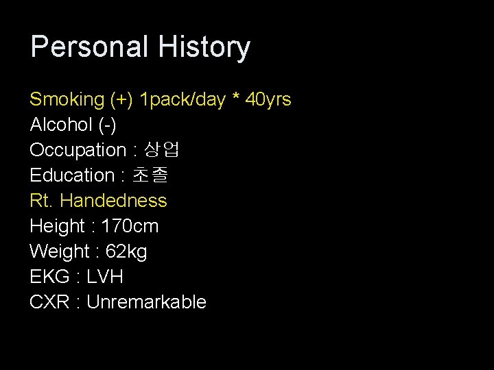
Personal History Smoking (+) 1 pack/day * 40 yrs Alcohol (-) Occupation : 상업 Education : 초졸 Rt. Handedness Height : 170 cm Weight : 62 kg EKG : LVH CXR : Unremarkable
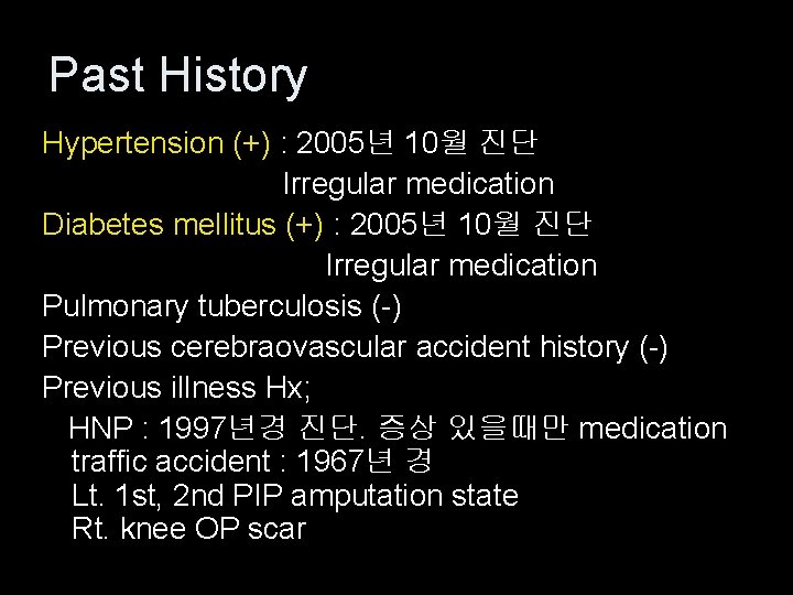
Past History Hypertension (+) : 2005년 10월 진단 Irregular medication Diabetes mellitus (+) : 2005년 10월 진단 Irregular medication Pulmonary tuberculosis (-) Previous cerebraovascular accident history (-) Previous illness Hx; HNP : 1997년경 진단. 증상 있을때만 medication traffic accident : 1967년 경 Lt. 1 st, 2 nd PIP amputation state Rt. knee OP scar
Family History Hypertension (-) Diabetes mellitus (-) Pulmonary tuberculosis (-) Cerebrovascular accident history (-)
Review of System 특이소견 없음
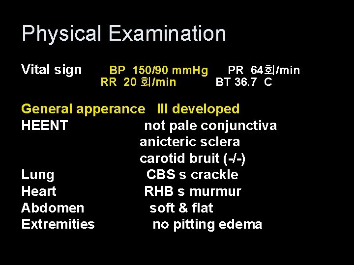
Physical Examination Vital sign BP 150/90 mm. Hg PR 64회/min RR 20 회/min BT 36. 7 C General apperance Ill developed HEENT not pale conjunctiva anicteric sclera carotid bruit (-/-) Lung CBS s crackle Heart RHB s murmur Abdomen soft & flat Extremities no pitting edema
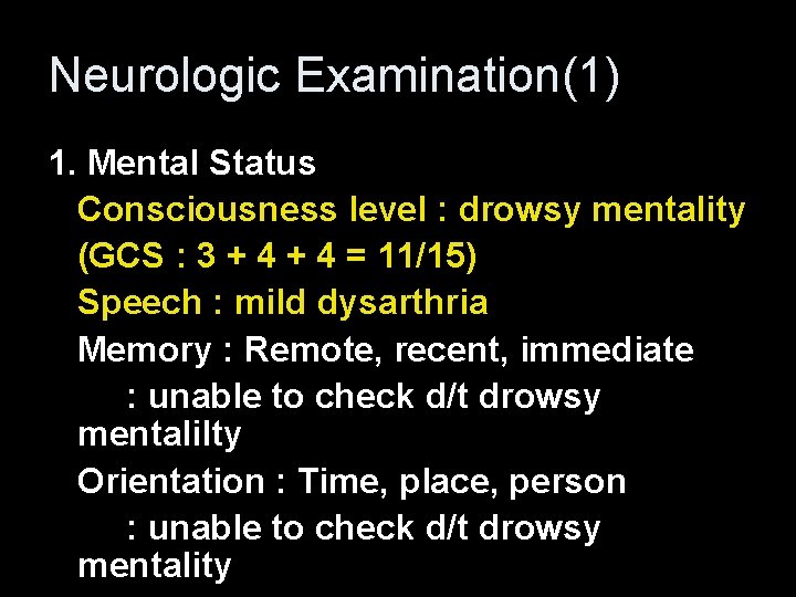
Neurologic Examination(1) 1. Mental Status Consciousness level : drowsy mentality (GCS : 3 + 4 = 11/15) Speech : mild dysarthria Memory : Remote, recent, immediate : unable to check d/t drowsy mentalilty Orientation : Time, place, person : unable to check d/t drowsy mentality
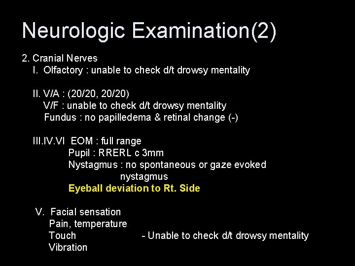
Neurologic Examination(2) 2. Cranial Nerves I. Olfactory : unable to check d/t drowsy mentality II. V/A : (20/20, 20/20) V/F : unable to check d/t drowsy mentality Fundus : no papilledema & retinal change (-) III. IV. VI EOM : full range Pupil : RRERL c 3 mm Nystagmus : no spontaneous or gaze evoked nystagmus Eyeball deviation to Rt. Side V. Facial sensation Pain, temperature Touch - Unable to check d/t drowsy mentality Vibration
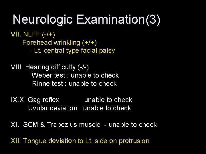
Neurologic Examination(3) VII. NLFF (-/+) Forehead wrinkling (+/+) - Lt. central type facial palsy VIII. Hearing difficulty (-/-) Weber test : unable to check Rinne test : unable to check IX. X. Gag reflex unable to check Uvular deviation unable to check XI. SCM & Trapezius muscle - unable to check XII. Tongue deviation to Lt. side on protrusion
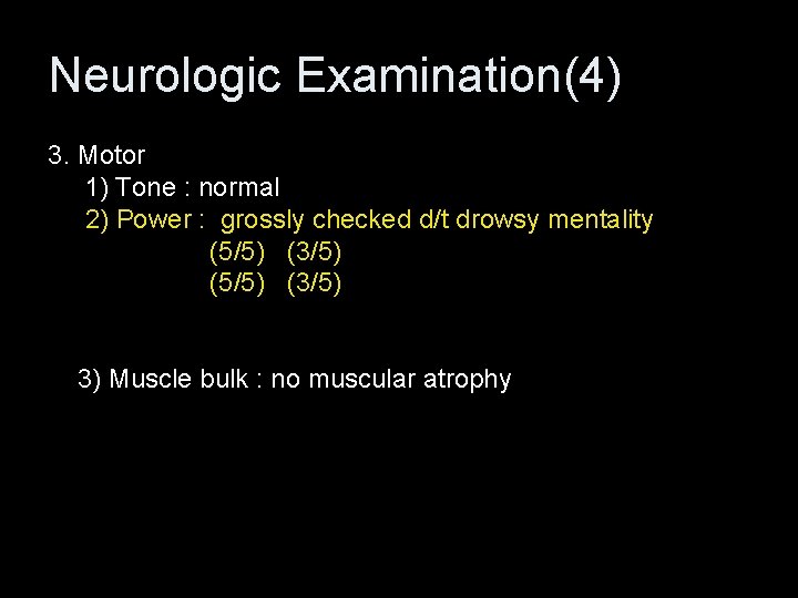
Neurologic Examination(4) 3. Motor 1) Tone : normal 2) Power : grossly checked d/t drowsy mentality (5/5) (3/5) 3) Muscle bulk : no muscular atrophy
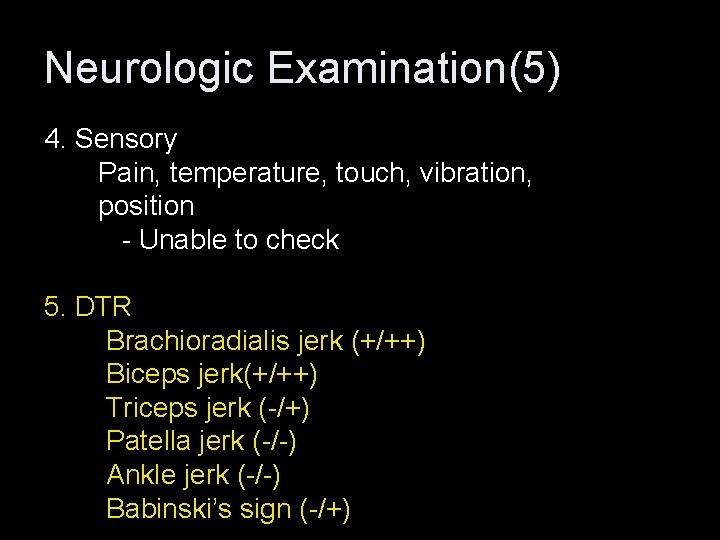
Neurologic Examination(5) 4. Sensory Pain, temperature, touch, vibration, position - Unable to check 5. DTR Brachioradialis jerk (+/++) Biceps jerk(+/++) Triceps jerk (-/+) Patella jerk (-/-) Ankle jerk (-/-) Babinski’s sign (-/+)
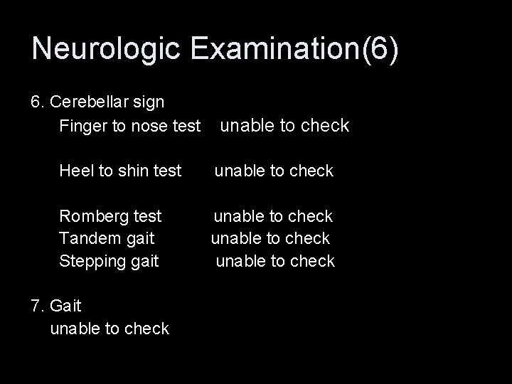
Neurologic Examination(6) 6. Cerebellar sign Finger to nose test unable to check Heel to shin test unable to check Romberg test unable to check Tandem gait unable to check Stepping gait unable to check 7. Gait unable to check
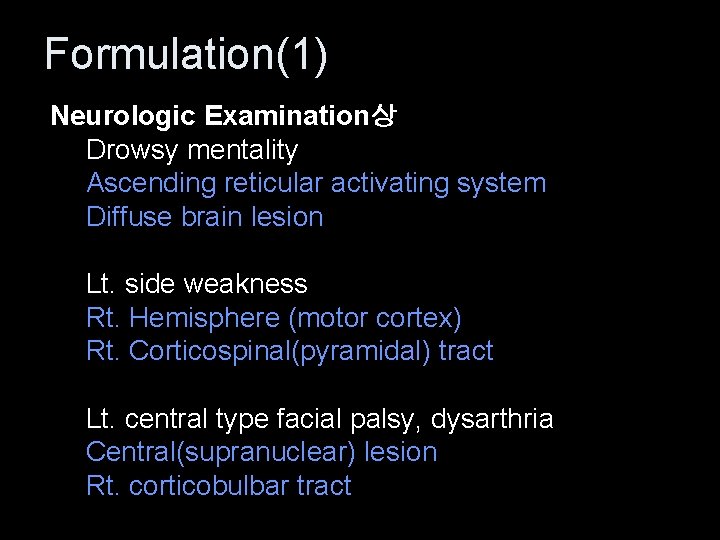
Formulation(1) Neurologic Examination상 Drowsy mentality Ascending reticular activating system Diffuse brain lesion Lt. side weakness Rt. Hemisphere (motor cortex) Rt. Corticospinal(pyramidal) tract Lt. central type facial palsy, dysarthria Central(supranuclear) lesion Rt. corticobulbar tract
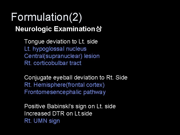
Formulation(2) Neurologic Examination상 Tongue deviation to Lt. side Lt. hypoglossal nucleus Central(supranuclear) lesion Rt. corticobulbar tract Conjugate eyeball deviation to Rt. Side Rt. Hemisphere(frontal cortex) Frontomesencephalic pathway Positive Babinski's sign on Lt. side Increased DTR on Lt. side Rt. UMN sign
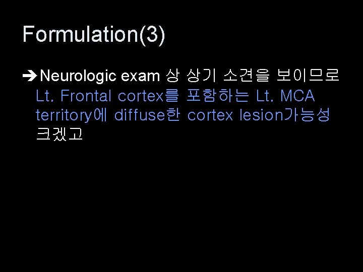
Formulation(3) Neurologic exam 상 상기 소견을 보이므로 Lt. Frontal cortex를 포함하는 Lt. MCA territory에 diffuse한 cortex lesion가능성 크겠고
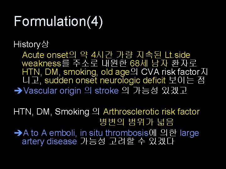
Formulation(4) History상 Acute onset의 약 4시간 가량 지속된 Lt. side weakness를 주소로 내원한 68세 남자 환자로 HTN, DM, smoking, old age의 CVA risk factor지 니고, sudden onset neurologic deficit 보이는 점 Vascular origin 의 stroke 의 가능성 있겠고 HTN, DM, Smoking 의 Arthrosclerotic risk factor 병변의 범위가 넓음 A to A emboli, in situ thrombosis에 의한 large artery disease 가능성 고려할 수 있겠다
Impression R/O Cerebral stroke - Rt. MCA infarct
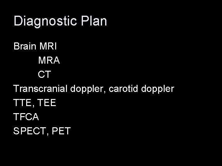
Diagnostic Plan Brain MRI MRA CT Transcranial doppler, carotid doppler TTE, TEE TFCA SPECT, PET
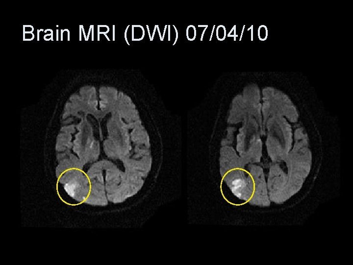
Brain MRI (DWI) 07/04/10
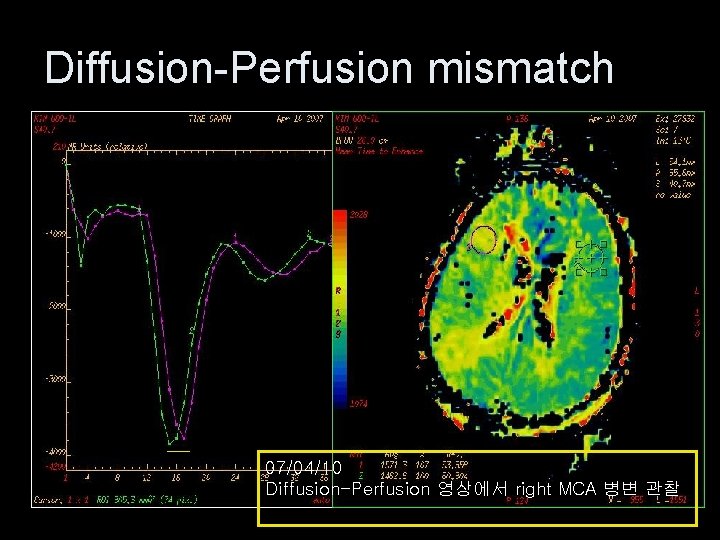
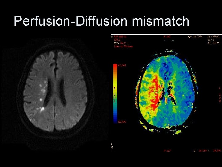
Diffusion-Perfusion mismatch 07/04/10 Diffusion-Perfusion 영상에서 right MCA 병변 관찰
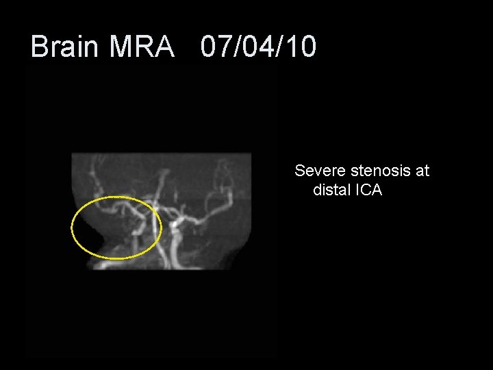
Brain MRA 07/04/10 Severe stenosis at distal ICA
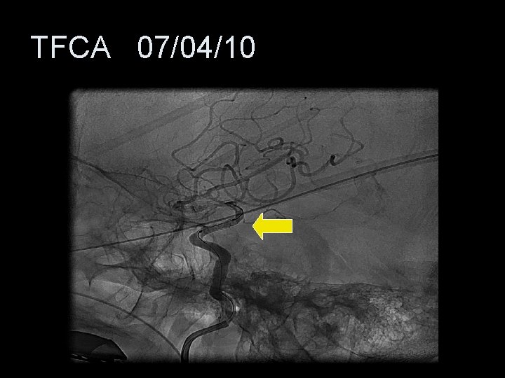
TFCA 07/04/10
Diagnosis # Cerebral stroke Rt. Distal ICA occlusion Rt. MCA stenosis MCA infarct
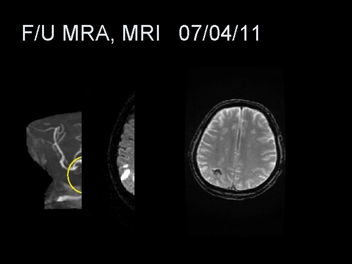
F/U MRA, MRI 07/04/11
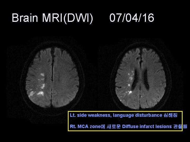
Brain MRI(DWI) 07/04/16 Lt. side weakness, language disturbance 심해짐 Rt. MCA zone에 새로운 Diffuse infarct lesions 관찰됨
Perfusion-Diffusion mismatch
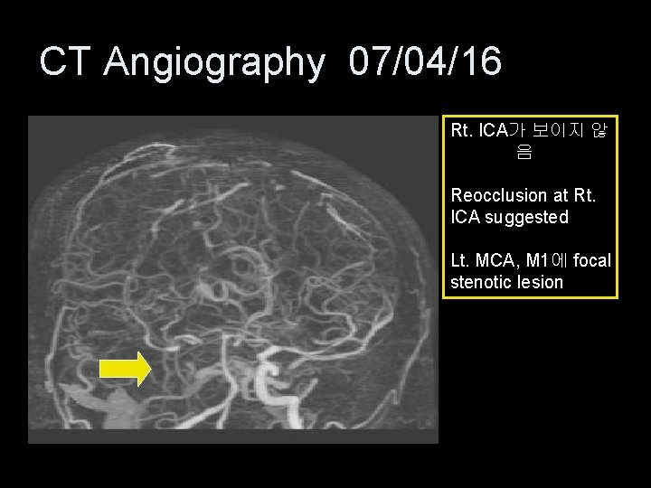
CT Angiography 07/04/16 Rt. ICA가 보이지 않 음 Reocclusion at Rt. ICA suggested Lt. MCA, M 1에 focal stenotic lesion
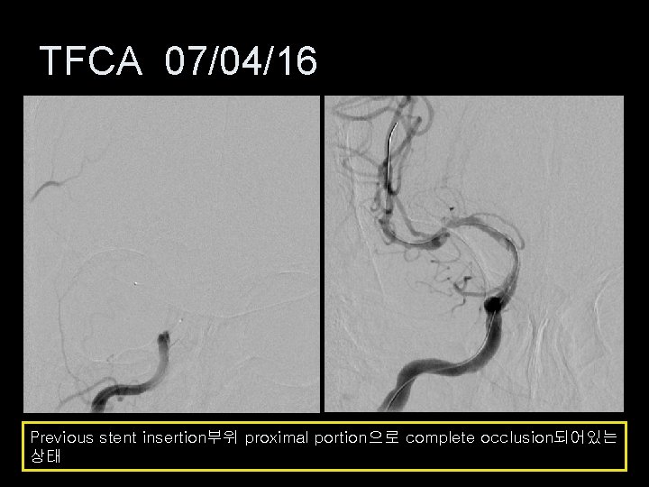
TFCA 07/04/16 Previous stent insertion부위 proximal portion으로 complete occlusion되어있는 상태
Progress 환자 Lt. side weakness 지속 Dysarthria 지속 Lt. side neglect
Disease review MCA occlusion의 Acute Management
Ischemic stroke care의 목표 • Permanent damage이전에 brain ischemia를 되돌림. • Secondary 또는 progressive stroke의 예 방 • 환자의 medical condition을 최적화 시키 고 stroke과 stroke intervention이후 나타 날 수 있는 후유증을 최소화 • 영구적인 손상이 일어난 이후에는 기능적 인 회복.
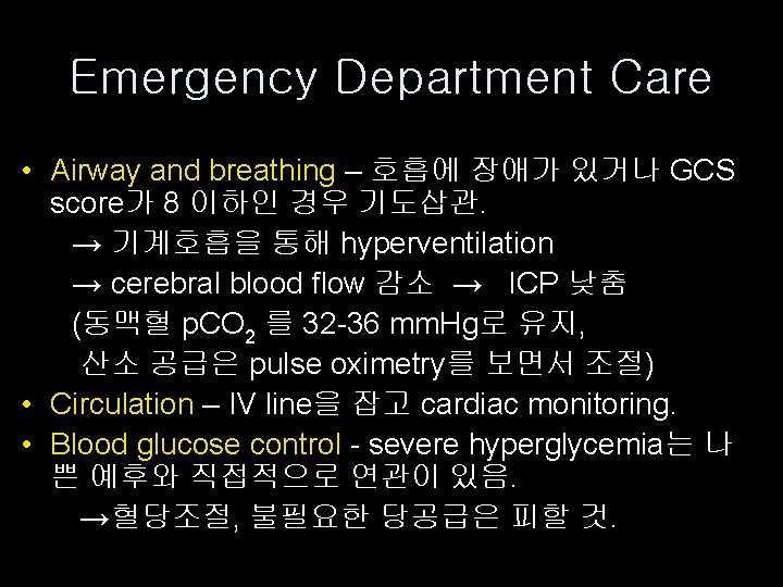
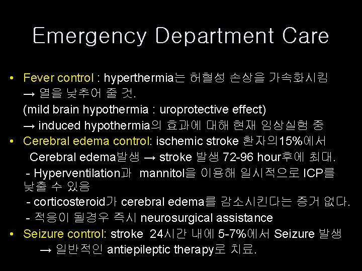
Emergency Department Care • Airway and breathing – 호흡에 장애가 있거나 GCS score가 8 이하인 경우 기도삽관. → 기계호흡을 통해 hyperventilation → cerebral blood flow 감소 → ICP 낮춤 (동맥혈 p. CO 2 를 32 -36 mm. Hg로 유지, 산소 공급은 pulse oximetry를 보면서 조절) • Circulation – IV line을 잡고 cardiac monitoring. • Blood glucose control - severe hyperglycemia는 나 쁜 예후와 직접적으로 연관이 있음. →혈당조절, 불필요한 당공급은 피할 것.
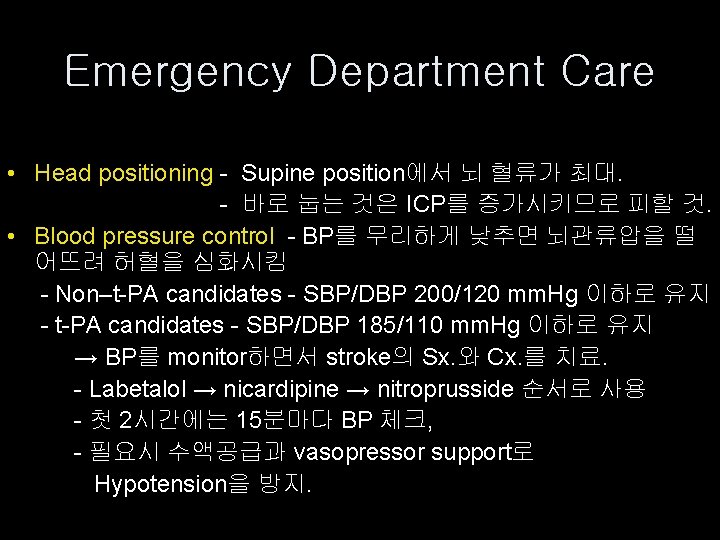
Emergency Department Care • Head positioning - Supine position에서 뇌 혈류가 최대. - 바로 눕는 것은 ICP를 증가시키므로 피할 것. • Blood pressure control - BP를 무리하게 낮추면 뇌관류압을 떨 어뜨려 허혈을 심화시킴 - Non–t-PA candidates - SBP/DBP 200/120 mm. Hg 이하로 유지 - t-PA candidates - SBP/DBP 185/110 mm. Hg 이하로 유지 → BP를 monitor하면서 stroke의 Sx. 와 Cx. 를 치료. - Labetalol → nicardipine → nitroprusside 순서로 사용 - 첫 2시간에는 15분마다 BP 체크, - 필요시 수액공급과 vasopressor support로 Hypotension을 방지.
Emergency Department Care • Fever control : hyperthermia는 허혈성 손상을 가속화시킴 → 열을 낮추어 줄 것. (mild brain hypothermia : uroprotective effect) → induced hypothermia의 효과에 대해 현재 임상실험 중 • Cerebral edema control: ischemic stroke 환자의 15%에서 Cerebral edema발생 → stroke 발생 72 -96 hour후에 최대. - Hyperventilation과 mannitol을 이용해 일시적으로 ICP를 낮출 수 있음 - corticosteroid가 cerebral edema를 감소시킨다는 증거 없다. - 적응이 될경우 즉시 neurosurgical assistance • Seizure control: stroke 24시간 내에 5 -7%에서 Seizure 발생 → 일반적인 antiepileptic therapy로 치료.
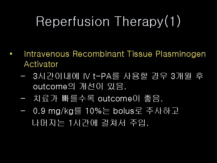
Reperfusion Therapy(1) • Intravenous Recombinant Tissue Plasminogen Activator - 3시간이내에 IV t-PA를 사용할 경우 3개월 후 outcome의 개선이 있음. - 치료가 빠를수록 outcome이 좋음. - 0. 9 mg/kg를 10%는 bolus로 주사하고 나머지는 1시간에 걸쳐서 주입.
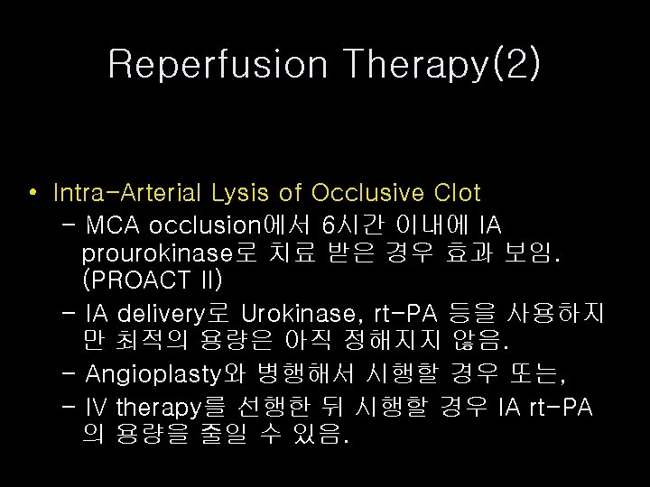
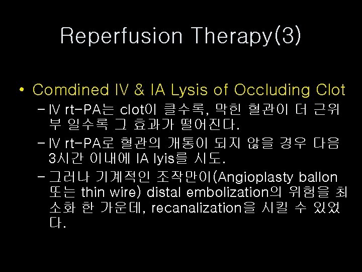
Reperfusion Therapy(2) • Intra-Arterial Lysis of Occlusive Clot - MCA occlusion에서 6시간 이내에 IA prourokinase로 치료 받은 경우 효과 보임. (PROACT II) - IA delivery로 Urokinase, rt-PA 등을 사용하지 만 최적의 용량은 아직 정해지지 않음. - Angioplasty와 병행해서 시행할 경우 또는, - IV therapy를 선행한 뒤 시행할 경우 IA rt-PA 의 용량을 줄일 수 있음.
Reperfusion Therapy(4) • Acute Revascularization - Perfusion image나 clinical exam 상 infarction의 위험이 있는 명백한 ischemic lesion이 있는 경우 emergent revascularization 시행. - Angioplasty 나 endovascular stenting 등 을 통해 revascularization