
Myopathy Primary Muscle Disease Functional Structural Lower vs

- Slides: 53
Myopathy • Primary Muscle Disease – Functional – Structural
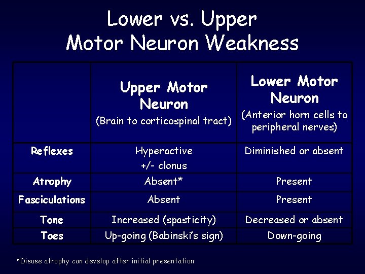
Lower vs. Upper Motor Neuron Weakness Upper Motor Neuron (Brain to corticospinal tract) Lower Motor Neuron (Anterior horn cells to peripheral nerves) Reflexes Hyperactive +/- clonus Diminished or absent Atrophy Absent* Present Fasciculations Absent Present Tone Increased (spasticity) Decreased or absent Toes Up-going (Babinski’s sign) Down-going *Disuse atrophy can develop after initial presentation
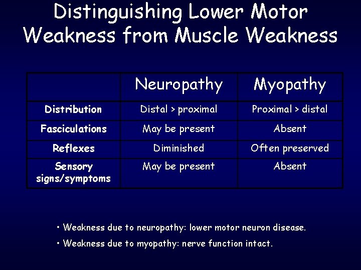
Distinguishing Lower Motor Weakness from Muscle Weakness Neuropathy Myopathy Distribution Distal > proximal Proximal > distal Fasciculations May be present Absent Reflexes Diminished Often preserved Sensory signs/symptoms May be present Absent • Weakness due to neuropathy: lower motor neuron disease. • Weakness due to myopathy: nerve function intact.
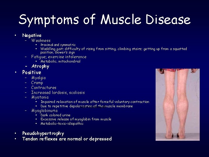
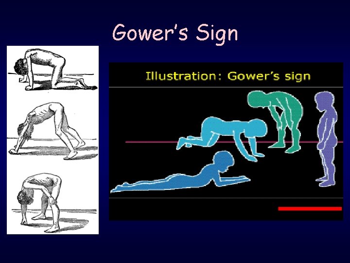
Symptoms of Muscle Disease • Negative – Weakness • • – • • • Proximal and symmetric Waddling gait; difficulty of rising from sitting, climbing stairs; getting up from a squatted position, Gower’s sign Fatigue; exercise intolerance • Metabolic, mitochondrial – Atrophy Positive – – – Myalgia Cramp Contractures Increased lordosis, scoliosis Myotonia – Myoglobinuria • • Impaired relaxation of muscle after forceful voluntary contraction Due to repetitive depolarization of the muscle membrane from A Kornberg MD • • • Dark colored urine Excessive release of myoglobin from muscle Metabolic-toxic-idiopathic Pseudohypertrophy Tendon reflexes are normal or depressed
Gower’s Sign
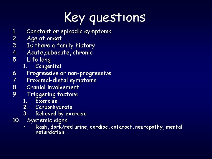
Key questions 1. 2. 3. 4. 5. 6. 7. 8. 9. 10. Constant or episodic symptoms Age at onset Is there a family history Acute, subacute, chronic Life long 1. Congenital 1. 2. 3. Exercise Carbonhydrate Relieved by exercise • Rash, dark/red urine, cardiac, cataract, neuropathy, mental retardation Progressive or non-progressive Proximal-distal symptoms Cranial involvement Triggering factors Systemic signs
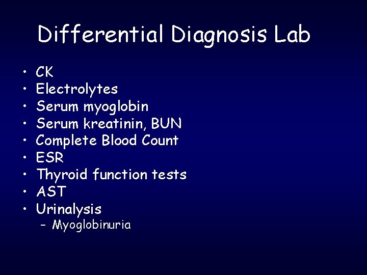
Differential Diagnosis Lab • • • CK Electrolytes Serum myoglobin Serum kreatinin, BUN Complete Blood Count ESR Thyroid function tests AST Urinalysis – Myoglobinuria
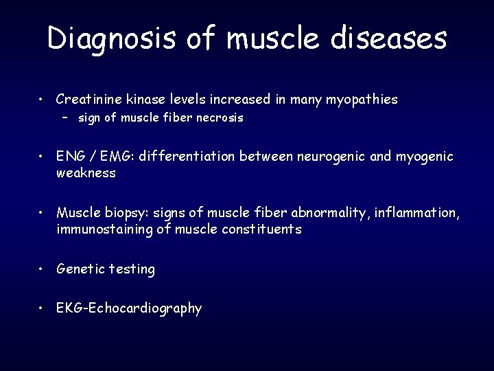
Diagnosis of muscle diseases • Creatinine kinase levels increased in many myopathies – sign of muscle fiber necrosis • ENG / EMG: differentiation between neurogenic and myogenic weakness • Muscle biopsy: signs of muscle fiber abnormality, inflammation, immunostaining of muscle constituents • Genetic testing • EKG-Echocardiography
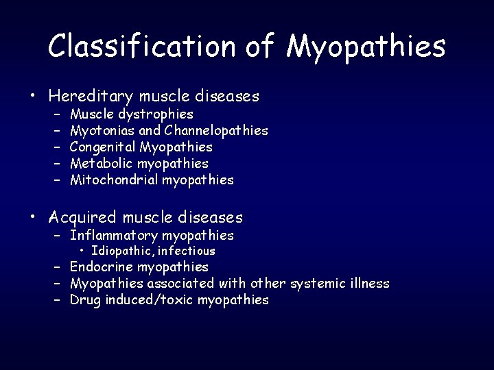
Classification of Myopathies • Hereditary muscle diseases – – – Muscle dystrophies Myotonias and Channelopathies Congenital Myopathies Metabolic myopathies Mitochondrial myopathies • Acquired muscle diseases – Inflammatory myopathies • Idiopathic, infectious – Endocrine myopathies – Myopathies associated with other systemic illness – Drug induced/toxic myopathies
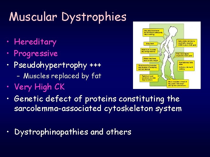
Muscular Dystrophies • Hereditary • Progressive • Pseudohypertrophy +++ – Muscles replaced by fat • Very High CK • Genetic defect of proteins constituting the sarcolemma-associated cytoskeleton system • Dystrophinopathies and others
Muscular Dystrophies • Dystrophinopaties – Duchenne, Becker • • • Limb Girdle dystrophies Fascioscapulohumeral dystrophy Oculopharyngeal dystrophy Distal muscular dystrophies Myotonic dystrophies
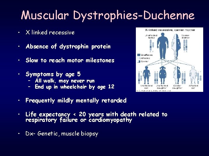
Muscular Dystrophies-Duchenne • X linked recessive • Absence of dystrophin protein • Slow to reach motor milestones • Symptoms by age 5 – All walk, may never run – End up in wheelchair by age 12 • Frequently mildly mentally retarded • Life expectancy < 20 years with death related to respiratory failure or cardiomyopathy • Dx- Genetic, muscle biopsy
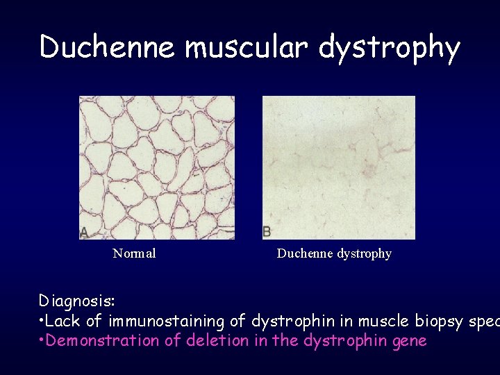
Duchenne muscular dystrophy Normal Duchenne dystrophy Diagnosis: • Lack of immunostaining of dystrophin in muscle biopsy spec • Demonstration of deletion in the dystrophin gene
Muscular Dystrophies-Becker • Less common than Duchenne • X-linked recessive • Later Onset – 10 years • Most patients ambulant until 3 rd-4 th decade • Death from heart failure due to dilated cardiomyopathy-late 40’s • Mental retardation is rare
Management of DMD/BMD • Annual echocardiography • Cardiomyopathy – ACE inhibitor and/or beta blocker. . digoxin • Cardiomyopathy and decreased pulmonary function + • Pneumoccal vaccine • Annual influenza vaccine • Anesthesia complications. . . – Consider TIVA – Susceptibility to malign hyperthermia » Rhabdomyolisis, hyperkalemia, cardiac arrest » Succinylcholine, halothane, isoflurane, sevoflurane – Reaction to inhaled anesthetics, muscle relaxants, hypoventilation, atelectasis, cardiac arrhytmias, heart failure, difficulty weaning from mechanical ventilation,
Management of DMD/BMD • Low dose daily prednol - deflazacort in DMD – – İncrease of muscle strength-maximum at 3 months Prolongs independent ambulation İmprovement of FVC Preserves cardiac function • Increases osteoporosis – AAN recommendations • Prednol 0, 75 mg/kg/day for patients over the age of 5 years – Watch for weight gain, cushingoid appearance, fractures, gastrointestinal symptoms. . . • Decrease to 0, 5 then 0, 3 mg/kg/day if weight gain occurs • Deflazacort 0, 9 mg/kg/day – Watch for weight gain and cataracts
Management of DMD/BMD • Vitamin D supplement – If serum vitamin D level less than 20 ng/ml • Nutritionist • Avoid obesity • Physical Therapy, orthopedic devices • Gene Therapy – Aminoglycosides, PTC 124, Creatine
Facioscapulohumeral dystrophy • • Prevalence: 1 in 20, 000 Most common after DMD and BMD Autosomal dominant Age of onset: before 20 • Sensorineural hearing loss
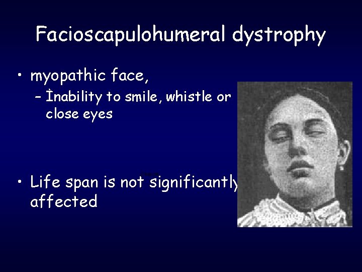
Facioscapulohumeral dystrophy • myopathic face, – İnability to smile, whistle or close eyes • Life span is not significantly affected Charcot
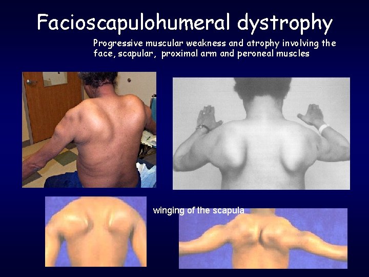
Facioscapulohumeral dystrophy Progressive muscular weakness and atrophy involving the face, scapular, proximal arm and peroneal muscles winging of the scapula
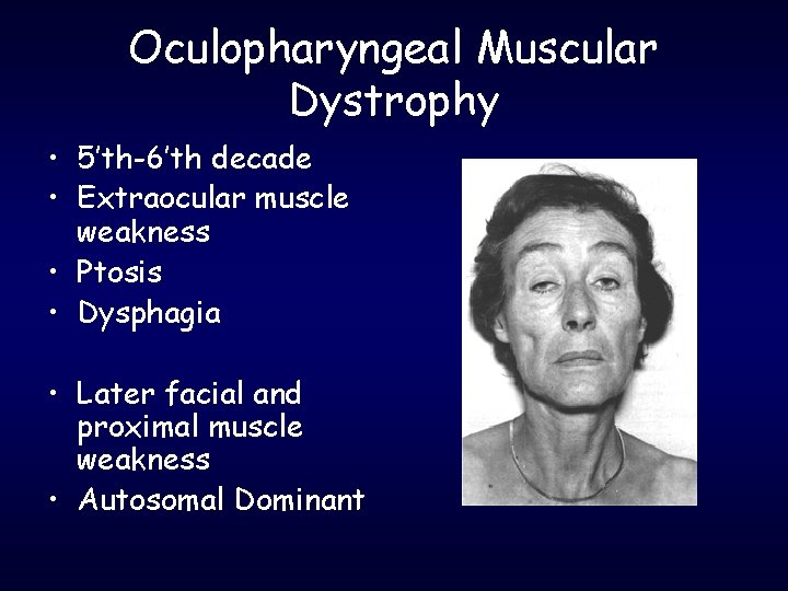
Oculopharyngeal Muscular Dystrophy • 5’th-6’th decade • Extraocular muscle weakness • Ptosis • Dysphagia • Later facial and proximal muscle weakness • Autosomal Dominant
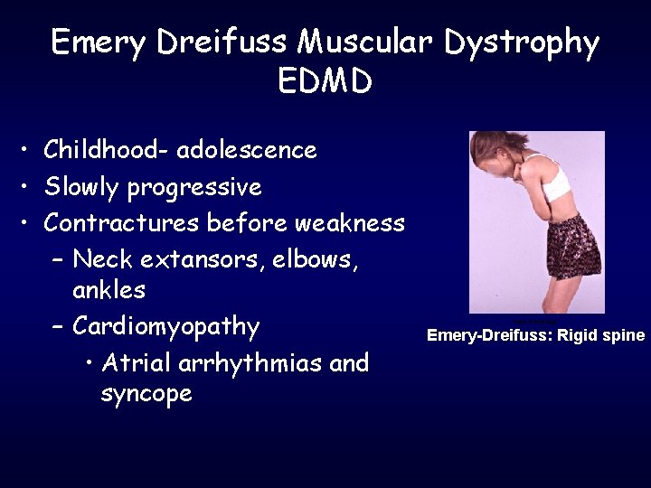
Emery Dreifuss Muscular Dystrophy EDMD • Childhood- adolescence • Slowly progressive • Contractures before weakness – Neck extansors, elbows, ankles – Cardiomyopathy • Atrial arrhythmias and syncope From: A Kornberg Emery-Dreifuss: Rigid spine
Limb-girdle dystrophies • Autosomal Dominant (10%) – LGMD-1 • Autosomal Recessive (90%) – LGMD-2 • • • Symptoms between 2 -40 High CK Slow progressive proximal muscle weakness Onset variable Spares the face
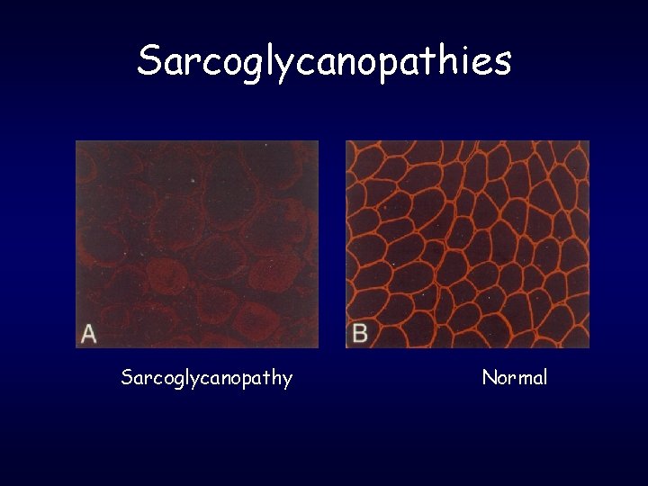
Limb-girdle dystrophies • Causes: – Sarcoglycanopathies • α, β, γ, δ sarcoglycans • Clinically often indistinguishable from Duchennedystrophy • scapular winging more pronounced, hamstrings and quadriceps equally affected – Calpain - 3 mutation – Dysferlin deficiency etc. – Caveolin deficiency (AD)
Sarcoglycanopathies Sarcoglycanopathy Normal
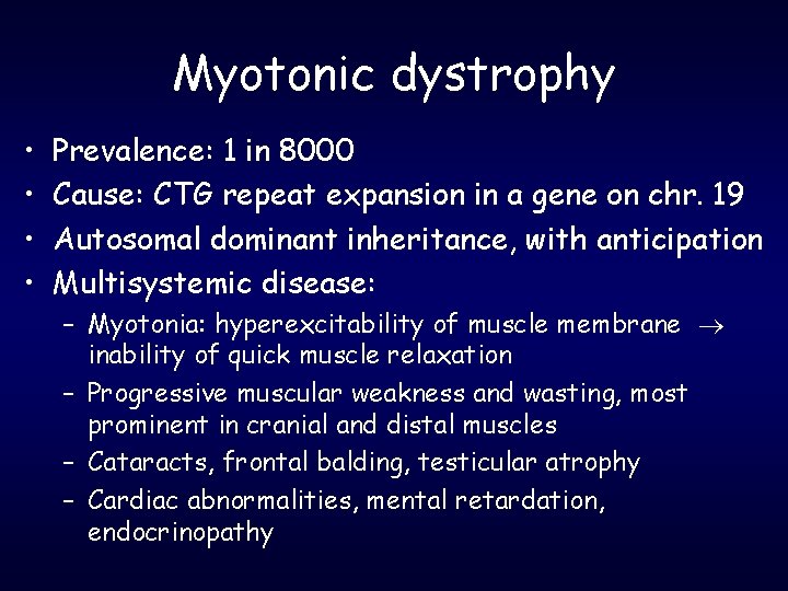
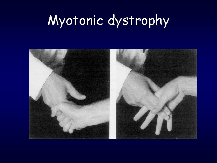
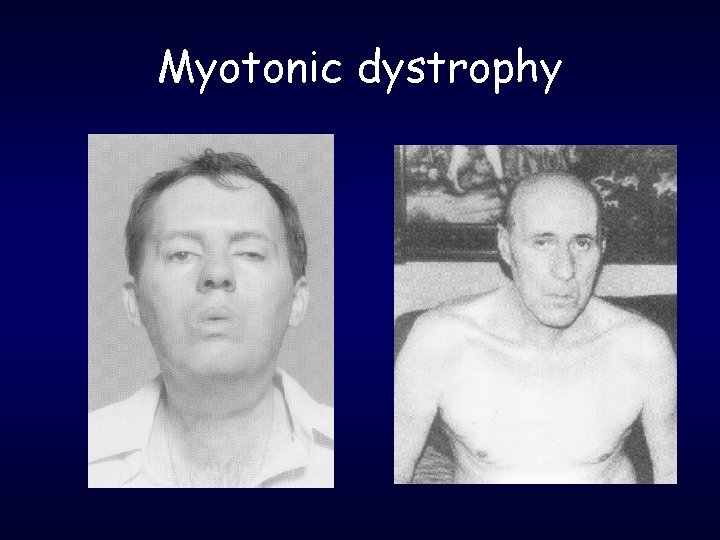
Myotonic dystrophy • • Prevalence: 1 in 8000 Cause: CTG repeat expansion in a gene on chr. 19 Autosomal dominant inheritance, with anticipation Multisystemic disease: – Myotonia: hyperexcitability of muscle membrane inability of quick muscle relaxation – Progressive muscular weakness and wasting, most prominent in cranial and distal muscles – Cataracts, frontal balding, testicular atrophy – Cardiac abnormalities, mental retardation, endocrinopathy
Myotonic dystrophy
Myotonic dystrophy
Muscle channelopathies
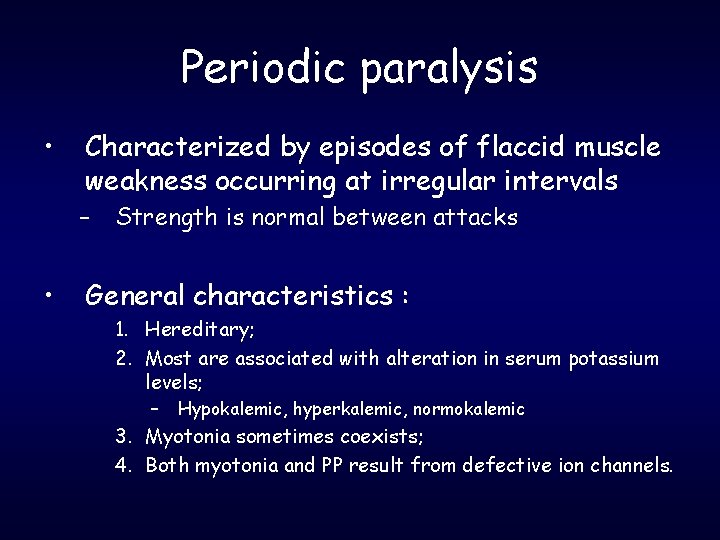
Periodic paralysis • Characterized by episodes of flaccid muscle weakness occurring at irregular intervals – • Strength is normal between attacks General characteristics : 1. Hereditary; 2. Most are associated with alteration in serum potassium levels; – Hypokalemic, hyperkalemic, normokalemic 3. Myotonia sometimes coexists; 4. Both myotonia and PP result from defective ion channels.
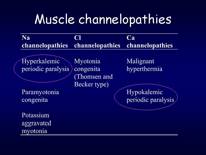
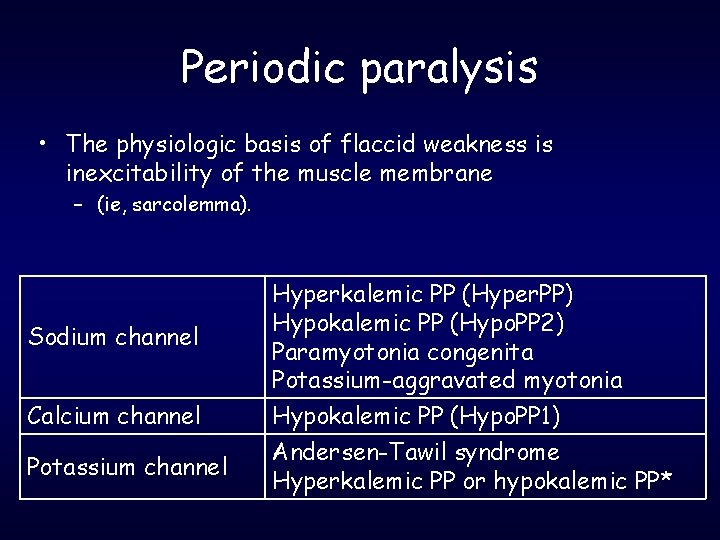
Periodic paralysis • The physiologic basis of flaccid weakness is inexcitability of the muscle membrane – (ie, sarcolemma). Sodium channel Calcium channel Potassium channel Hyperkalemic PP (Hyper. PP) Hypokalemic PP (Hypo. PP 2) Paramyotonia congenita Potassium-aggravated myotonia Hypokalemic PP (Hypo. PP 1) Andersen-Tawil syndrome Hyperkalemic PP or hypokalemic PP*
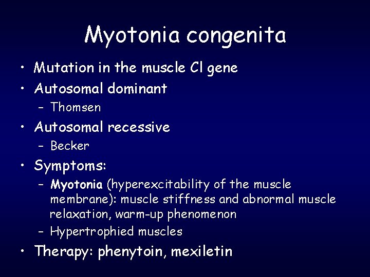
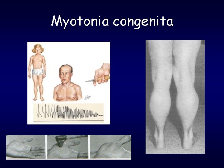
Myotonia congenita • Mutation in the muscle Cl gene • Autosomal dominant – Thomsen • Autosomal recessive – Becker • Symptoms: – Myotonia (hyperexcitability of the muscle membrane): muscle stiffness and abnormal muscle relaxation, warm-up phenomenon – Hypertrophied muscles • Therapy: phenytoin, mexiletin
Myotonia congenita
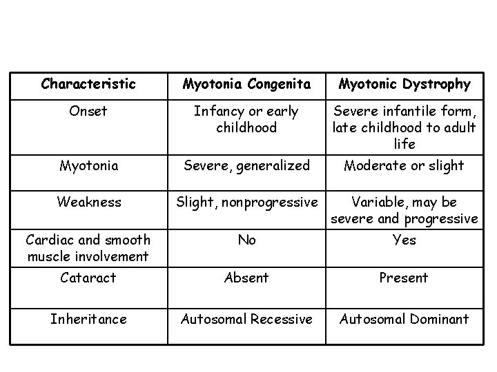
Characteristic Myotonia Congenita Myotonic Dystrophy Onset Infancy or early childhood Severe infantile form, late childhood to adult life Myotonia Severe, generalized Moderate or slight Weakness Slight, nonprogressive Variable, may be severe and progressive Cardiac and smooth muscle involvement No Yes Cataract Absent Present Inheritance Autosomal Recessive Autosomal Dominant
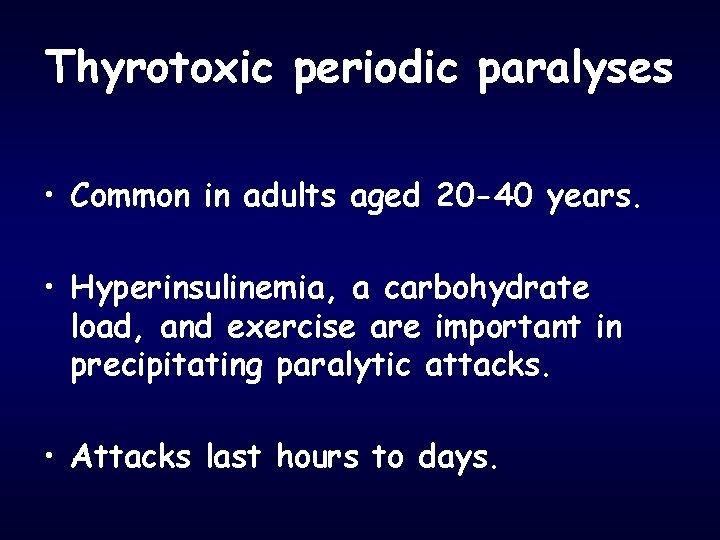
Thyrotoxic periodic paralyses • Common in adults aged 20 -40 years. • Hyperinsulinemia, a carbohydrate load, and exercise are important in precipitating paralytic attacks. • Attacks last hours to days.
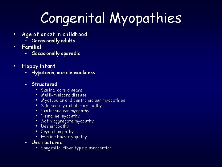
Congenital Myopathies • Age of onset in childhood • Familial • Floppy infant – Occasionally adults – Occasionally sporadic – Hypotonia, muscle weakness – Structered • • • Central core disease Multi-minicore disease Myotubular and centronuclear myopathies X-linked myotubular myopathy Centronuclear myopathy Nemaline myopathy Actin aggregate myopathy Desminopathy Crystallinopathy Hyaline body myopathy – Unstructured • Congenital fiber type disproportion
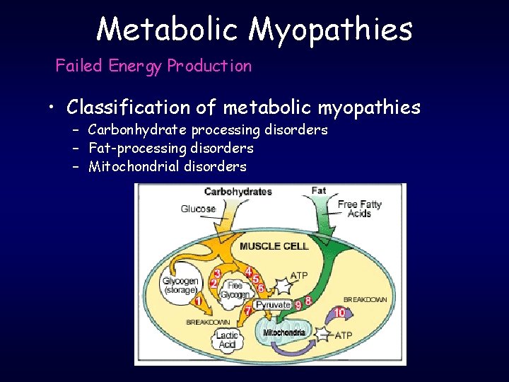
Metabolic Myopathies Failed Energy Production • Classification of metabolic myopathies – Carbonhydrate processing disorders – Fat-processing disorders – Mitochondrial disorders
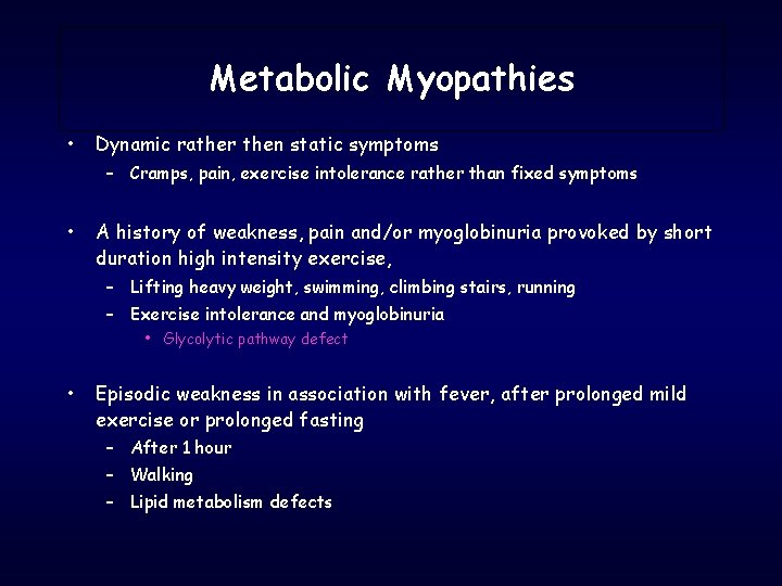
Metabolic Myopathies • Dynamic rather then static symptoms – Cramps, pain, exercise intolerance rather than fixed symptoms • A history of weakness, pain and/or myoglobinuria provoked by short duration high intensity exercise, – Lifting heavy weight, swimming, climbing stairs, running – Exercise intolerance and myoglobinuria • Glycolytic pathway defect • Episodic weakness in association with fever, after prolonged mild exercise or prolonged fasting – After 1 hour – Walking – Lipid metabolism defects
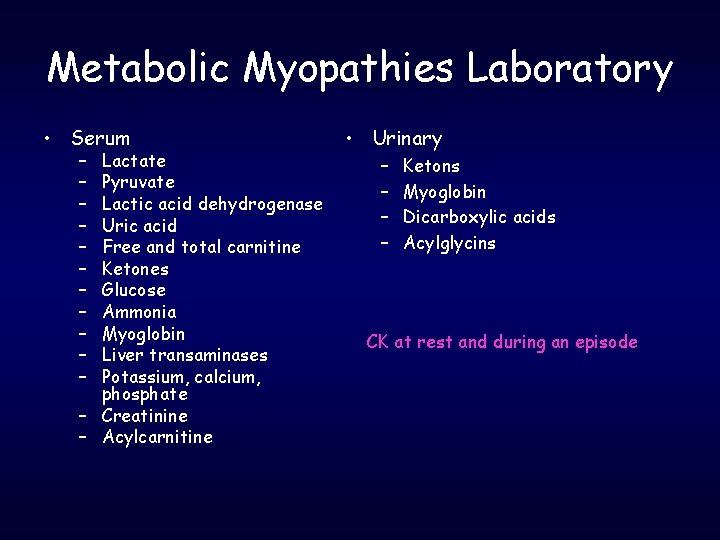
Metabolic Myopathies Laboratory • Serum – – – Lactate Pyruvate Lactic acid dehydrogenase Uric acid Free and total carnitine Ketones Glucose Ammonia Myoglobin Liver transaminases Potassium, calcium, phosphate – Creatinine – Acylcarnitine • Urinary – – Ketons Myoglobin Dicarboxylic acids Acylglycins CK at rest and during an episode
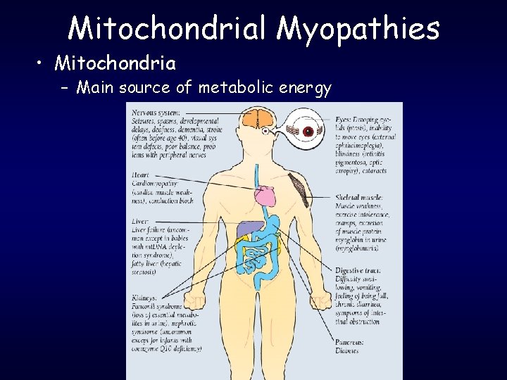
Mitochondrial Myopathies • Mitochondria – Main source of metabolic energy
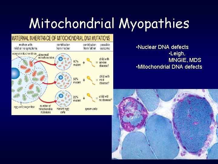
Mitochondrial Myopathies • Nuclear DNA defects • Leigh, MNGIE, MDS • Mitochondrial DNA defects
Idiopathic Inflammatory Myopathies • Clinical Classification – Polymyositis (PM) – Dermatomyositis (DM) – Inclusion Body Myositis (IBM) • Juvenile Dermatomyositis • Myositis associated with malignancy • Myositis associated with collagen vascular disease Bohan & Peter (1975). NEJM. Tanimoto et al. (1995). J Rheumatology.
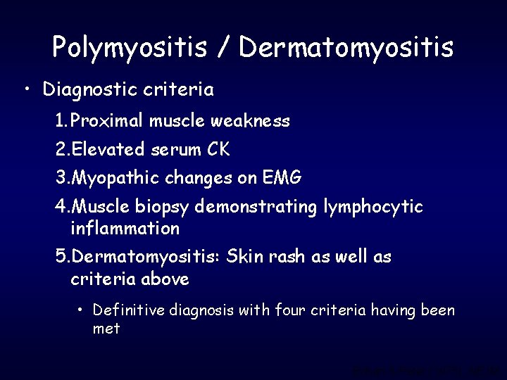
Polymyositis / Dermatomyositis • Diagnostic criteria 1. Proximal muscle weakness 2. Elevated serum CK 3. Myopathic changes on EMG 4. Muscle biopsy demonstrating lymphocytic inflammation 5. Dermatomyositis: Skin rash as well as criteria above • Definitive diagnosis with four criteria having been met Bohan & Peter (1975). NEJM.
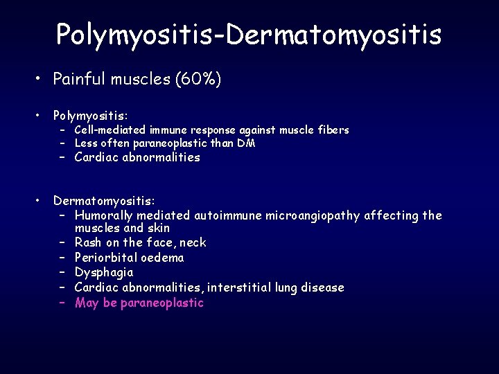
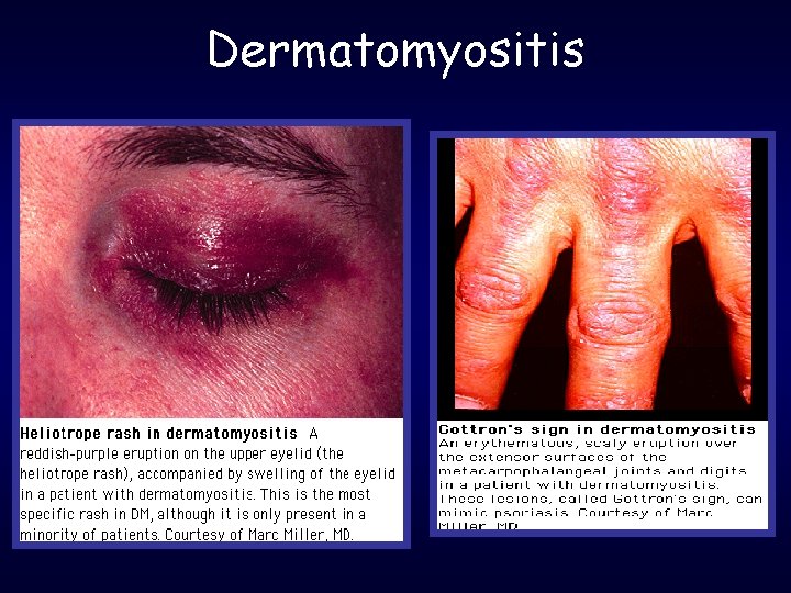
Polymyositis-Dermatomyositis • Painful muscles (60%) • Polymyositis: – Cell-mediated immune response against muscle fibers – Less often paraneoplastic than DM – Cardiac abnormalities • Dermatomyositis: – Humorally mediated autoimmune microangiopathy affecting the muscles and skin – Rash on the face, neck – Periorbital oedema – Dysphagia – Cardiac abnormalities, interstitial lung disease – May be paraneoplastic
Dermatomyositis
Treatment for DM, PM • Immunosuppression – long-term treatment with corticosteroids (1 mg/day) – Mtx-Imuran. . .
Inclusion Body Myositis • Mainly affects older males • Symptoms begin insidiously and progress slowly • Symptoms are often present 5 -6 years before diagnosis • Finger flexors-quadriceps • May include focal, distal or asymmetric weakness • Neurogenic or mixed neurogenic / myopathic changes on EMG • Dysphagia is noted in more than 20% of patient Wortmann RL. Primer on Rheum Dis. 12 th edition. 2001: 369– 376.
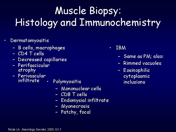
Muscle Biopsy: Histology and Immunochemistry • Dermatomyositis • IBM – B cells, macrophages – CD 4 T cells – Same as PM; also: – Decreased capillaries – Rimmed vacuoles – Perifascicular atrophy – Eosinophilic – Perivascular cytoplasmic infiltrate • Polymyositis inclusions – Mononuclear cells – CD 8 T cells – Endomysial infiltrate – Myonecrosis – Patchy, focal Rolak LA. Neurology Secrets. 2005: 63 -7.
Infectious causes of myopathy • • Trichinosis Cysticercosis Toxoplasmosis HIV Coxsackie A and B Influenza Lyme Staphylococcus aeurus
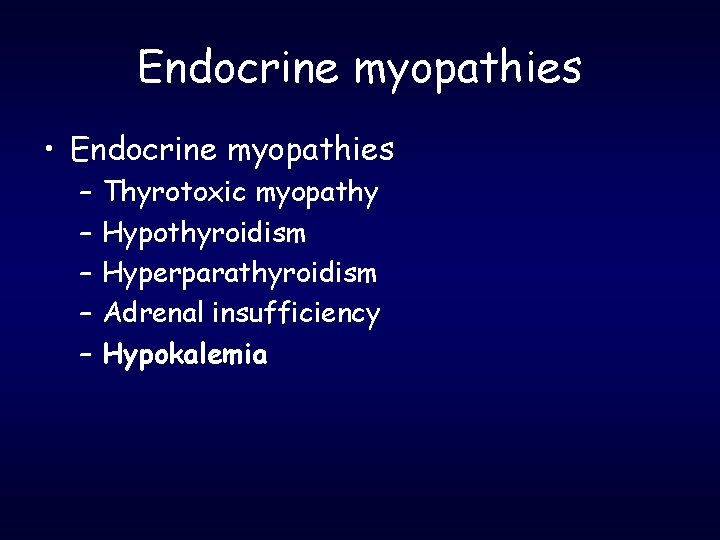
Endocrine myopathies • Endocrine myopathies – – – Thyrotoxic myopathy Hypothyroidism Hyperparathyroidism Adrenal insufficiency Hypokalemia
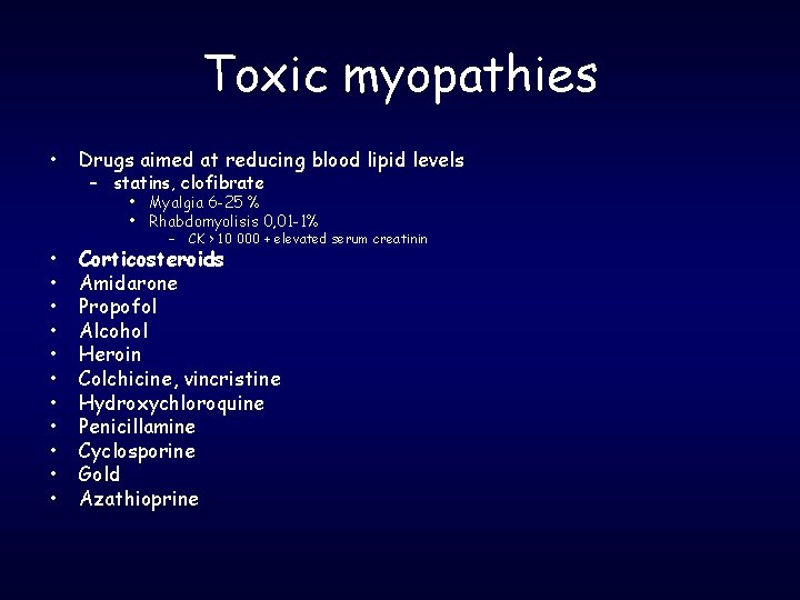
Toxic myopathies • Drugs aimed at reducing blood lipid levels – statins, clofibrate • Myalgia 6 -25 % • Rhabdomyolisis 0, 01 -1% • • • – CK > 10 000 + elevated serum creatinin Corticosteroids Amidarone Propofol Alcohol Heroin Colchicine, vincristine Hydroxychloroquine Penicillamine Cyclosporine Gold Azathioprine
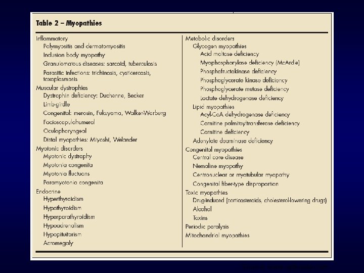
Source: www. uptodate. com
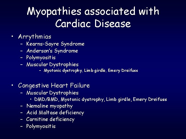
Myopathies associated with Cardiac Disease • Arrythmias – – Kearns-Sayre Syndrome Anderson’s Syndrome Polymyositis Muscular Dystrophies – Myotonic dystrophy, Limb girdle, Emery Dreifuss • Congestive Heart Failure – Muscular Dystrophies – – • DMD/BMD, Myotonic dystrophy, Limb girdle, Emery Dreifuss Nemaline myopathy Acid Maltase deficiency Carnitine deficiency Polymyositis