The Antiglobulin Test Hemolytic disease of newborn Objectives

The Antiglobulin Test Hemolytic disease of newborn

Objectives • – – – – Understand types of Coomb`s test Indications, Steps and interpretation Hemolytic Disease of the Newborn State the testing to be performed on the mother to monitor the severity of HDN. List the laboratory tests and values State the treatment options State the requirements of blood to be used for transfusion of the fetus and newborn.

Antigen antibody reactions
 affixed to RBCs or")
ANTIGLOBULIN TEST • Detection of antibodies- (Ig. G or complement) affixed to RBCs or free in plasma – in vivo-Direct antiglobulin test (DAT) – in vitro -Indirect antiglobulin test (IAT)

Types of Coomb`s Test DAT- Direct antiglobulin test IAT- Indirect antiglobulin test
 bind to human globulins either free")
ANTIGLOBULIN TEST • Principle - Antihuman globulins (AHG) bind to human globulins either free in serum or attached to RBCs

ANTIGLOBULIN TEST – Pentameric Ig. M Abs are so large that, when bound to RBC Ags, the RBCs agglutinate (usually at RT) – Ig. G Abs usually need a little help, a bridge molecule, to agglutinate RBCs – AHG acts as a bridge molecule
 was discovered by Coombs etal in 1945.")
The Antiglobulin Test �Antiglobulin serum (Coombs’Serum) was discovered by Coombs etal in 1945.
 Reagent • Preparation – Anti-human globulin reagent is produced by immunizing")
Anti-Human Globulin (AHG) Reagent • Preparation – Anti-human globulin reagent is produced by immunizing rabbits, goats or sheep with human serum or purified type antigen. – Animals are bled after a specified period and the reagent is purified by absorbing unwanted antibodies.

Types of AHG reagent �Polyspecific antiglobulin reagent human Ig. G, C 3 and C 4 �Monospecific antiglobulin reagent � Any one- human Ig. M, Ig. D, Ig. A, C 3 or C 4
")
DIRECT ANTIGLOBULIN TEST (DAT)

DAT • detects sensitized red cells with Ig. G and/or complement components C 3 b and C 3 d in vivo. • In vivo coating may occur when any immune mechanism is attacking the patient's own RBC's. – Autoimmunity – Alloimmunity – Drug-induced immune-mediated mechanism.

Examples of alloimmune hemolysis • Hemolytic transfusion reaction • Hemolytic disease of the newborn (also known as HDN or erythroblastosis fetalis) – Rhesus D – ABO – Anti-Kell – Rhesus c, E – Other -Rh. C, Rhe, Kidd, Duffy, MN, P or others

Examples of autoimmune hemolysis – Warm antibody autoimmune hemolytic anemia – Idiopathic – Systemic lupus erythematosus – Cold antibody autoimmune hemolytic anemia – Infectious mononucleosis – Paroxysmal cold hemoglobinuria (rare)

Drug-induced immune-mediated hemolysis • Methyldopa • Penicillin • Quinidine • Cephalosporins

Blood Sample �fresh �EDTA vial

Procedure of DAT 1 drop of EDTA sample Wash the red cells 3 -4 times in saline- to remove free globulin molecules. Add 2 drops of polyspecific AHG serum Mix, Centrifuge at 1000 rpm for 1 minute Check for agglutination Add Check (Ig. G coated) cells to a negative test. If agglutination is obtained, the result is valid.
 �Indications- to determine the presence of free antibodies in")
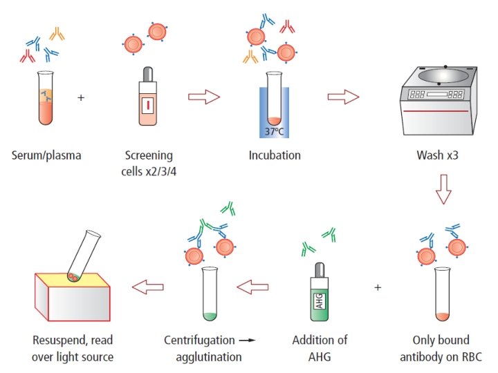
Indirect Antihuman globulin Test (IAT) �Indications- to determine the presence of free antibodies in serum. �in vitro sensitization of red cells with Ig. G and/or complement 1. Compatibility testing. 2. Unexpected antibodies in serum.

Indirect antiglobulin test

Blood Sample �fresh �Plain vial

Procedure: 2 -3 drops of the test serum Add 1 drop of 3 -5% suspension of washed O Rh (D) positive red cells Mix and incubate at 37°C for 30 -40 minutes. Centrifuge at 1000 rpm for 1 minutes. Examine for hemolysis and/or agglutination-complete antibodies. If not wash cells 3 -4 times Add 2 drops of AHG serum to the cells. Mix and centrifuge at 1000 rpm for 1 minutes immediately. Examine for agglutination- incomplete antibody

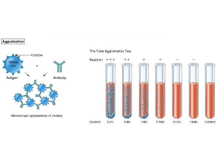
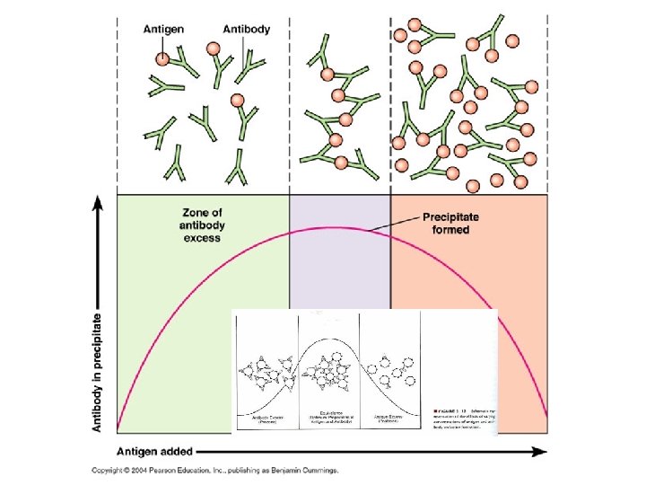
Antigen-Antibody Ratio • Prozone - antibody excess: Antibodies saturating all antigen sites; no antibodies forming cross-linkages between cells; no agglutination • Zone of equivalence: antibodies and antigens present in optimum ratio, agglutination formed • Zone of antigen excess (Post-zone): too many antigens - any agglutination is hidden by masses of unagglutinated antigens

COOMB’S CELLS • Antibody-coated cells are used as a positive indicator – To show that test cells were properly washed – No reagent deterioration has occurred • Failure to agglutinate-test result is not valid

Hemolytic Disease of the Newborn
")
Cause of Hemolytic Disease Pregnancy with fetal red blood cells having antigen(of paternal origin) Exposure to red blood cells during transfusion. Maternal Ig. G antibodies produced

Cause of Hemolytic Disease Antigen of paternal origin present on the fetal red blood cells Maternal Ig. G antibodies cross the placenta to coat fetal antigens Decreased red blood cell survival which can result in anemia
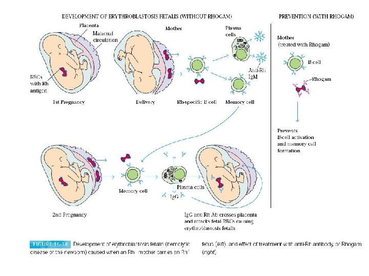

Three Classifications of HDN • Rh – anti-D • ABO • “Other” –anti-C, c, E, e, Jk, K, Fy, S etc.

Rh Hemolytic Disease • Anti-D is the commonest form of severe HDN • mild to severe.

ABO Hemolytic Disease • Mother group O-anti-A, -B and –A, B in their plasma • Fetal group A or B- RBCs attacked by antibodies • Occurs in only 3%, is severe in only 1%

“Other” Hemolytic Disease • Uncommon, occurs in ~0. 8% of pregnant women. • Anti-K – mild to severe – usually caused by multiple blood transfusions – is the second most common form of severe HDN

Hemolysis of fetal red blood cells Results in anemia As the red blood cells break down, bilirubin is formed Baby's responds by trying to make more red blood cells in the bone marrow, liver and spleen Hyperbilirubinemia results in jaundice Hepatosplenomegaly New red blood cells released prematurely from bone marrow and are unable to do the work of mature red blood cells

Complications During Pregnancy • Severe anemia • Hydrops Fetalis – Baby's organs are unable to handle the anemia – The heart begins to fail – Fluid build up in the baby's tissues and organs • A fetus with hydrops is at great risk of being stillborn.
 – Edema")
Postnatal problems – Asphyxia – Pulmonary hypertension – Pallor (due to anemia) – Edema (hydrops, due to low serum albumin) – Respiratory distress – Coagulopathies (↓ platelets & clotting factors) – Jaundice – Kernicterus (from hyperbilirubinemia) – Hypoglycemia (due to hyperinsulinemnia from islet cell hyperplasia)
 • High levels of indirect bilirubin (>20 mg/d. L) – crosses")
Kernicterus (bilirubin encephalopathy) • High levels of indirect bilirubin (>20 mg/d. L) – crosses the blood-brain barrierunbound unconjugated bilirubin – penetrates neuronal and glial membranes- lipid soluble – toxic to nerve cells • Patients who survive kernicterus have severe permanent neurologic symptoms – Choreoathetosis, spasticity, muscular rigidity, ataxia, deafness, mental retardation).
 ↑ nucleated RBC")
Laboratory Findings • • • Anemia Hyperbilirubinemia Reticulocytosis (6 to 40%) ↑ nucleated RBC count (>10/100 WBCs) Thrombocytopenia Leukopenia Positive Direct Antiglobulin Test Hypoalbuminemia Rh negative blood type or ABO incompatibility Smear: polychromasia, anisocytosis, no spherocytes

MCA Doppler study • Reliable non-invasive screening tool to detect fetal anemia. – The vessel can be easily visualized with color flow Doppler as early as 18 weeks’ gestation. – In cases of fetal anemia, an increase in the fetal cardiac output and a decrease in blood viscosity contribute to an increased blood flow velocity

Blood Bank Testing

Management • Measure bilirubin in cord blood and at least every 4 hours for the first 12 to 24 hours • Transcutaneous Monitoring
 • To prevent hydrops fetalis and fetal death. • Transfusions done")
Intrauterine Transfusion (IUT) • To prevent hydrops fetalis and fetal death. • Transfusions done every 1 to 4 weeks until the fetus is mature enough to be delivered safely. • A compatible blood type (usually type O, Rh-negative) is delivered into the fetus's abdominal cavity or into an umbilical cord blood vessel.

Selection of Blood • CPD, as fresh as possible, preferably <5 days old. • A hematocrit of 80% or greater is desirable to minimize the chance of volume overload in the fetus. • The volume transfused- 75 -175 m. L depending on the fetal size and age. • CMV negative • IRRADIATED • O negative, lack all antigens to which mom has antibodies and Coomb’s compatible.

Treatment of Mild HDN • Phototherapy is the treatment of choice.

Exchange Transfusion • • • If the total serum bilirubin level is approaching 20 mg/d. L Continues to rise despite intense in-hospital phototherapy. Removes • • Correct anemia Restores albumin and coagulation factors – sensitized cells – Reduces level of maternal antibody. – Removes about 60 percent of bilirubin from the plasma
- Slides: 46