Nervous System Functions of the Nervous System Sensory


 inside and")
 – Includes the Brain and Spinal")
 Division – Sends impulses to the CNS")
 Motor = Efferent (comes second, brain")


 lumped together (term is commonly used to")


 by exocytosis to send signals to neighboring neurons.")


")

: gaps between neurons • Myelin Sheath:")


: carry impulses from sensory receptors to CNS")


 • Sulci: shallow")


 – Where your")
 – Responsible for")


 –")


 – Mammillary body (smell recognition)")
")


 containing CSF in the brain –")


 – voluntary movement of muscles • Norepinephrine –")


 because of an obstruction • “Water on the")


 • Concussion – Slight or mild brain injury – Bleeding")
 • TBI can often result in either temporary or permanent")
- Slides: 107
Nervous System
Functions of the Nervous System • Sensory Input – Monitors changes (stimuli) inside and outside the body • Integration – Processes and interprets sensory input to decide what should be done • Motor Output – Supplies response by activating muscles or glands
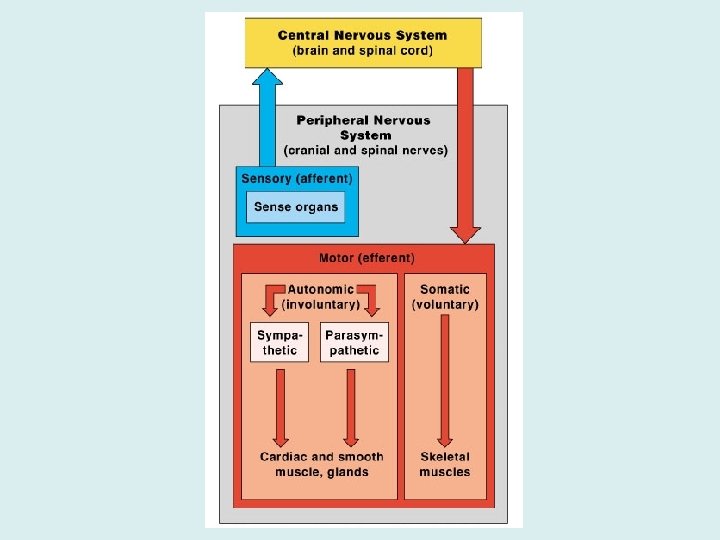
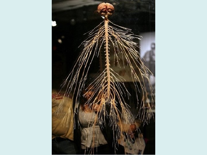
Nervous System Divisions • Central Nervous System (CNS) – Includes the Brain and Spinal Cord • Peripheral Nervous System (PNS) – Includes Spinal nerves & Cranial nerves
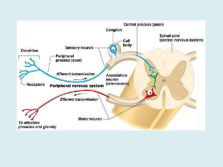
Further Breakdown of PNS • Sensory (afferent) Division – Sends impulses to the CNS from sensory receptors in body – Somatic sensory fibers: messages from skin, muscles and joints – Visceral sensory fibers: messages from internal organs • Motor (efferent) Division – Carries impulses from CNS to effector organs (muscles and glands) to cause a response
SAME Sensory = Afferent (comes first, body brain) Motor = Efferent (comes second, brain body)
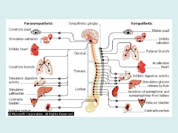
Further Breakdown of Motor Division • Somatic System – Voluntary control of skeletal muscles • Autonomic System – Involuntary body responses, such as smooth & cardiac muscles, and glands
Nervous Tissue • Made up of two types of cells – Supporting Cells: these structures do not directly transmit any signals, but they help the neurons to function properly; aka “glia” – Neurons: actually pass signals
Supporting Cells • Neuroglia: supporting cells (glia) lumped together (term is commonly used to reference all types of supporting cells)
Neuron Cell Body Neuroglia Cells
Types of Supporting Cells in CNS • Astrocytes – Form barrier between capillaries and neurons – Help control chemical environment in brain – “To Make Mice Smarter, Add A Few Astrocytes!” – Scroll to bottom of website to see how astrocytes communicate • Microglia – Dispose of debris, such as dead brain cells and bacteria (immune system)
Astrocytes can release gliotransmitters (like glutamate) by exocytosis to send signals to neighboring neurons. Each astrocyte has its own territory (they don't overlap), and each may interact with several neurons and hundreds to thousands of synapses to properly integrate information. "End-feet" connect to blood vessels in the brain. By signaling blood vessels to expand or narrow, astrocytes regulate local blood flow to provide oxygen and nutrients to neurons in need.
Neurons Astrocytes
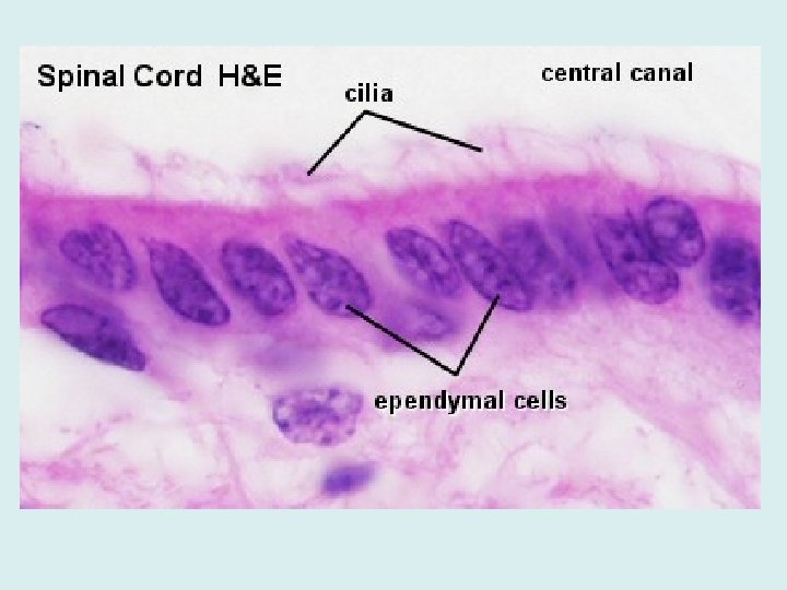
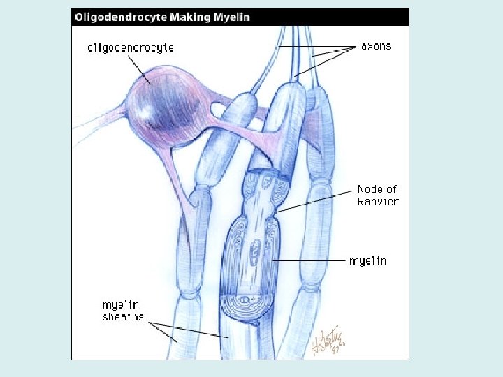
Types of Supporting Cells in CNS • Ependymal – Lines cavity of brain & spinal cord – Circulate cerebrospinal fluid • Oligodendrocytes – Wrap around nerve fibers, forming myelin sheaths
Types of Supporting Cells in PNS • Schwann cells: form myelin sheaths in PNS • Satellite Cells: protective, cushioning cells in PNS
Neuron Cell Body Neuroglia Cells
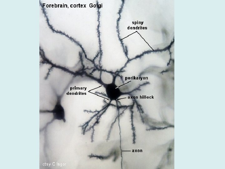
Parts Of A Neuron • Cell Body: metabolic center, contains rough ER (Nissl substance) and neurofibrils (maintain shape) • Dendrites: convey incoming messages toward cell body
Parts Of A Neuron • Axons: conduct outgoing messages away from cell body (has only one originating at axon hillock) • Axon Terminal: branching at end of axon which contain neurotransmitters
Parts Of A Neuron • Synaptic Cleft (Synapse): gaps between neurons • Myelin Sheath: protects and insulates nerve fibers (axon), increases rate of nerve impulses; made of proteins and lipids
Parts Of A Neuron • Schwann cells: cells forming myelin sheath in PNS – CNS = oligodendrocytes • Nodes of Ranvier: gaps between Schwann cells on axon Did you know Einstein’s brain was STOLEN? Here the story here.
FYI: Multiple Sclerosis • Myelin sheaths around the fibers are destroyed, and converted into hardened sheaths called scleroses • Person loses ability to control muscles
Neuron Classification • Functional – Sensory (afferent): carry impulses from sensory receptors to CNS – Motor (efferent): carry impulses from CNS to body – Association (interneurons): connect motor and sensory neurons in neural pathways of CNS
Neuron Classification • Structural – Multipolar: several extensions from cell body (common for motor neuron) – Bipolar: one axon and one dendrite from cell body (common for interneurons) – Unipolar: single process from cell body, process divides (common for sensory neuron)
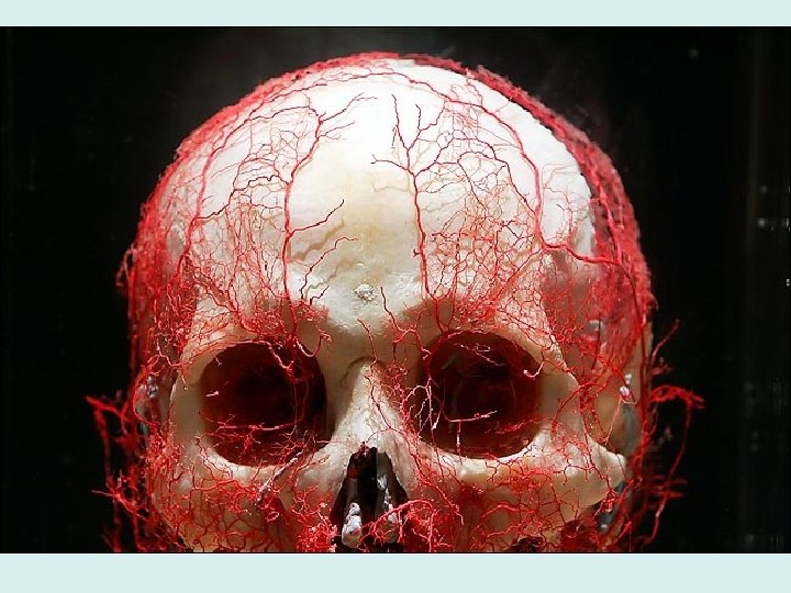
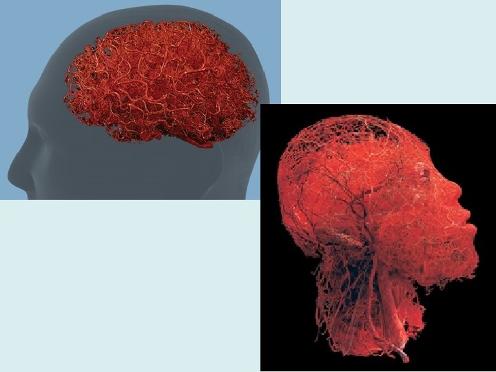
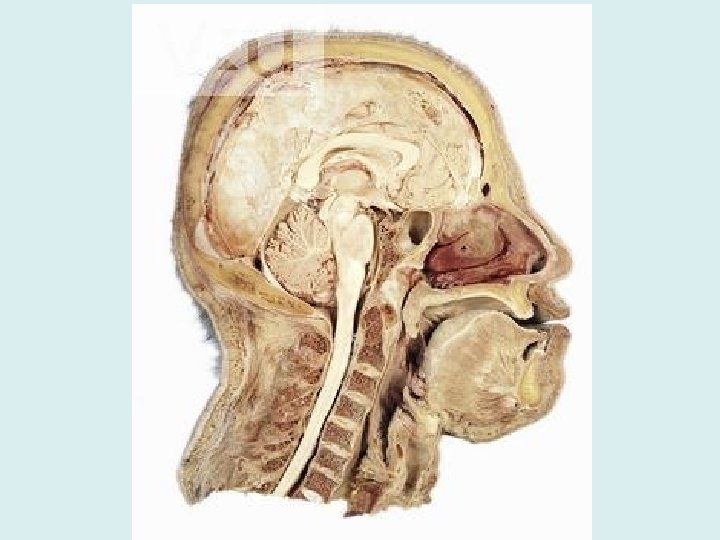
Central Nervous System
http: //www. youtube. com/watch? v=sn. O 68 a. J TOp. M
Central Nervous System …also called the CEREBRUM
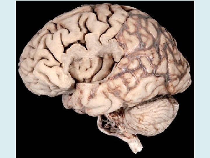
Brain Characteristics • Gyri: elevated ridges of tissue (gyrus is singular) • Sulci: shallow grooves separating gyri (sulcus is singular) • Fissures: deep grooves separating large regions of brain • Lobes: separations of hemispheres of brain
Senses & Controls of Brain - Cerebrum • White matter: collections of myelinated nerve fibers, usually found in deeper brain matter; carries the impulses (axons) • Gray matter: collections of UNmyelinated nerve fibers and cell bodies; usually found in the outer areas; contains the neuron cell bodies
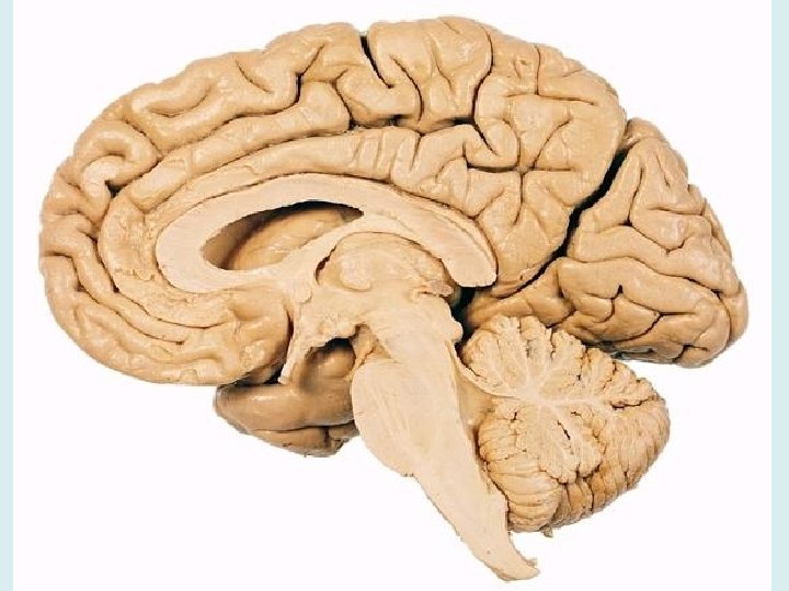
Cerebrum • The cerebrum is divided into right and left hemispheres by the longitudinal fissure • Controls consciousness • Senses and motor control occurs here! • Each hemisphere divided into 4 lobes
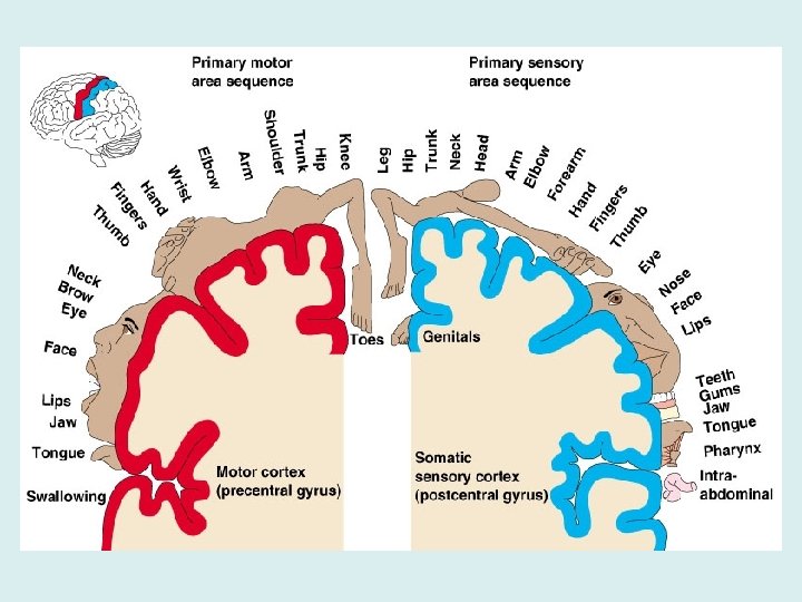
Funny thing about the brain… • The somatic sensory area (cortex) – Where your senses are picked up & processed – Upside down • Signals from body parts toward the head are picked up at the base of the area, while signals from lower (inferior) body parts are picked up at the top of the area – Crossed pathways • Left side of the sensory area receives impulses from the right side of the body, and vice versa
Funny thing about the brain… • The primary motor area (cortex) – Responsible for sending out signals for movement – Also upside down with crossed pathways
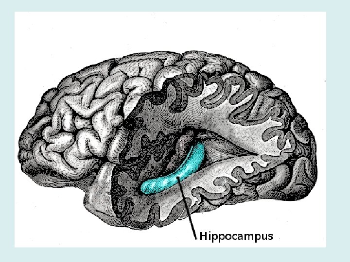
Senses & Controls of Brain - Cerebrum • Occipital Lobe: contains visual sense • Temporal Lobe: contains smell & auditory senses – Also includes the hippocampus, which is responsible for longterm memory (including forming new memories about events) and spatial navigation
Senses & Controls of Brain - Cerebrum Frontal Lobe… • Broca’s area: ability to speak (vocalize), usually more developed in left hemisphere Language Comprehension • Language comprehension area: word meanings Broca’s Area
Senses & Controls of Brain - Cerebrum • Speech Area (aka Wernicke’s Area) – located at the junction of the temporal, parietal, and occipital lobes – Allows one to context spoken words (use them correctly) – Usually more developed in left hemisphere Speech Area
Senses & Controls of Brain - Cerebrum • The Corpus Callosum is a very large nerve fiber tract (bundle) that connects the cerebral hemispheres & allows them to communicate
Cerebellum • Provides timing for skeletal muscle activity • Controls balance and equilibrium • Are you clumsy? Uncoordinated? Blame your cerebellum!
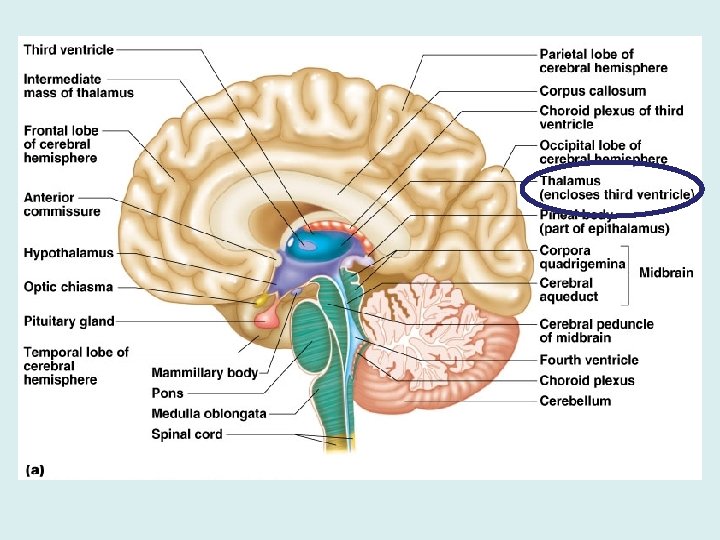
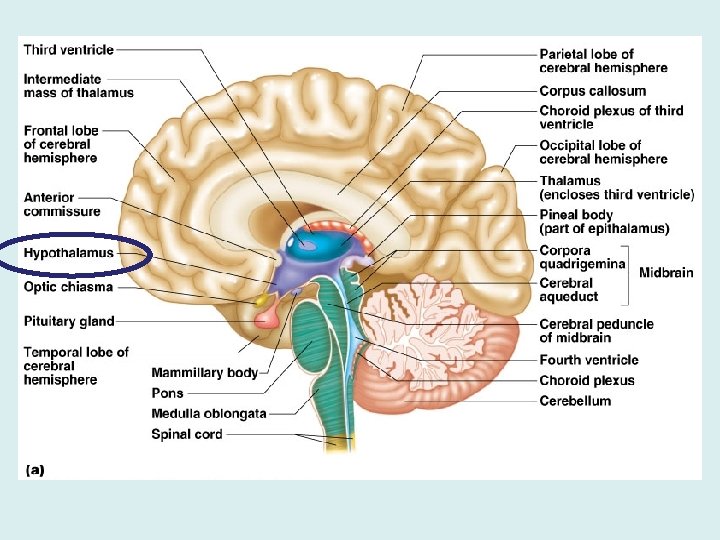
Sections of Diencephalon • Thalamus – Relay station for sensory impulses up to correct part of sensory cortex – Like an e-mail server… sends the message to the right place!
Sections of Diencephalon • Hypothalamus – Helps regulate body temp, water balance, and metabolism (homeostasis) – Involved with emotions; contains thirst, appetite, sex, pain, fear, rage, affection, and pleasure centers
Sections of Diencephalon – Regulates the pituitary gland (attached) – Mammillary body (smell recognition) and optic chiasma (optic nerves crossing) attached ANATOMY HUMOR… The hypothalamus is one of the most important parts of the brain, involved in many kinds of motivation, among other functions. The hypothalamus controls the “Four F’s”: fighting, fleeing, feeding, and mating. -Unknown psychology professor in neuropsychology course
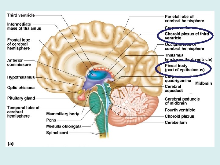
Sections of Diencephalon • Epithalamus – Pineal body: assists in biological clock (daily/seasonal/life cycles) by releasing melatonin – Choroid plexus: forms cerebrospinal fluid (there is also another one in the brainstem)
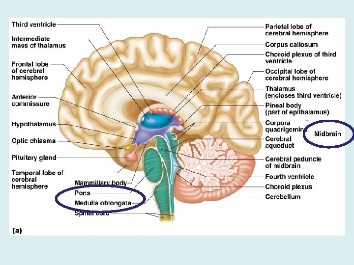
Sections of Brainstem • Midbrain – Conveys ascending and descending impulses • Pons – Conveys ascending and descending impulses – Helps to regulate breathing
Sections of Brainstem • Medulla Oblongata – Conveys ascending and descending impulses – Regulates autonomic activities such as heart rate, blood pressure, breathing, swallowing, vomiting, defecation, sneezing, coughing… the list goes on • As soon as it passes out of the skull (through the foramen magnum), the brainstem is then known as the spinal cord
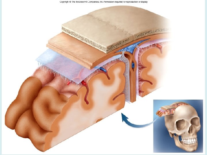
Meninges of the CNS • Dura Mater: thick, outermost • Arachnoid: spiderweb-like • Pia Mater: innermost, clings to brain
Cerebrospinal Fluid • A watery broth that circulates and protects the brain & spinal cord from trauma • Formed by choroid plexuses, circulated by ependymal cells • A very weak salt solution with low amounts of protein (200 m. L total; 135 -150 m. L around the brain) • Any change in composition (levels of protein, presence of blood, glucose levels) may be a sign of meningitis, tumors, infection, or something else • Tested through a spinal tap
• Ventricles – Set of structures (openings) containing CSF in the brain – Drains into the central canal of the spinal cord
Central Nervous System • Brain • Spinal Cord – Extends from medulla oblongata (at foramen magnum) to the T 12 vertebrae (last thoracic vertebrae) – Below T 12 is the cauda equina (a collection of spinal nerves)
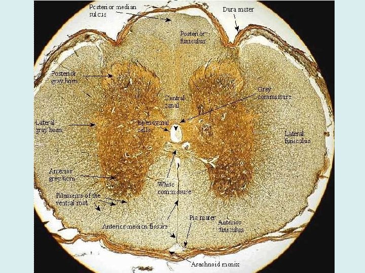
Spinal Cord – 3 Basic Parts • Inner gray matter columns – Mostly cell bodies • Outer white matter columns – Conduction tracts – Composes most of spinal cord • Central canal filled with CSF
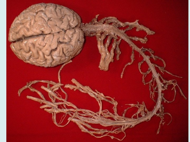
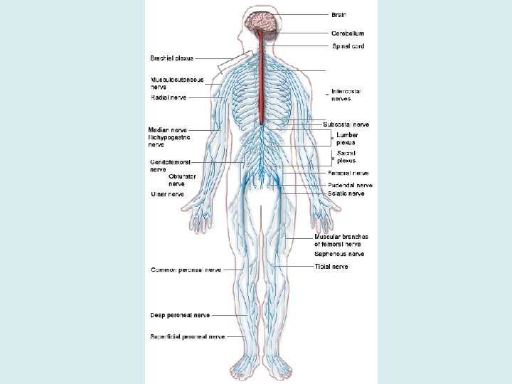
Peripheral Nervous System • Composed of Nerves: bundles of nerve fibers • Neuron fibers surrounded by connective tissue (work similar to muscle fibers!) • Two Types of Peripheral Nerves: Spinal & Cranial Nerves
Nerves • Endoneurium surrounds each fiber • Groups of fibers are bound into fascicles by perineurium • Fascicles are bound together by epineurium
Spinal Nerves • Spinal Nerves – 31 originating from spinal cord – Named for segments • Cervical – 8 (not 7) • Thoracic – 12 • Lumbar – 5 • Sacral – 5 • Coccygeal – 1 (not 4) • Spinal nerves continue on to form the rest of the nerves in your body!
Nerves • Endoneurium surrounds each fiber • Groups of fibers are bound into fascicles by perineurium • Fascicles are bound together by epineurium
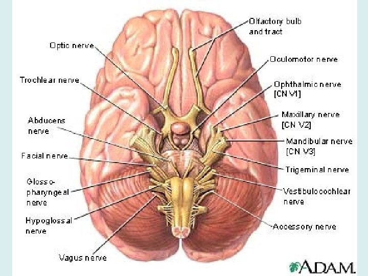
Nerves • Cranial Nerves – 12 originating from brainstem • Olfactory – smell • Optic – vision • Oculomotor – eye lens shape, pupil size (moving eyeball) • Trochlear – follow moving objects with eye
Nerves • Trigeminal – skin senses from face, chewing muscles • Abducens – roll eye laterally • Facial – facial expression muscles, lacrimal (tear) and salivary (spit) glands, taste • Vestibulocochlear – sense of balance, hearing • Glossopharyngeal – swallowing and saliva production
Nerves • Vagus – sense & motor of pharynx & larynx • Accessory – activate sternocleidomastoid and trapezius muscles (turn neck, shrug shoulders) • Hypoglossal – control tongue movements
Transmitting Impulses • Nerve impulses have a domino effect • Each neuron receives an impulse and must pass it on to the next neuron – Dendrites pick up the impulse – Shuttled through the axon – Transmitted to next neuron at axon terminal
Sending The Signal
Impulse through a neuron… • Polarized: the neuron at rest – More sodium ions outside the cell, less potassium ions on the inside (in comparison to the # of Na ions on outside) – Sodium/Potassium pumps keep it this way – Because the abundance of sodium ions outside the cell is way higher than the abundance of potassium ions on the inside, we would say that there is a partially positive charge on the outside of the cell, and a partially negative charge on the inside of the cell – This balance of charges is called the Resting Potential
Through a neuron… • Depolarization: A stimulus comes along, and Na+ moves into the membrane – The stimulus (neurotransmitter) opens sodium channels, allowing Na+ to rush in – The neuron continues to open channels all along membrane
Through a neuron… • Action Potential: The depolarization wave continues to move down the membrane – Once this process is started, it continues to move all the way down the membrane • Once you “pop”, you can’t stop!
Through a neuron… • Repolarization: K+ ions move outside, and Na+ ions stay inside the membrane – Potassium channels on inside of membrane open to allow K+ to move out
Through a neuron… • Refractory period: Everything gets put back to normal… K+ returns to inside, Na+ returns to outside – All thanks to Na+/K+ pump – Can’t respond to another stimulus during this period • The cell is then returned to the polarized state until another impulse comes along • Remember: the process takes less time if it has a myelin sheath around the neuron processes!
• http: //highered. mcgrawhill. com/sites/0072495855/student_view 0/ chapter 14/animation__the_nerve_impulse. html
Between neurons… • Remember that action potential that was created? Well, eventually, it reaches the end of the axon (axon terminals) • When it does, it goes through steps to pass the signal on to the next neuron…
Between neurons • Calcium gates in axon terminals open, allowing Ca+2 in • Due to Ca+2 entering the axon terminal, a specific neurotransmitter gets released • Neurotransmitter diffuses across synapse • Neurotransmitter binds with specific receptors on the next neuron
Returning to normal • The neurotransmitter can do one of two things… – If received by another neuron, it will open Na+ gates on the next neuron, beginning a new action potential on the new neuron – If received by some body part, it will stimulate some sort of change (muscles, glands, etc. ) • After neurotransmitter does its job, the receptor releases it back into synapse, and finds its way back to the neuron so it can be re-released
• http: //highered. mcgrawhill. com/sites/0072495855/student_view 0/ chapter 14/animation__chemical_synapse_ _quiz_2_. html
• There approximately 20 known neurotransmitters
Types of neurotransmitters • Acetylcholine (ACh) – voluntary movement of muscles • Norepinephrine – wakefulness or arousal • Dopamine – motivation, pleasure, associated with addiction and love • Serotonin – memory, emotions, temperature regulation • Histamine – wakefulness • Endorphins – natural pain killer ** Many neurotransmitters also have hormonal effects.
Nervous System Conditions
Meningitis • Inflammation of the meninges • Can then spread to actual nervous tissue of CNS, and inflame the brain (encephalitis)
Hydrocephalus • CSF accumulates (most commonly) because of an obstruction • “Water on the Brain”
Myelitis • Swelling of the spinal cord • Disrupts CNS functions linking the brain and limbs
Multiple Sclerosis • Myelin sheaths around the fibers are destroyed, and converted into hardened sheaths called scleroses • Person loses ability to control muscles • Victims experience muscle weakness, abnormal muscle spasms, and difficulties in movement, coordination, & balance
Cerebrovascular Accident • Commonly called a stroke • The result of a ruptured blood vessel supplying a region of the brain • Brain tissue supplied with oxygen from that blood source dies • Loss of some functions or death may result
Alzheimer’s Disease • Progressive degenerative brain disease • Structural changes in the brain include abnormal protein deposits, twisted fibers within neurons, and atrophy of the brain • Victims experience memory loss, irritability, confusion, and hallucinations
Parkinson’s Disease • Loss of dopamine-secreting cells • Causes other transmission problems, which leads to symptoms • Victims experience tremors, rigidity, and gait issues (posture and walking)
Traumatic Brain Injury (TBI) • Concussion – Slight or mild brain injury – Bleeding & tearing of nerve fibers happened – Recovery likely with possible memory loss • Contusion – A more severe TBI – Nervous tissue destruction occurs – Nervous tissue does not regenerate
Traumatic Brain Injury (TBI) • TBI can often result in either temporary or permanent amnesia – Anterograde amnesia: new events are not transferred to the permanent as long-term memory – Retrograde amnesia: inability to recall memories of the past