
Neuronal Function in the Nervous System Learning Objectives
Neuronal Function in the Nervous System
Learning Objectives n n n n 1. Explain parts of a typical nerve cell and describe their functions. 2. Discuss common types of nerve and glia cells. 3. Describe functions of nerve and glia cells. 4. Explain electrical and chemical properties of nerve cells. 5. Describe mechanism of impulse generation and its conduction. 6. Explain nerve cell responses to injuries in the nervous system. 7. Explain differential regenerative processes between central and peripheral nervous system. 8. Discuss common neurotransmitters and their functions.
Nerve Cells n Basic anatomic and functional unit of the nervous system Primary Function: n Two Primary Types n
Neuron n Three Basic Elements n General Process
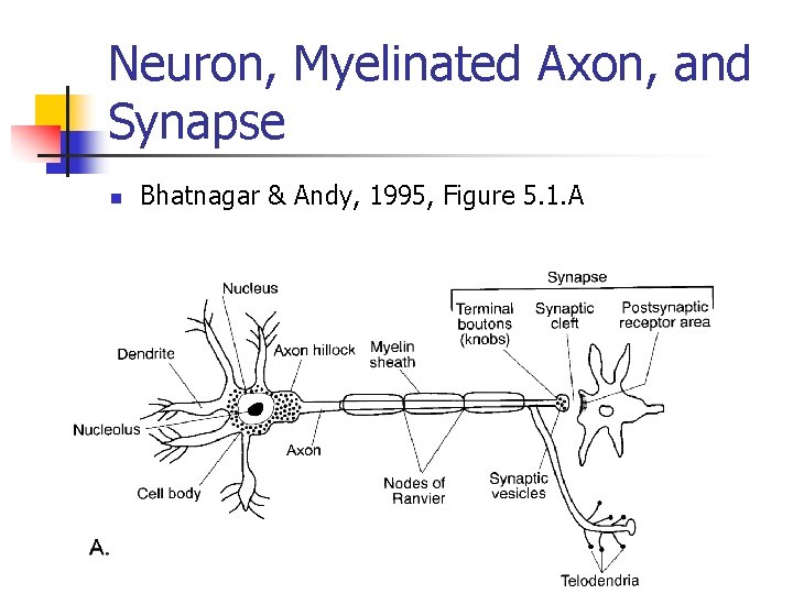
Neuron, Myelinated Axon, and Synapse n Bhatnagar & Andy, 1995, Figure 5. 1. A
Nerve Cell Structure of Neurons: Cell Body n Two major components n 1. Cytoplasm
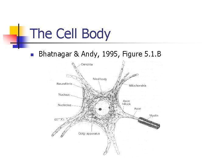
The Cell Body n Bhatnagar & Andy, 1995, Figure 5. 1. B
Nerve Cell Structure of Neurons: Cell Body n 2. Nucleus
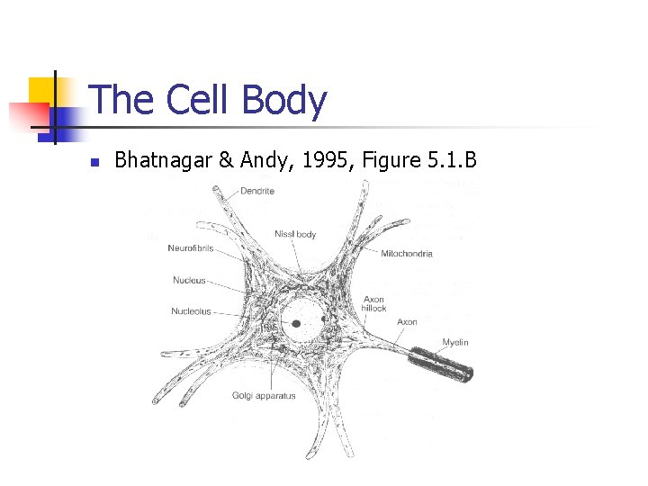
The Cell Body n Bhatnagar & Andy, 1995, Figure 5. 1. B
Nerve Cell Structure of Neurons: Dendritic and Axonal Processes n Cytoplasmic extensions n Dendrites n Nerve fibers
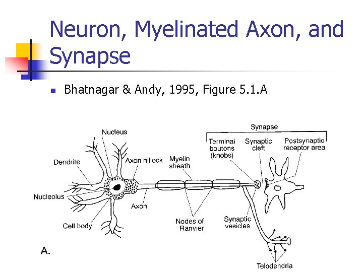
Neuron, Myelinated Axon, and Synapse n Bhatnagar & Andy, 1995, Figure 5. 1. A
Nerve Cell Structure of Neurons: Myelin Sheath n n Speed of nerve conduction is determined by: Myelin
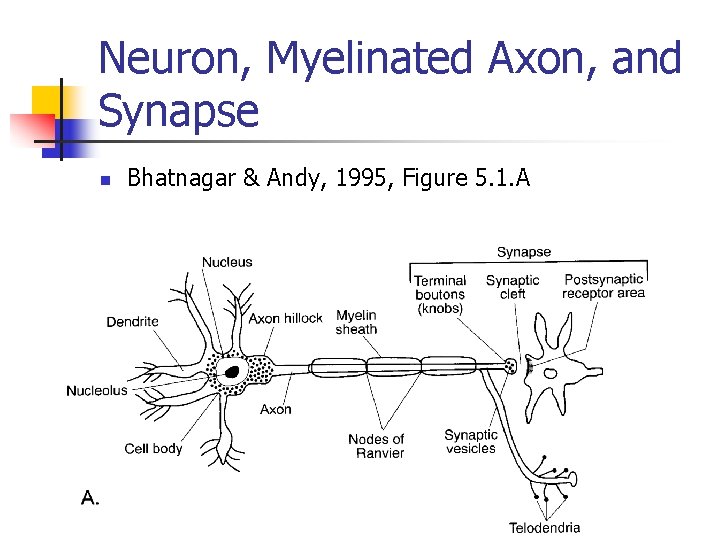
Neuron, Myelinated Axon, and Synapse n Bhatnagar & Andy, 1995, Figure 5. 1. A
Nerve Cell Structure of Neurons: Myelin Sheath n Myelin
Nerve Cell Structure of Neurons: Synapse n n Connection point between neurons Three Parts: n 1. Knob (Synaptic vesicles) n 2. Synaptic Cleft n 3. Receptive Sites of Connecting Nerve Cells
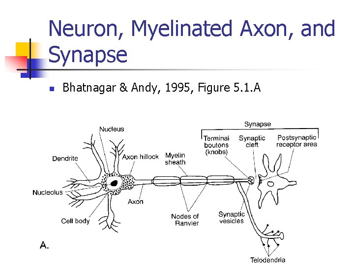
Neuron, Myelinated Axon, and Synapse n Bhatnagar & Andy, 1995, Figure 5. 1. A
Nerve Cell Structure of Neurons: Synapse n Process of Nerve Impulses
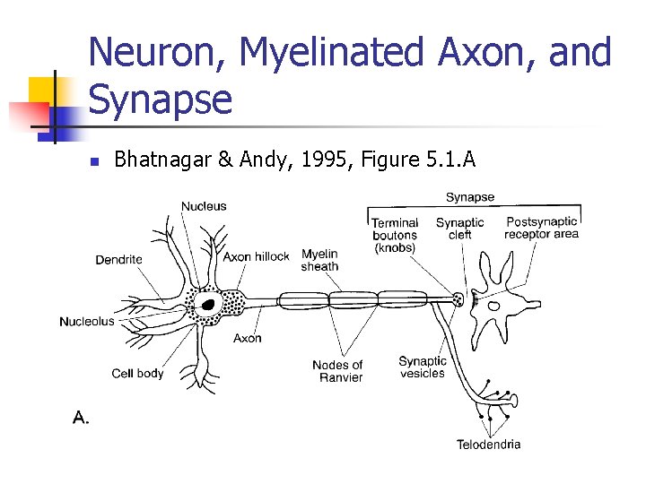
Neuron, Myelinated Axon, and Synapse n Bhatnagar & Andy, 1995, Figure 5. 1. A
Nerve Cell Types n Classification: n Three Cell Types: n 1. Multipolar n 2. Bipolar n 3. Unipolar
Nerve Cell Types n Bhatnagar & Andy, 1995, Figure 5. 2
Neuroglia Cells n Function: n Location:
Neuroglia Cells in the Central Nervous System n Four Types of Glia Cells in the CNS n 1. Astrocytes n Location: n Function:
Neuroglia Cells in the Central Nervous System n 2. Ogliodendroglia n 3. Ependymal n 4. Microglia
Neuroglial Cells in the Peripheral Nervous System n Schwann cells n Function: n Impairments: n Demyelinating Neuropathologies n Benign Tumors of Schwann Cells
Central and Peripheral Nervous Systems n Cytological Differences n 1. Different myelin forming cells n PNS n CNS
Central and Peripheral Nervous Systems n Cytological Differences n 2. Presence of endoneurium n PNS n CNS
Nerve Impulse n Communication n Principles of Process n Chemical component n Excitability of nerve cells n Action potential
Nerve Impulse n Process n Excitability of nerve cells n An action potential n Activation releasing neurotransmitter n Opening of channels in postsynaptic receptors
Nerve Impulse Process n Resting State n Cell is in resting state n In this resting state
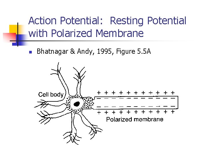
Action Potential: Resting Potential with Polarized Membrane n Bhatnagar & Andy, 1995, Figure 5. 5 A
Nerve Impulse Process n Resting Membrane Potential n Voltage inside the cell membrane n Outside the cell membrane n Inside the cell membrane n Ionic channels
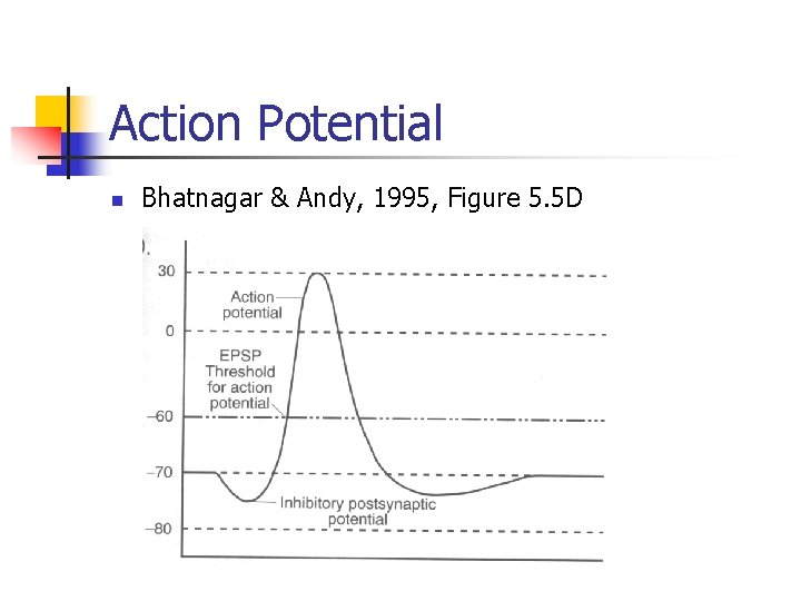
Action Potential n Bhatnagar & Andy, 1995, Figure 5. 5 D
Membrane Channels n Membrane channels are gated n Flow of ions through the membrane n Depends on: n n n 1. The density of the channels 2. The size of the opening 3. The ion concentration gradient across the membrane
Membrane Channels n Distribution of sodium and potassium across the cellular membrane n n Is constantly adjusted by the sodiumpotassium pump Because of the membrane pore size
Membrane Channels n n With the attraction of opposite ions and the repulsion of identical ions With this tug of war n An electrochemical gradient forms along the membrane
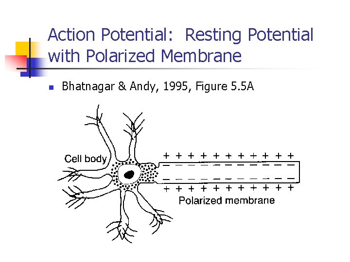
Action Potential: Resting Potential with Polarized Membrane n Bhatnagar & Andy, 1995, Figure 5. 5 A
Nerve Excitability n Refers to:
Nerve Excitability n Stimuli can include:
Nerve Excitability n During the resting state n n The neuron undergoes several short changes in the intracellular potentials Triggering an action potential
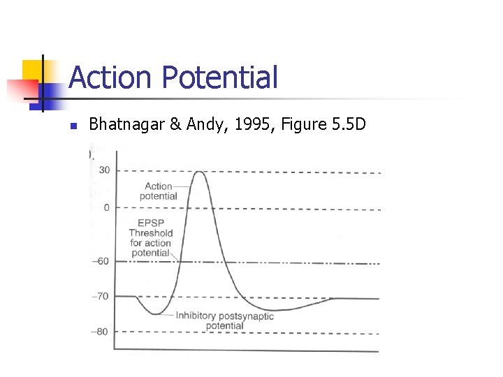
Action Potential n Bhatnagar & Andy, 1995, Figure 5. 5 D
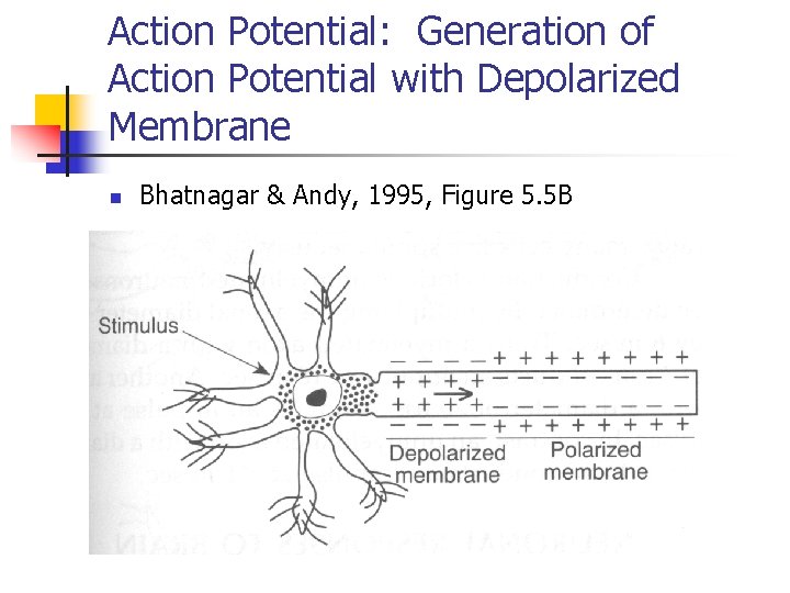
Action Potential: Generation of Action Potential with Depolarized Membrane n Bhatnagar & Andy, 1995, Figure 5. 5 B
Nerve Excitability n In membrane depolarization
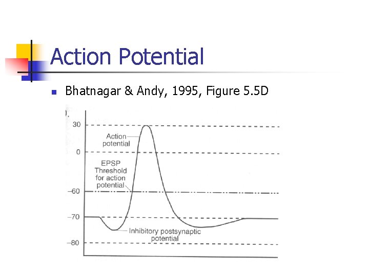
Action Potential n Bhatnagar & Andy, 1995, Figure 5. 5 D
Nerve Excitability n Membrane potential from this peak returns to the absolute refractive period
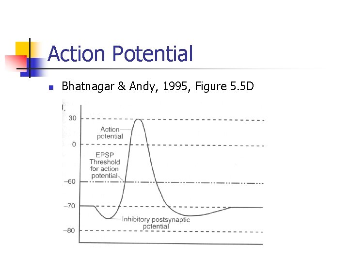
Action Potential n Bhatnagar & Andy, 1995, Figure 5. 5 D
Nerve Excitability n Not all stimuli n n Are strong enough to change the membrane potentials to 10 m. V Many weak stimuli with subthreshold strength n If temporally and spatially summated n n Can initiate a nerve impulse Each weak stimulus arrives in a sequence and their cumulative effect is strong enough to initiate an impulse
Impulse Conduction n Nerve impulse is passively conducted a short distance in the axon
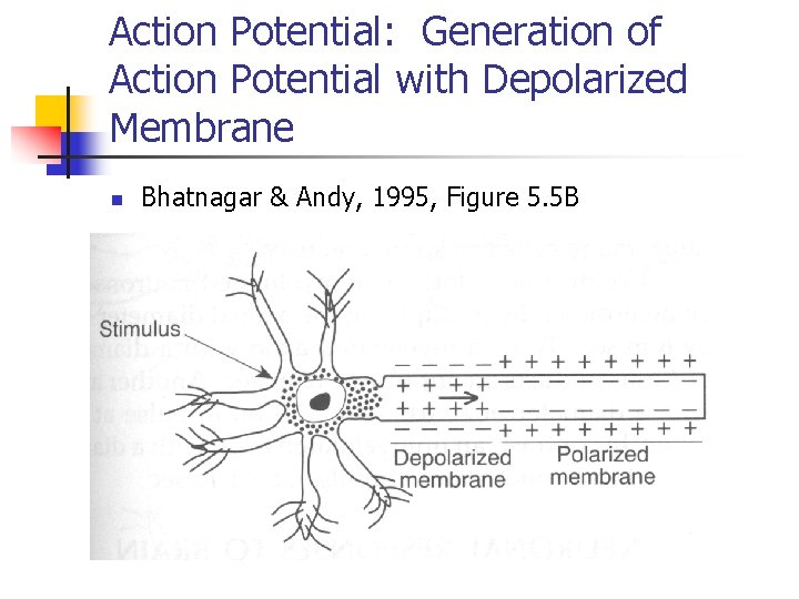
Action Potential: Generation of Action Potential with Depolarized Membrane n Bhatnagar & Andy, 1995, Figure 5. 5 B
Impulse Conduction n This gradually changes the membrane potential in the neighboring area
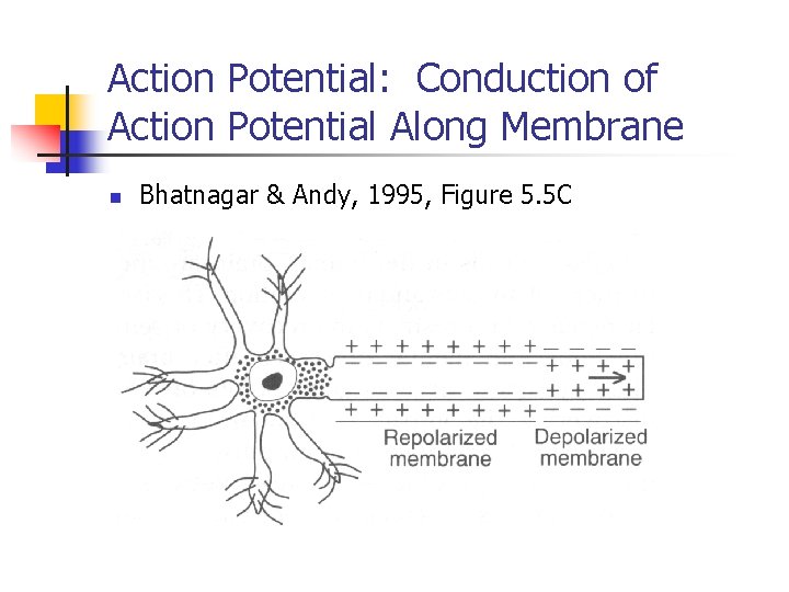
Action Potential: Conduction of Action Potential Along Membrane n Bhatnagar & Andy, 1995, Figure 5. 5 C
Impulse Conduction n Saltatory Conduction in Myelinated Axons
Impulse Conduction n Action potential or nerve impulse n Excitatory Postsynaptic Potential (EPSP) n Inhibitory Postsynaptic Potential (IPSP)
Neuronal Responses to Brain Injuries n Nerve Cells in the Human Brain n n Are incapable of further cell division and regeneration Synapses serve as good points of reference for discovering the impact of cellular injuries
Neuronal Responses to Brain Injuries n n Understanding the Processes of Spontaneous Recovery Two Types of Degenerative Changes Occur After Axonal Sectioning n 1. Axonal or Retrograde Reaction n 2. Wallerian (Anterograde) Degeneration
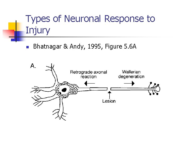
Types of Neuronal Response to Injury n Bhatnagar & Andy, 1995, Figure 5. 6 A
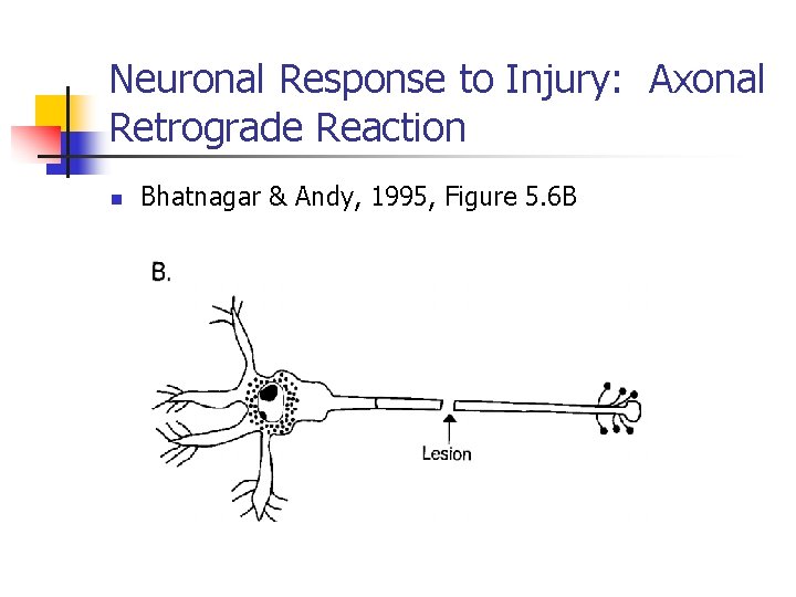
Neuronal Response to Injury: Axonal Retrograde Reaction n Bhatnagar & Andy, 1995, Figure 5. 6 B
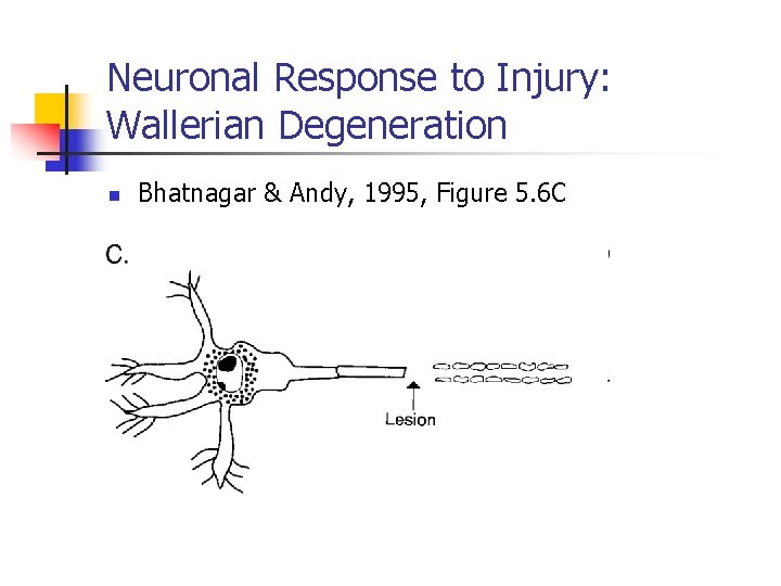
Neuronal Response to Injury: Wallerian Degeneration n Bhatnagar & Andy, 1995, Figure 5. 6 C
Axonal Regeneration in Peripheral Nervous System n Regeneration of Fibers in the PNS
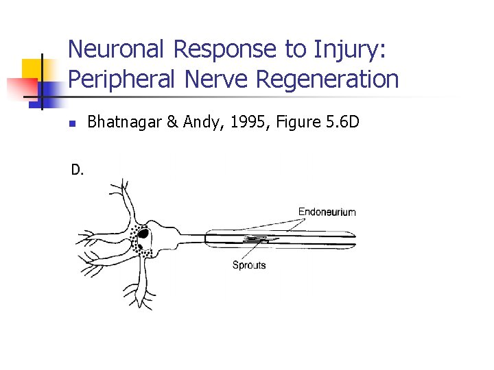
Neuronal Response to Injury: Peripheral Nerve Regeneration n Bhatnagar & Andy, 1995, Figure 5. 6 D
Axonal Regeneration in Central Nervous System n Axons severed in the CNS
Neurotransmitters n Along with projections from the reticular formation n Regulate brain mechanisms that control: n n n n n Cognition Language Speech Hearing Brain Tuning Moods Attention Memory Personality Motivation
Neurotransmitters n Two types of transmitters in the nervous system: n 1. Small molecules n Include: n n n n Acetylcholine Dopamine Norepinephrine Serotonin Glutamate y-aminobutyric acid (GABA) 2. Larger molecules n Peptides
Neurotransmitters: Acetylcholine n Synthesis and Dissolution n Acetylcoenzyme A and choline n Acetylcholinesterase
Neurotransmitters: Acetylcholine in the PNS n Location of Cells in PNS: n Function: n Impairments:
Neurotransmitters: Acetylcholine in the CNS n Location of Cells In CNS: n Function: n Impairments:
Sites of Cell Bodies and Their Projections in the Brain: Acetylcholine n Bhatnagar & Andy, 1995, Figure 5. 7 A
Neurotransmitters: Dopamine n Location of Cells: n Two Important Dopaminergic Projections n 1. Mesostriatal System (Midbrain and Striatum) n Pathways: n Impairments:
Neurotransmitters: Dopamine n Two Important Dopaminergic Projections n 2. Mesocortical System (Midbrain and Cortical) n Pathways: n Function: n Impairments:
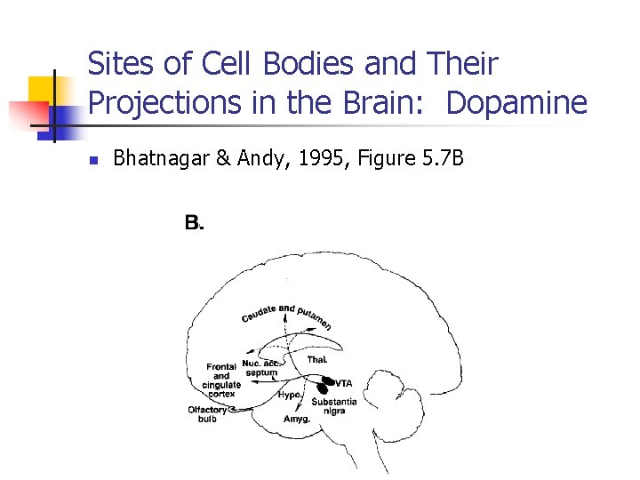
Sites of Cell Bodies and Their Projections in the Brain: Dopamine n Bhatnagar & Andy, 1995, Figure 5. 7 B
Neurotransmitters: Norepinephrine n Location of Cells: n Pathways: n Ascending Fibers: n Descending Fibers n Function: n Impairments:
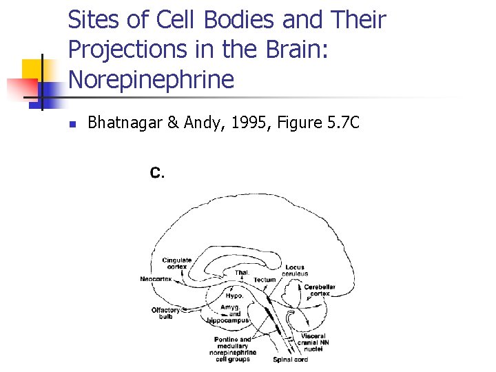
Sites of Cell Bodies and Their Projections in the Brain: Norepinephrine n Bhatnagar & Andy, 1995, Figure 5. 7 C
Neurotransmitters: Serotonin n Location of Cells: n Pathways: n Function: n Impairment:
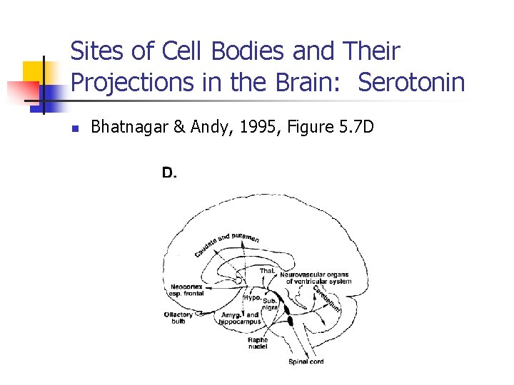
Sites of Cell Bodies and Their Projections in the Brain: Serotonin n Bhatnagar & Andy, 1995, Figure 5. 7 D
Neurotransmitter: y-Aminobutyric Acid (GABA) n Location of Cells: n Location of Projections: n Function: n Impairment:
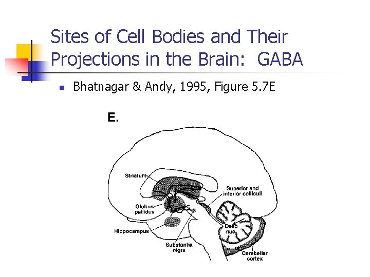
Sites of Cell Bodies and Their Projections in the Brain: GABA n Bhatnagar & Andy, 1995, Figure 5. 7 E
Neurotransmitters: Peptides n Characteristics: n Function:
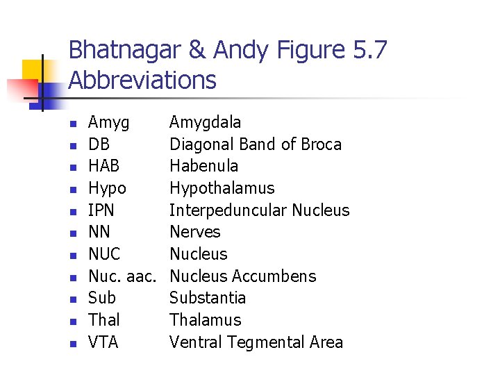
Bhatnagar & Andy Figure 5. 7 Abbreviations n n n Amyg DB HAB Hypo IPN NN NUC Nuc. aac. Sub Thal VTA Amygdala Diagonal Band of Broca Habenula Hypothalamus Interpeduncular Nucleus Nerves Nucleus Accumbens Substantia Thalamus Ventral Tegmental Area
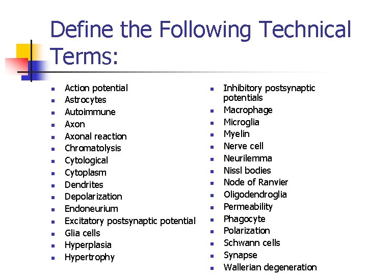
Define the Following Technical Terms: n n n n Action potential Astrocytes Autoimmune Axonal reaction Chromatolysis Cytological Cytoplasm Dendrites Depolarization Endoneurium Excitatory postsynaptic potential Glia cells Hyperplasia Hypertrophy n n n n Inhibitory postsynaptic potentials Macrophage Microglia Myelin Nerve cell Neurilemma Nissl bodies Node of Ranvier Oligodendroglia Permeability Phagocyte Polarization Schwann cells Synapse Wallerian degeneration
Review Questions n n n 1. With a diagram of a typical nerve cell, identify the major structures and describe their functions. 2. List the major glia cells and describe their functions. 3. Explain how the following terms are related to impulse generation: n Action potential n Depolarization n Membrane excitability n Polarized membrane n Repolarized membrane n Resting potential n Subthreshold stimulus n Summation
Review Questions n n n 4. Describe chemical and electrical events that are related to impulse transmission beginning with resting potentials and ending in generation of action potentials. 5. Describe the effects of inhibitory and excitatory postsynaptic potentials on a postsynaptic neuron. 6. Discuss how nerve and glia cells respond to injuries. Specifically discuss chromatolysis, wallerian degeneration, axonal reaction, and regeneration of axonal fibers.
Review Questions n n n 7. Describe how axonal growth in the CNS is different from that in the PNS. 8. Name primary neurotransmitters in the central nervous system and briefly discuss their functions. 9. Describe the pathophysiology of multiple sclerosis and myasthenia gravis.
Neuronal Function in the Nervous System Graphics
THINGS TO DO BEFORE LECTURE n n Reticular Formation Cytological
- Slides: 83