Chapter 8 Nervous System 5 Functions 1 Sensory





- Slides: 55
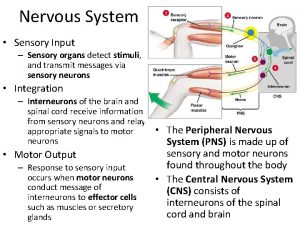
Chapter 8 Nervous System 5 Functions 1. Sensory function = receptors monitor external & internal body changes; providing input
Functions continued: 2. Integration function = brain & spinal cord process sensory information from receptors & initiate a proper response
Functions continued: 3. Motor function = output response from brain & spinal cord by way of nerve impulses to effectors (muscles, glands, etc)
Functions continued 4. Homeostasis-Depends on the ability of the NS to detect, interpret, and respond to change in internal and external conditions. In response, the NS can stimulate or inhibit the activities of other systems to maintain homeostasis.
Functions continued 5. Mental Activity-The brain is the center of mental activity, including consciousness, memory, and thinking.
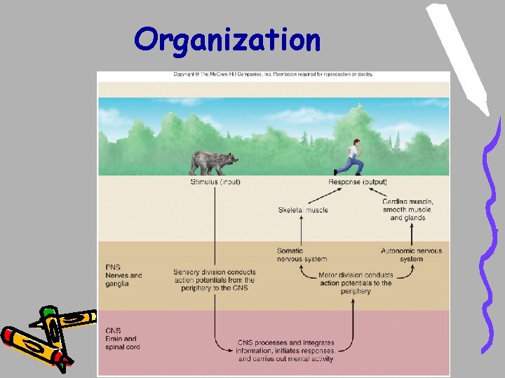
Organization
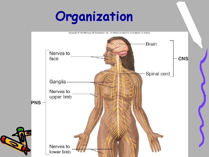
Organization
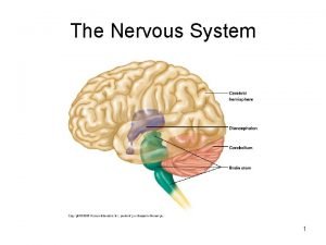
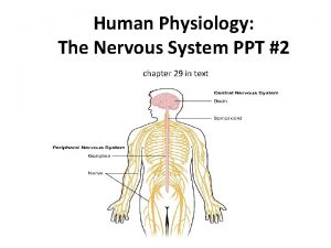
Organization - 2 Major Division 1. CNS (central nervous system) = brain , spinal cord, nuclei
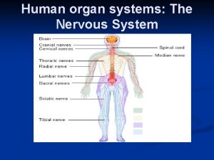
Organization - 2 Major Division 2. PNS (peripheral nervous system) = nerves (cranial & spinal nerves) & ganglia
Organization: Divisions of PNS • PNS (peripheral nervous system) is further divided into 2 divisions: 1. Sensory division = nerves which send impulses to CNS from receptors
Organization: Divisions of PNS • PNS 2. Motor Division = nerves which carry impulses away from CNS to effectors (muscles, glands)
Organization: Divisions of PNS • PNS Motor Division is further divided into 1. Somatic Nervous System = (SNS) nerves that carry impulses away from CNS to voluntary effectors (skeletal muscles)
Organization: Divisions of PNS • PNS Motor Division is further divided into 2. Autonomic Nervous System (ANS) = nerves which carry impulses away from CNS to involuntary effectors (glands, heart, smooth muscle)
Organization: Divisions of PNS - PNS Motor Division, Autonomic Nervous System (ANS) is further divided into 1. Parasympathetic Division = carries impulses away from CNS to involuntary effectors under normal conditions
Organization: Divisions of PNS 2. Sympathetic Division = carries impulses away from CNS to involuntary effectors under stressful conditions - Sometimes called “fight or flight”
Nervous System CNS PNS Sensory Somatic Parasympathetic Motor Autonomic Sympathetic
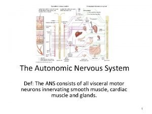
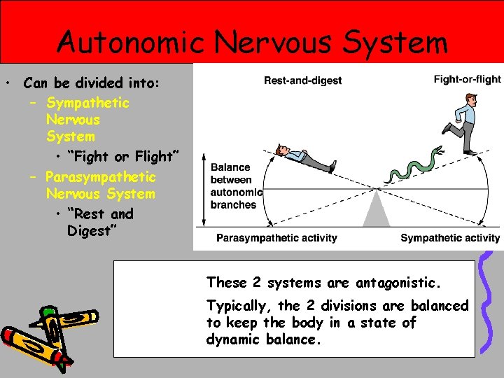
Autonomic Nervous System • Can be divided into: – Sympathetic Nervous System • “Fight or Flight” – Parasympathetic Nervous System • “Rest and Digest” These 2 systems are antagonistic. Typically, the 2 divisions are balanced to keep the body in a state of dynamic balance.
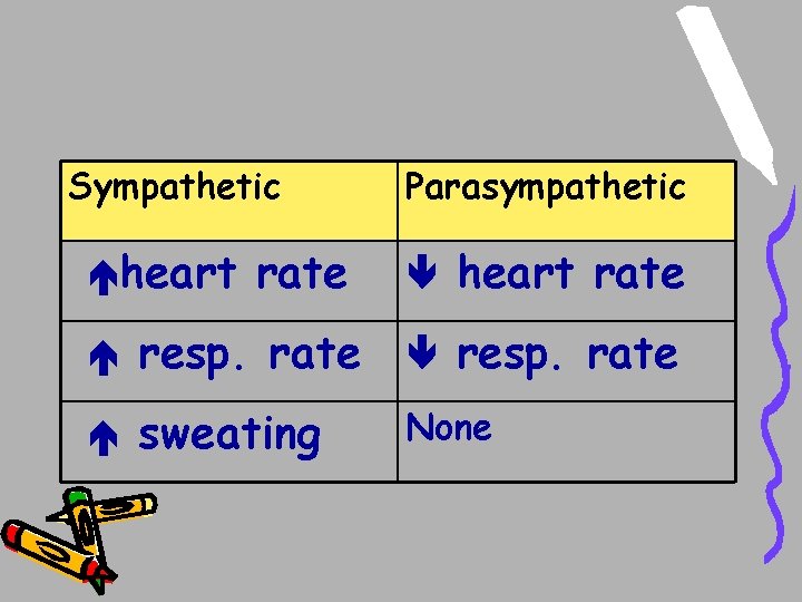
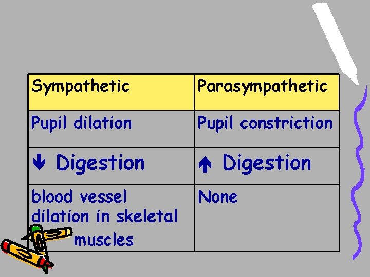
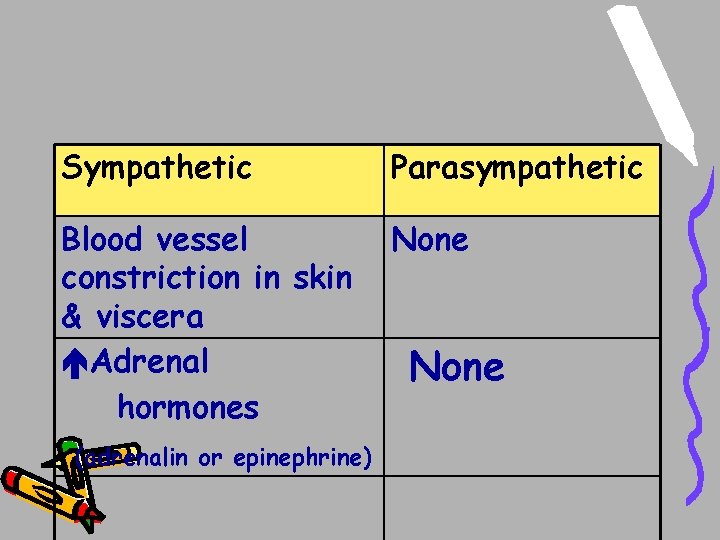
ANS: -Sympathetic & Parasympathetic tend to produce opposite results on their effectors
Sympathetic heart rate Parasympathetic heart rate resp. rate sweating None
Sympathetic Parasympathetic Pupil dilation Pupil constriction Digestion blood vessel dilation in skeletal muscles Digestion None
Sympathetic Parasympathetic Blood vessel constriction in skin & viscera Adrenal hormones None (adrenalin or epinephrine) None
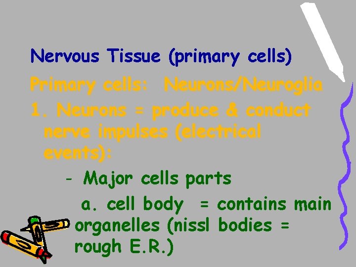
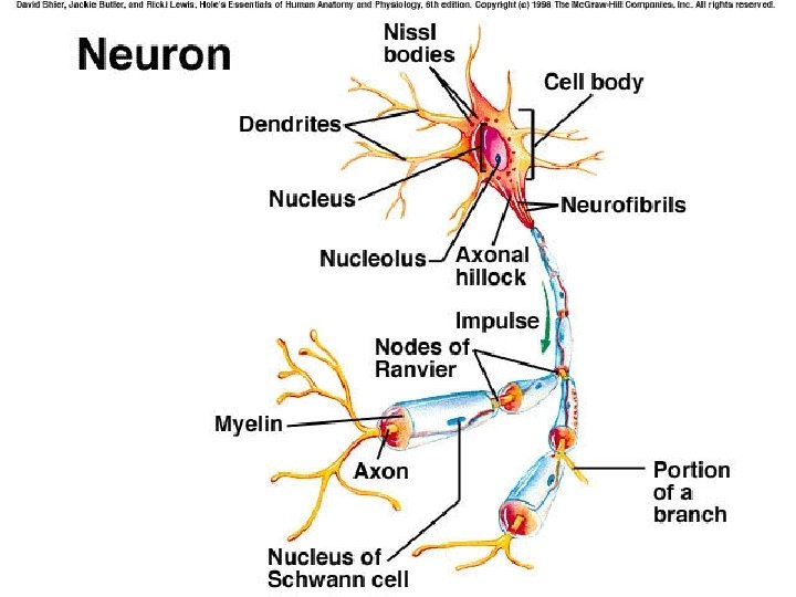
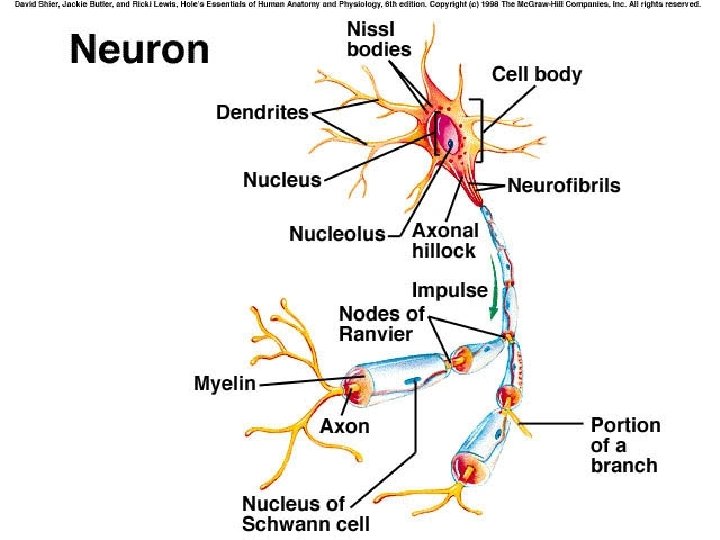
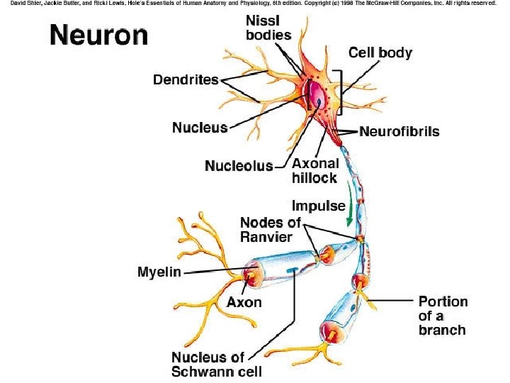
Nervous Tissue (primary cells) Primary cells: Neurons/Neuroglia 1. Neurons = produce & conduct nerve impulses (electrical events): - Major cells parts a. cell body = contains main organelles (nissl bodies = rough E. R. )
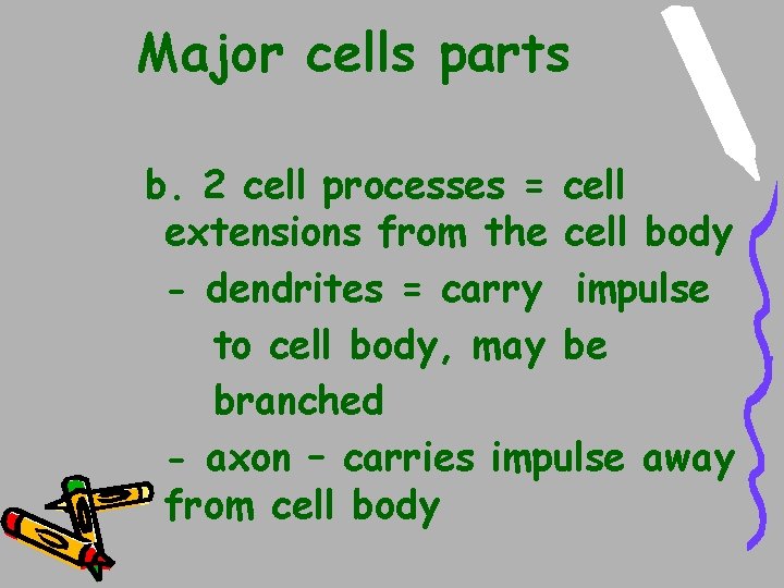
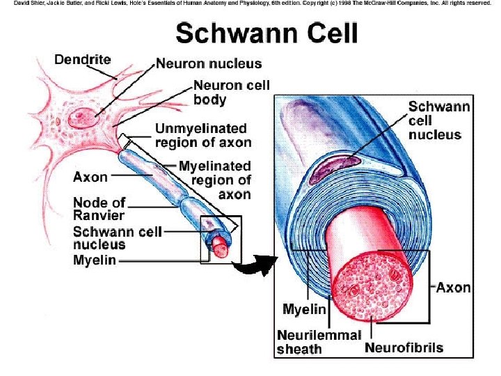
Major cells parts b. 2 cell processes = cell extensions from the cell body - dendrites = carry impulse to cell body, may be branched - axon – carries impulse away from cell body
c. Axon terminal = swollen ending of axons contain synaptic vesicles with chemicals (neurotransmitters)
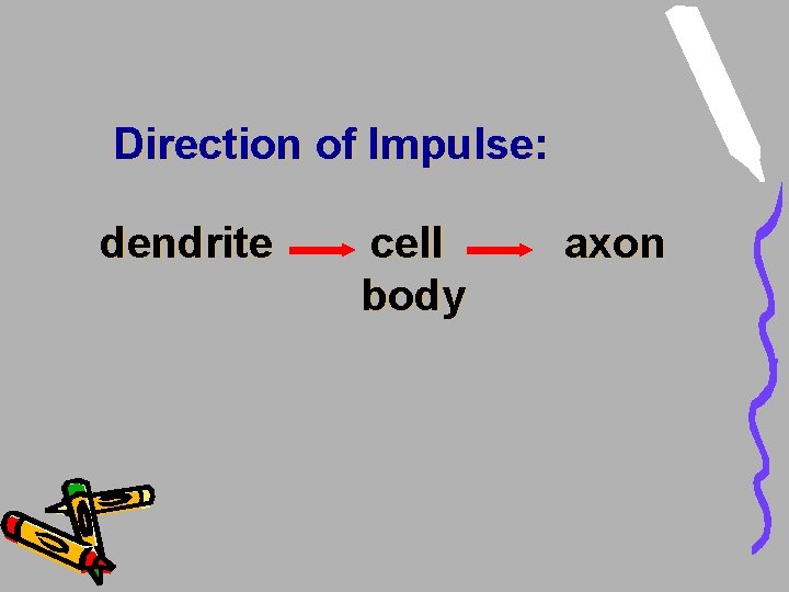
Direction of Impulse: dendrite cell body axon
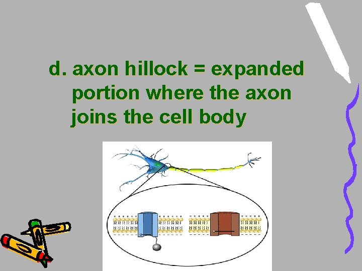
d. axon hillock = expanded portion where the axon joins the cell body
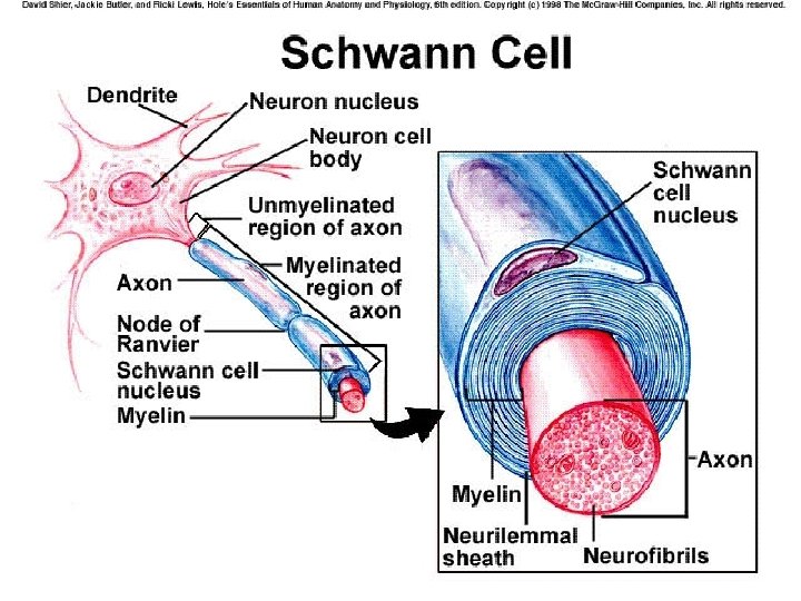
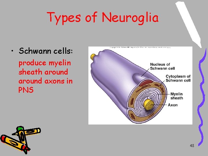
e. myelin sheath = whitish insulation layer surrounding axon, protects & increases speed of nerve impulses -actually formed by Schwann cells (a neuroglial cell)
2 types neurons - myelinated neurons (white matter) - unmyelinated neurons (gray matter)
f. Neurilemmal sheath = surrounds myelin sheath, used in repair of axon
g. Nodes of Ranvier = small areas on myleinated axon where no myelin sheath is formed.
Nuclei in the CNS By definiton: 1. clusters of cell bodies in the CNS (brain & spinal cord)
Types of Neurons By Function : (what they do? ) 1. sensory neuron = carries impulses to CNS 2. motor neuron = carries impulses away from the CNS to effectors
Types of Neurons 3. interneuron = neurons between sensory & motor neurons; located only in CNS
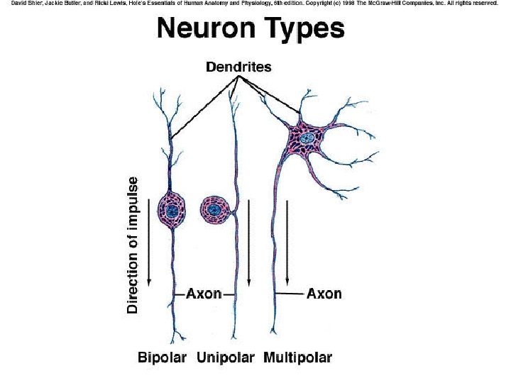
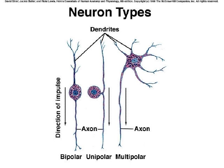
Types of Neurons By structure (what they look like? ) 1. multipolar neuron = one axon & many highly branched dendrites
2. bipolar = one axon & one dendrite on opposite sides of cell body
3. unipolar neuron = one axon & one dendrite connected to cell body by a small T-like process (ie one process extends from the cell body)
multipolar neurons = motor neuron & interneurons bipolar neurons = sensory neurons, usually in eye unipolar neurons = sensory neurons in skin
Nervous Tissue (2 primary cell types) 2. Neuroglia = 5 types of supportive cells: - Oligodendrocytes, Schwann cells. Microglia, astrocytes, ependymal
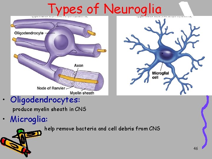
Types of Neuroglia • Oligodendrocytes: produce myelin sheath in CNS • Microglia: help remove bacteria and cell debris from CNS 46
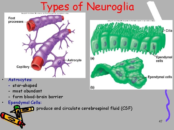
Types of Neuroglia • • Astrocytes: - star-shaped - most abundant - form blood-brain barrier Ependymal Cells: produce and circulate cerebrospinal fluid (CSF) 47
Types of Neuroglia • Schwann cells: produce myelin sheath around axons in PNS 48
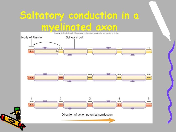
Saltatory conduction in a myelinated axon
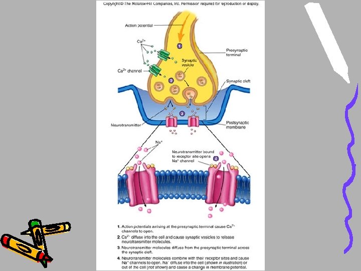
Synapse • What is it? - where an axon comes in close contact to a muscle, gland, organ, or other neuron - involved with release of neurotransmitters - Ex. Neuromuscular junction 51
Figure 8. 13
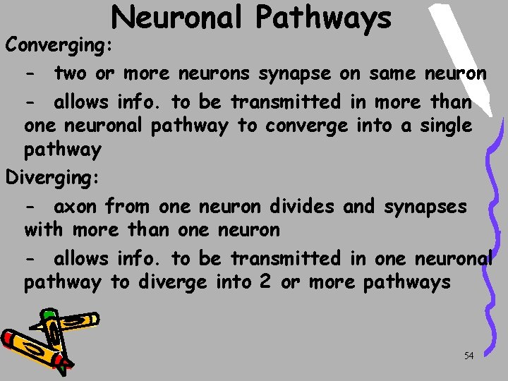
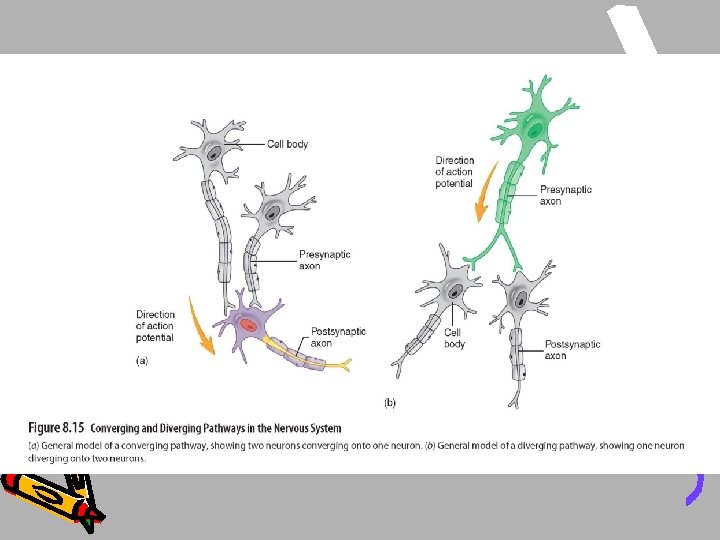
Neuronal Pathways Converging: - two or more neurons synapse on same neuron - allows info. to be transmitted in more than one neuronal pathway to converge into a single pathway Diverging: - axon from one neuron divides and synapses with more than one neuron - allows info. to be transmitted in one neuronal pathway to diverge into 2 or more pathways 54