Special Senses Ch 10 Sensory functions 1 Sensory


 l Stretch, tension, position l muscle, tendons, ear • Muscle")


")

 tube l l connect middle ear to throat helps maintain")


 - contains hair like hearing receptors - stimulated by")


 and visual")
- Slides: 40
Special Senses Ch. 10
Sensory functions 1. Sensory pathways begin with l l l Stimulus Receptor CNS 2. Receptors l l Vary in structure They all excite Stimulus specific Sensory adaptation: decrease rates of stimuli
3. Receptor types: classified by sensitivity a. Mechanoreceptors • • Mechanical or physical change Touch. Pressure, tension, hearing, equilibrium, & BP 1. Meissner’s corpuscles (touch) -Found in fingers, palms, soles, lips external genitalia 2. Merkel’s disc’s (touch) -End in epidermis 3. Pacinian Corpuscles (pressure) -Joints, tendons, visceral organs
• Proprioceptors (positional) l Stretch, tension, position l muscle, tendons, ear • Muscle spindles • Golgi tendon organs
• Thermo receptors l l l Temperature changes Free nerve endings in the skin <10 ° C - >45° C pain is also triggered
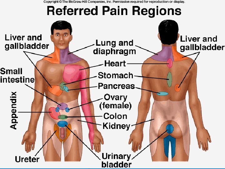
Nociceptors l • • • Pain from chemical or physical damage Protective function Free nerve endings in skin, muscle, organs Can be generated by other receptors Referred pain
Photoreceptors l • • Changes to amount of light Only in retina of eye Chemoreceptor l • • • Detect chemicals dissolved in fluid/air Smell & taste Provide detection of O 2 & CO 2 in blood
4. Sensory pathway a. General: skin or visceral organs • 1 st order neuron-2 nd-3 rd order to cortex for processing b. Special: special sensory organs( eye/ear) • • More variation& # of neurons used Route impulse to specialized areas of brain
Special Senses 5. Smell: olfaction a. Chemoreceptor in nasal cavity b. Olfactory bulb located high in nasal cavity c. Detect ~50 smells d. Neuron travels through cribiform plate
e. How it works • • • Chemicals dissolve in mucous & cause nerve impulse Olfactory nerve to frontal lobe/interpretation Closely linked to limbic system l l emotional expression smells can trigger memories/emotions
6. Taste: gustation a. Associated with smell b. Both chemoreceptors c. receptor: taste bud • • 10, 000 on tongue infants all over mouth located on surface of pappilae gustatory cells/supportive epithelial cells 1. taste hairs( microvilli) 2. taste pores 3. 4 tastes • sweet • sour • salty • bitter
7. Sight a. Eyes & accessory structures • • • Eye lids: protection Lashes: protection Conjunctiva: inner membrane of lid & eye 1. secretes mucous to lubricate eye • Lacrimal apparatus/gland-duct-sac 1. secrete tears 2. lubrication/antibacterial • Nasolacrimal duct: empty tears into nasal cavity
muscles Lateral rectus VI Lateral eye movement Medial rectus III Medial eye movement Superior rectus III Elevates/rolls eye upward Inferior rectus III Depresses/rolls eye downward Superior oblique III Elevates & turns eye laterally Inferior oblique IV Depresses and turns eye laterally
b. Eye structure • Fibrous tunic: thick outermost layer of eye 1. sclera: white of eye 2. cornea: clear front of eye • Vascular tunic 1. choroids: thin, dark brown membrane a. absorbs light/minimize reflection b. vessels nourish the retina 2. ciliary body: connects to lens by suspensory ligaments 3. iris: attached to ligaments a. colored part of eye b. controls light 4. pupil: opening in center of iris 5. lens: elastic structure that helps focus image a. accomodation
• Cavities 1. Anterior Cavity: cornea & lens a. Contains aqueous humor: watery fluid bathes eye b. Anterior chamber: cornea to iris c. Posterior chamber: iris to lens 2. Posterior Cavity: behind lens a. contains vitreous humor: gel-like support of eye 2
• Nervous tunic 1. retina: inner lining of posterior wall a. detects light- optic nerve 2. photoreceptor cells a. rods: light intensity - low light/ shades of grey - rhodopsin: visual purple b. cones: color sensitive -need more light/sharper vision -red(erythrolabe), green(chlorolabe), blue(cyanolabe
3. optic disc: “blind spot” a. area where optic nerve leave back of eye 4 4. macula lutea: yellowish spot on back of eye 4. /5 5. fovea centralis: high concentration of cone cells only 3. a. sharpest vision area
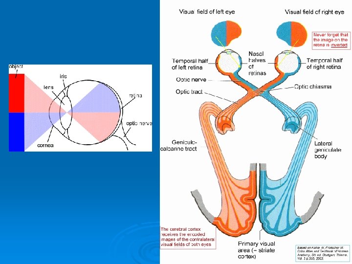
c. eye function • path of light 1. light is refracted as is passes into the eye 2. image is focused on the retina 3. image is upside down, smaller and reversed left from right ( due to refracting light) 4. brain coordinates proper image in visual interpretation center
vision problems 1. myopia: nearsightedness a. image focused in front of retina b. too much refraction c. eye is too long 2. hyperopia: farsightedness A, image focus is behind retina b. weak or lazy lens c. eye that is too short 3. astigmatism a. unequal curvatures of the cornea or lens b. blurred vision near & far Because of 2 focal points
Vision disease 4. Color blindness a. Absence of one or more cone cells b. hereditary condition • -sex-linked. • - 1 -30 people, 1 -12 men affected • - most common is red-green
Vision Problems 5. Cataract a. Lens or cornea become cloudy or opaque b. Light is scattered and causes blurred vision
Vision disease 6. Glaucoma a. Aqueous humor formation exceeds rate of removal -usually from blockage/restriction of drain b. Increases eye pressure c. Decreases blood supply to retina d. Can cause permanent blindness
Visual Acuity Snellen Eye chart 20/20: what a normal person can see from 20 feet. 20/10: what you can see @ 20 feet a normal person has to see @ 10 feet. 20/100: what a normal person can see @ 100 feet you see @ 20
Astigmatism Test : curvature of the eye is unequal If lines appear darker in one direction there is an astigmatism.
The ear Ø The ear l l Ø - has external, middle and inner parts organ of hearing & equilibrium II. External Ear 1. auricle or pinna l l -outer funnel-like structure -collects sound waves 2. External auditory meatus l l l S- shaped tube that passes through the temporal Bone carries sound waves to ear drum hair & ceruminous glands (Cerumen) protect ear
III. Middle Ear 1. Typanic membrane “ear drum” l l outer surface- Thin skin layer inner surface- mucous membrane oval-cone shape; apex is attached to auditory ossicles ( malleous) sound waves cause pressure changes- which results in movement of the ear drum and the auditory ossicles
2. Auditory ossicles l l l thin bones in the inner ear Malleous (hammer) attach to ear drum , attaches to incus (anvil) & stapes (stirrup) attaches to the oval window on tympanic cavity lever system that increases/amplifies vibration=22 x@ oval window
l tympanic reflex- muscles that make middle ear more rigid reducing vibration= muffles loud sounds
3. Auditory (eustachian ) tube l l connect middle ear to throat helps maintain air pressure balance on each side of the tympanic membrane (ear drum) needed for normal hearing
IV. Inner ear 1. Labyrinth • complex system of interconnected tubes & chambers • osseous labyrinth- bony canal in temporal b. • membranous labyrinth- tube within the osseous labyrinth filled with liquid = endolymph
Ø A. Semicircular canals l l Ø provide sense of equillibrium 3 axis B. Cochlea l l coiled, bony canal connected to oval window & round window dissipates vibrations through cochlea
i. Organ of Corti (microphone) - contains hair like hearing receptors - stimulated by vibrations through endolymph - receptor frequency differentiated - receptors move against tectoral membrane - epithelial cells that act like neurons - transmit to vestibulocochlear N. (VIII) cochlear branch- hearing vestibular branch- equilibrium
How we hear Ø http: //msjensen. cehd. umn. edu/1135/Links/ Animations/Flash/0019 swf_effect_of_soun. swf Ø http: //www. sumanasinc. com/webcontent/a nimations/content/soundtransduction. html
V. Equilibrium 1. Static : position of head & body when motionless A. Structures - receptors located in vestibule (bony chamber between cochlea & semicircular canals) -utricle & saccule chambers inside vestibule - contain hair cells & supporting cells called _macula -cells have vertical or horizontal orientation inside chambers - otolithic membrane- gelatinous material - otoliths-calcium carbonate crystals give weight to O. M.
b. How it works 1. head moves • • O. M. moves across hair cells & macula causing them to bend nerve impulses sent via the vestibulocochlear n. to the CNS
2. Dynamic- sense of position when body is moving a. structures -semicircillar canals occupy the 3 planes - ampulla -swelling at end of canal communicates with utricle - crista ampullaris sensory hair cells (similar to macula) -cupula - gelatinous mass in canal
b. How it works 1. movement of head causes canal to move with head 2. Cupula stays stationary because of inertia 3. Movement bends hair cells in opposite direction of movement 4. Impulse sent via vestibulocochlear n.
Ø Side note: Brain gains other information about equilibrium from Proprioceptors (mechanoreceptors) and visual input from the _eyes. How it works http: //www. sumanasinc. com/webcontent/animations/content/vestibular. html