CHAPTER 9 The General and Special Senses General

")
- Free nerve endings that are found in the superficial portions")

free nerve scattered right below the surface of the skin, in")


 � Olfactory organs are located in the nasal cavity and contain the")

 � Taste buds: sensory structures formed from taste receptors and specialized epithelial")


 � Functions as some physical protection, attachment site for extrinsic")


 � The inner tunic consists of the retina, which contains")


- Slides: 82
CHAPTER 9 The General and Special Senses
General Senses � Six general senses �Temperature �Pain �Touch �Pressure �Vibration �Proprioception (body position) � Receptors for each sense, classified according to the nature of the stimulus that excites them
� Nociceptors (pain receptors)- Free nerve endings that are found in the superficial portions of the skin, joint capsules, and around blood vessel walls � Three types of pain �Fast pain: very localized sensations, reach CNS very quick, and relayed to primary sensory cortex �Slow pain: only allow you to determine the general area involved �Referred pain: perception of pain coming from parts of the body that are not actually stimulated
Referred Pain
� Thermoreceptors (Temperature receptors)free nerve scattered right below the surface of the skin, in skeletal muscles, liver, and hypothalamus �Very active when the temperature is changing but quickly adapts to a stable temperature � Mechanoreceptors- sensitive to stimuli that distort their cell membranes �Tactile: sensation of touch, pressure, and vibration �Baroreceptors: changes in pressure �Proprioceptors: monitors position of joints, tension in tendons and ligaments, and the state of muscular contractions
Tactile receptors � Fine touch and pressure receptors: detailed information � Crude touch and pressure receptors: poor localization and little information about stimulus � Six types of tactile receptors: �Free nerve endings �Root hair plexus �Tactile discs (Merkel’s disc) �Tactile corpuscles (Meissner’s corpuscles) �Lamellated corpuscles �Ruffini corpuscles
Tactile Receptors in the Skin
Baroreceptors � Free nerve endings that branch within the elastic tissues in the wall of distensible organs � Respond immediately to changes in pressure but adapt rapidly to the change and return to a state or “normal” Proprioceptors � Do not adapt to constant stimulation and each receptor continuously sends information to CNS � How you know your current body position
Chemoreceptors � Only respond to water-soluble and lipidsoluble substances that dissolve in surrounding fluid � Sends information to the brain stem centers that deal with autonomic control of respiratory and cardiovascular functions � Monitor the bloods p. H, CO₂, and oxygen levels
Concept Check What would happen to you if the information from proprioceptors in your legs were blocked from reaching the CNS? Ø Proprioceptors relay information about limb position and movement to the CNS, especially the cerebellum. Blockage of this information would result in uncoordinated movements, and the individual probably would be unable to walk. �
The Special Senses � The five special senses �Smell �Taste �Vision �Equilibrium �Hearing � Receptors from these senses are distributed to specific areas of the cerebral cortex and brain stem
Smell (Olfaction) � Olfactory organs are located in the nasal cavity and contain the olfactory receptors plus epithelial supporting cells � Receptor cells are bipolar neurons with hairlike cilia covering that dendrites. The cilia project into the nasal cavity � Inhaled air reaches the olfactory organs, water and lipid soluble chemicals (Chemoreceptors) must diffuse into the mucus in order for the olfactory receptors to stimulate
� Information from the receptors are relayed to the CNS, which interprets the smell on the basis of the particular pattern of receptor activity Olfactory Pathway 1. When olfactory receptors are stimulated, their fibers synapse with neurons in the olfactory bulbs 2. Sensory impulses are first analyzed in the olfactory blubs, then travel along the olfactory tracts to the limbic system, and then arrive to the olfactory cortex
Taste (Gustatory) � Taste buds: sensory structures formed from taste receptors and specialized epithelial cells � Papillae: epithelial projections �Circumvallate papillae � Taste cells (gustatory cells) are modified epithelial cells that function as receptors �Contain the taste hairs that are the portions sensitive to taste. These hairs protrude from openings called taste pores
Circumvallate papillae
Taste Sensations � Specific taste receptors are concentrated on different areas of the tongue �Sweet- tip of the tongue �Sour- along lateral edges of the tongue �Salt- abundant in the tip and upper portion of the tongue �Bitter- back of the tongue � Two additional tastes discovered �Umami- taste of chicken and beef broth �Water
Taste Receptors
Taste Pathways � Taste impulses travel on the facial, glossopharyngeal, and vagus nerves to the medulla oblongata and then to the gustatory cortex of the cerebrum � Olfactory receptors play an important role in taste perception
Concept Check If you completely dry the surface of your tongue and then place salt or sugar crystals on it, you cannot taste them. Why not? ? Ø Drying the surface of the tongue removes moisture needed to dissolve the sugar molecules or salt ions. Because taste bud (receptors) are sensitive only to molecules and ions that are in a solution, the taste buds will not be stimulated. �
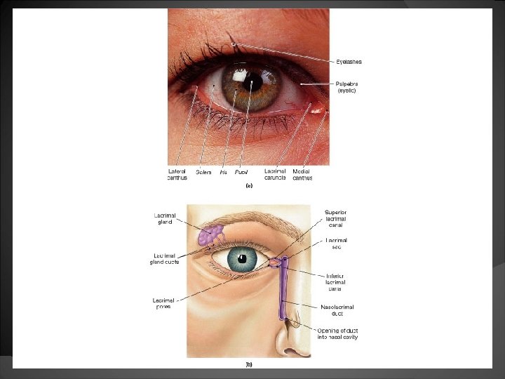
Vision � Accessory organs: lacrimal apparatus, eyelids, and extrinsic muscles aid the eye in its function �Provide protection, lubrication, and support
Visual Accessory Organs � The eyelid: protects the eye from foreign objects and is made up of the thinnest skin of the body lined with conjunctiva �Eyelashes: very robust hairs that also help prevent foreign particles from reaching the eye surface
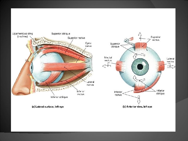
� The lacrimal apparatus produces tears that lubricate and cleanse the eye �Two small ducts (nasolacrimal ducts) drain tears into the nasal cavity �Tears also contain lysozyme, which is a antibacterial enzyme � The extrinsic muscles of the eye attach to the sclera and allow the eye to move in all directions
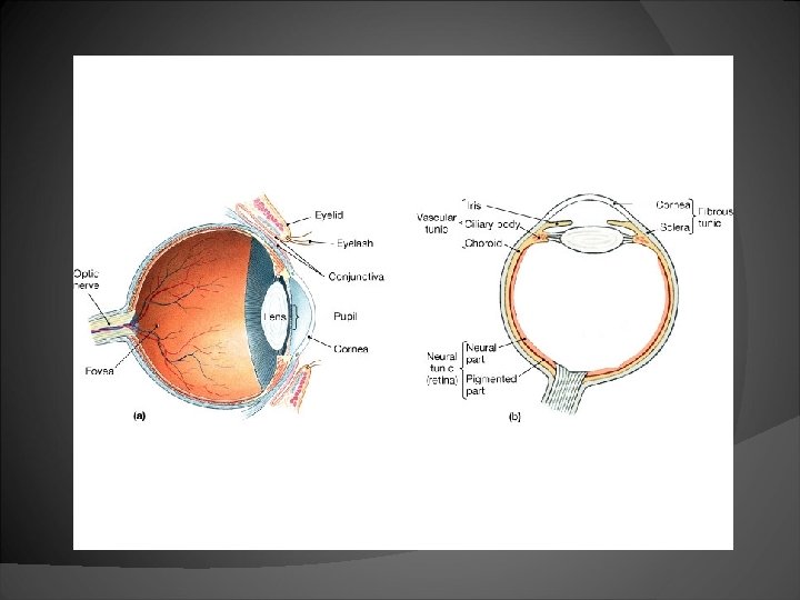
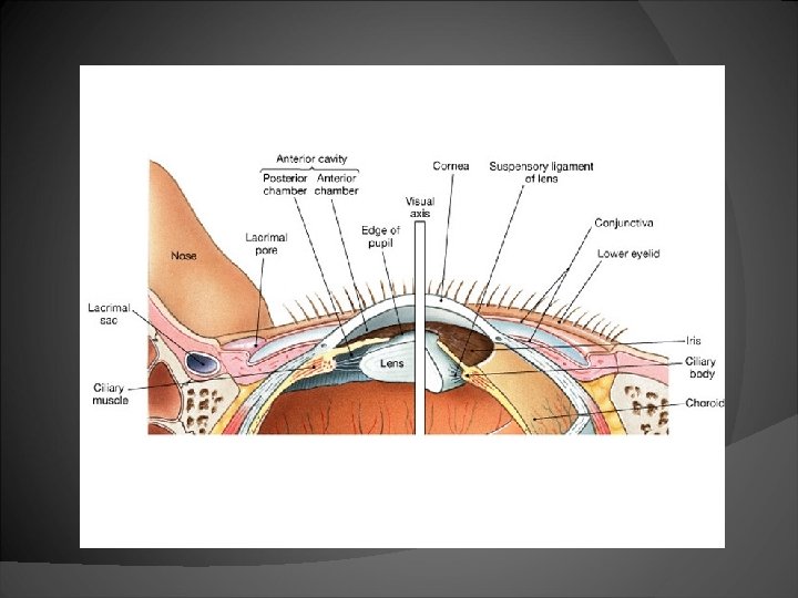
Structure of the Eye � The eye is a fluid-filled hallow sphere with three distinct layers or tunics �The fibrous tunic (Outer) �The vascular tunic (Middle) �The Neural tunic (Inner)
The Fibrous Tunic (Outermost) � Functions as some physical protection, attachment site for extrinsic muscles, and assists in the focusing process � Consists of the transparent cornea at the front of the eye, and the white sclera of the anterior eye.
The Vascular Tunic � The middle, vascular tunic includes the iris, ciliary body, and choroid � The choroid coat is vascular and darkly pigmented. Performs two functions: nourish other tissues of the eye and keep the inside of the eye dark. �Separates the fibrous and neural tunics posterior to the ciliary body
� The ciliary body is a ring of smooth muscle around the front of the eye, contains suspensory ligaments that position the lens so light passing through the pupil passes through the center of the lens � The lens can adjust its shape to facilitate focusing by accommodation
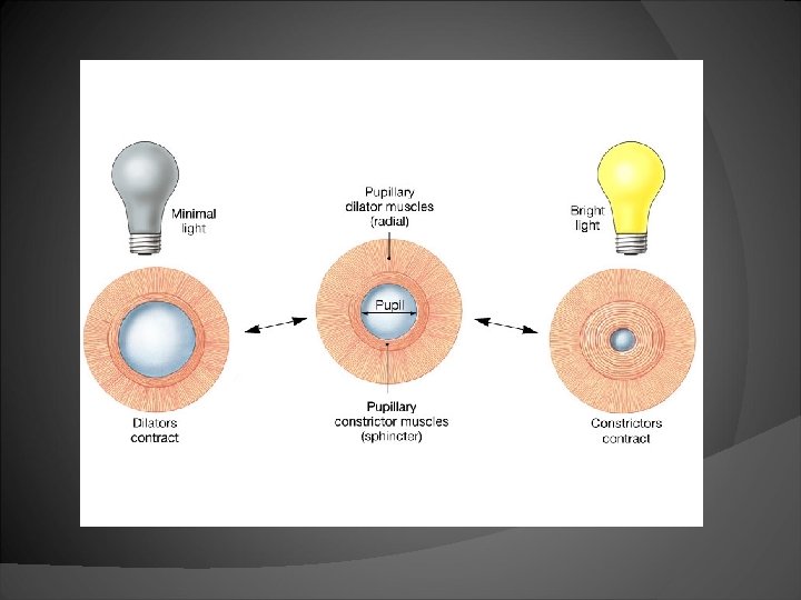
� The iris is a thin, smooth muscle that adjusts to the amount of light that is coming through the pupil, a hole in its center �Has a set of muscles that either contract or dilate depending on light intensity ○ Pupillary dilator muscles (radial) ○ Pupillary constrictor muscles (sphincter)
Eye Color Eye color is determined by the amount of melonocytes that the iris contains and the pigmented epithelium on its posterior surface � When melonocytes are absent, light can pass through which makes the eye appear blue � The more melonocytes the iris has, less light can pass throught, which makes the eye become darker like gray, brown, or black �
The Neural Tunic (Innermost) � The inner tunic consists of the retina, which contains photoreceptors. It covers the back side of the eye to the ciliary body � Retina can be divided into two layers: the pigmented and neural part �Pigmented part- absorbs light �Neural part- photoreceptors that respond to light, preliminary processing and integration of visual information
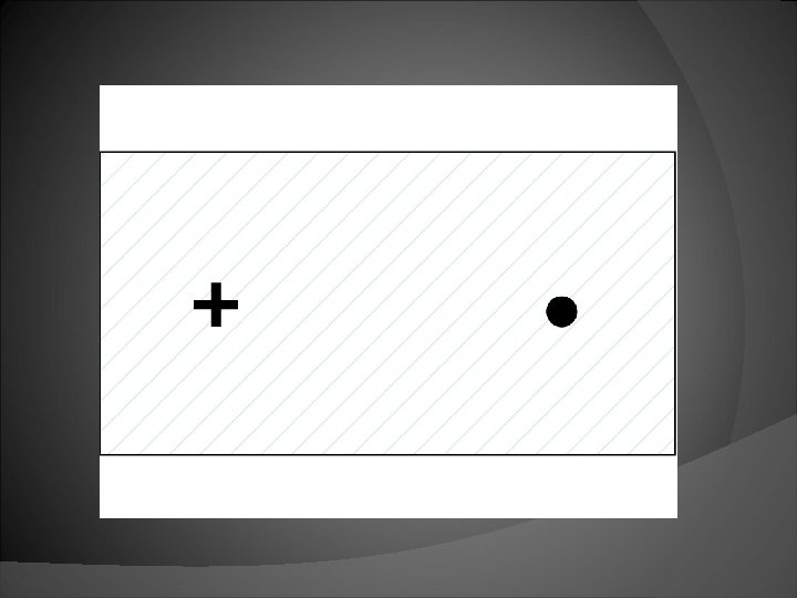
� In the center of the retina is the macula lutea with the fovea centralis in its center. The center for color blindness and sharpest vision � Medial to the fovea centralis is the optic disc, where nerve fibers leave the eye and there is a blind spot � The large cavity of the eye is filled with vitreous humor. Which helps maintain the shape of the eye and holds the retina against choroid.
Blind spot
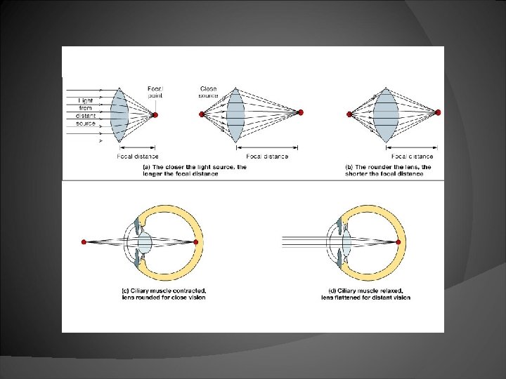
The Lens � The function of the lens to focus the visual image on the photoreceptors. This occurs by changing the shape of the lens called accommodation � The lens can either become rounder to focus on nearby images or flattens to focus on distant images
Concept Check A man in his early 60 s comes to the clinic complaining of fuzzy vision. An eye examination reveals clouding of his lens. What is his problem and what factors might have contributed to it? Ø The man would be diagnosed with cataracts. This could be caused from smoking, UV radiation, aging, or an injury. As aging occurs, the lens becomes less elastic, and begins to lose its transparency. �
Light Refraction � When light waves pass from one medium to another they bend because of the different densities. This occurrence is called refraction. � The cornea and lens both bend the light waves � The lens provides the extra refraction needed to focus the light rays from an object to a focal point, a specific point of interaction on the retina.
Image Formation The Brain compensates for the upside down and backwards image, without our Conscious awareness
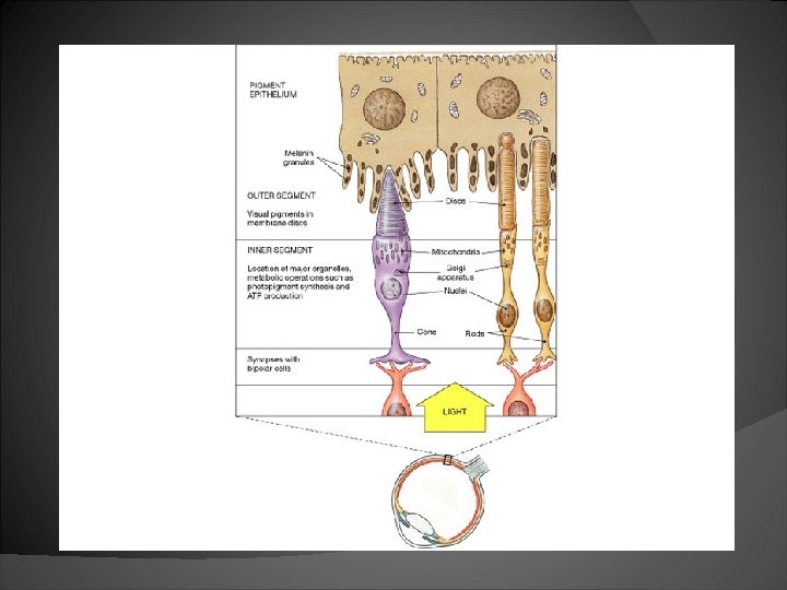
Visual Receptors � Two kinds of modified neurons comprise the visual receptors; elongates rods and blunt-shaped cones � Rods are more sensitive to light and are able to function in dim light; they do not discriminate among colors of light
� Cones are less sensitive than rods, which allow them only to function in bright light. They also allow us to see images in color � To see things in detail, a person moves the eyes so the image falls on the fovea centralis, which contains the greatest amount of cones
Visual Pigments � The discs of the outer segment in rods and cones contain the visual pigments � Light sensitive pigment in rods is rhodopsin, which breaks down into a protein, opsin, and retinal (from vitamin A) in the presence of light
� Breaking down of rhodopsin activates an enzyme that initiates changes in the rod cell membrane, creating a nerve impulse � These nerve impulses travel away from the retina and are interpreted as vision
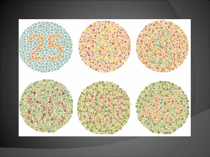
� Light sensitive pigments in cones are also proteins. There are three different types of cones, each containing a different visual pigment. � Color-blindness occurs if one or more classes of cones are absent or not functional
Concept Check A man claiming to have difficulty seeing at night decides to go to his eye doctor. What is the name of the disorder that he has? What dietary supplement will be recommended? If this condition progressed too far, what retinal structures will be deteriorated? Ø The man is suffering from night blindness, a condition when an insufficient amount of vitamin A is in the body. Which effects the amount of visual pigment in the photoreceptors, causes it to drop. He will need to intake more vitamin A. If he does not get an adequate amount, his rods will become affected. �
� The wavelength of light determines the color perceived from it; each of the three pigments is sensitive to different wavelengths of light � The color perceived depends on which set of cones the light stimulates; if all three sets are stimulated, the color is white; if none are stimulated, the color is black.
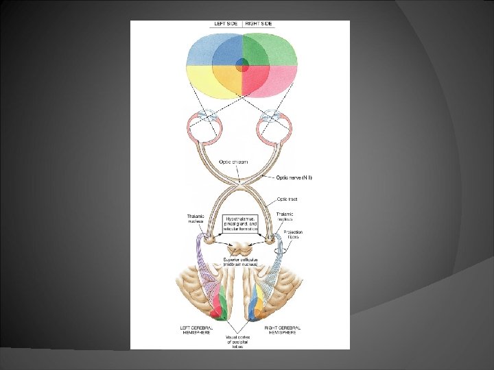
Visual Pathway The axons of ganglion cells converge on the optic disc, penetrate the wall of the eye, and go to the optic nerve 2. The two optic nerves, one from each eye, cross over in the optic chiasm 3. Impulses are transmitted to the thalamus and then to the visual cortex 1.
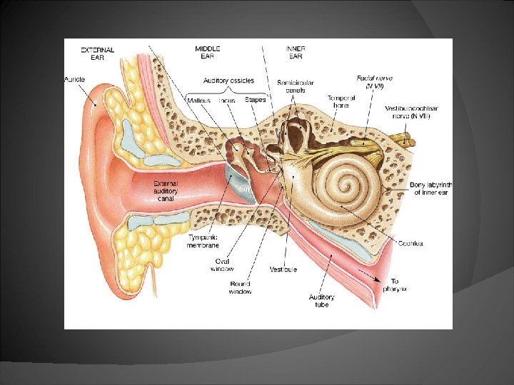
Hearing � The ear is divided into three regions: the external, middle, and inner ear. These three parts allow us to have the sense of hearing and equilibrium. � External ear: consists of the auricle which collects the sound and travels down the external auditory meatus. �Auricle- supported by elastic cartilage, that protects the opening of the canal.
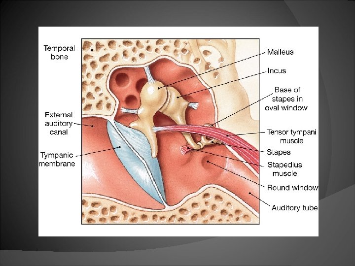
� Middle ear: begins with the tympanic membrane, and is an air-filled space that houses the auditory ossicles. � There are three auditory ossicles, the malleus, the incus, and the stapes. � The tempanic membrane converts arriving sound energy into mechanical movements of the auditory ossicles.
� The stapes vibrates the fluid inside the oval window of the inner fluid � The auditory ossicles both transmit and amplify sound waves
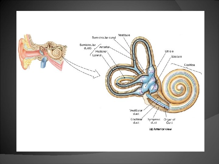
� Inner ear: consists of the membranous labyrinth, bony labyrinth, endolymph, and perilymph �Between the bony and membranous labyrinths is a fluid called, perilymph. �Inside the membranous labyrinth is a fluid called, endolymph.
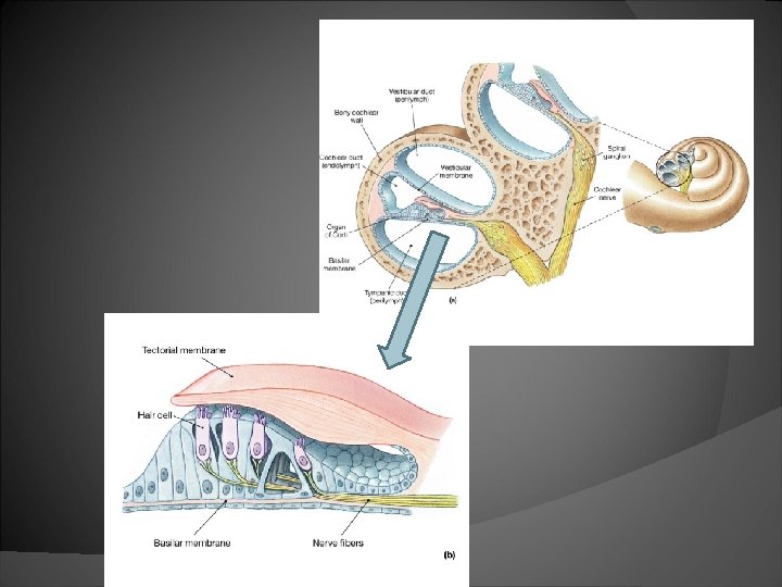
� The bony labyrinth can be divided into 3 parts: �Vestibule- consists of the saccule and utricle. Receptors in these sacs provide sense of gravity and acceleration. �Semicircular canals- enclose semicircular ducts, the receptors are stimulated by rotation of the head. �Cochlea- contains the cochlear duct which provides the sense of hearing ○ the cochlea duct lies between 2 chambers called the vestibular duct and the tympanic duct
� The organ of Corti consists of hair cells of the cochlea duct, which lie on the basilar membrane �The hair cells come into contact with the tectorial membrane, which is attached to the inner wall of the cochlear duct �Vibrations in the fluid of the inner ear cause the hair cells to bend �This stimulates sensory neurons nearby
Concept Check � How would the loss of stereocilia from the hair cells of the organ of Corti affect hearing? Ø Loss of stereocilia (as a result from constant exposure to loud sounds) would reduce hearing sensitivity and could eventually result in deafness.
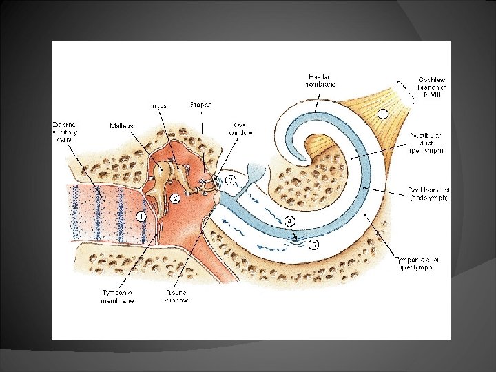
The Hearing Process: Sound waves arrive at the tympanic membrane. 2. The vibration of the tympanic membrane causes movement of the auditory ossicles. 3. The movement of the stapes at the oval window establish pressure waves in the perilympth of the vestibular duct. 1.
The pressure waves distort the basilar membrane on their way to the round window of the tympanis duct. 5. The vibration of the basilar membrane cause the vibration of hair cells against the tectorial membrane. 6. Information about the region and intensity of stimulation is relayed to the CNS. 4.
� Auditory Nerve Pathway: �Nerve fibers carry impulses to the auditory cortices of the temporal lobes where they are interpreted.
Equilibrium � There are two aspects of equilibrium, static and dynamic equilibrium. �Static equilibrium- these organs help us maintain the position of the head when the head and body are still �Dynamic equilibrium- these organs help us maintain balance when the head and body suddenly move and rotate.
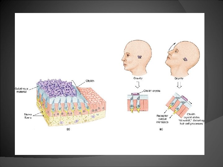
Static Equilibrium � These organs are located within the bony vestibule of the inner ear, inside the utricle and saccule � Receptors in the utricle and saccule respond to gravity and linear acceleration. � A macula, consists of hair cells and supporting cells
� The hair cells contact gelatinous material holding otoliths, which is the whole complex consisting of gelatinous mass and crystals. � Gravity causes the gelatin and crystals to shift, bending hair cells and generates a nervous impulse.
� Impulses travel to the brain by a vestibule branch of the vestibulocochlear nerve, indicating the position of the head.
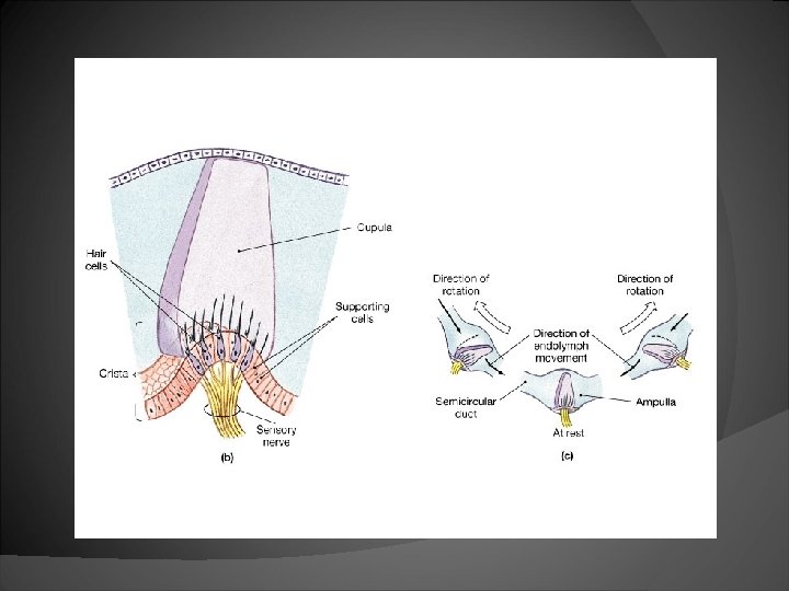
Dynamic Equilibrium � The three semicircular ducts detect motion of the head, and aid in balancing the head and sudden movements of the body. �The three ducts are; anterior, posterior, and lateral semicircular. �Each duct contains an ampulla, which is a swollen region containing sensory receptors.
� Hair cells attached to the wall of the ampulla form a raised structure known as crista. � The stereocilia of the hair cells are embedded in cupula, which is a gelatinous structure. � When the head rotates, movement of the endolymph pushes against the structure and stimulates the hair cells.
Aging and the Senses � The general lack of replacement of neurons in the nervous system leads to a decline in sensory function with age. � Some of this decline can be compensated by increase in stimuli strength or concentration, but the loss of axons cannot be increased in a like manner.
� Smell: olfactory receptor cells are replaced regularly but the total number does decline with age. These receptors also become less sensitive. This is why elderly individuals have trouble detecting odors in small concentrations. � Taste: Mucous membranes begin to thin and the number and sensitivity of taste buds decline. Elderly people find their food to be bland, whereas children find their food to be too spicy.
� Vision: there are various disorders that are associated with aging �The lens loses its elasticity with age and stiffens. They also become farsighted, a condition called presbyopia. �Cataracts develop �Gradual loss of rods �Macular degeneration-growth and proliferation of blood vessels in the retina which causes blindness. Loss of photoreceptors and retinal scarring.
SS Macular Degeneration
� Hearing: the tympanic membrane loses some of its elasticity, making it more difficult to hear high pitched sounds. The progressive loss of hearing is called, presbycusis.
Concept Check � Why does it increasingly get more difficult to see close object as we get older? Ø With age, the lens loses it elasticity and stiffens. These changes result in a flatter lens that cannot round up to focus the image of a close object on the retina. This can be corrected with a converging lens.