Special Senses What are the 5 special senses
 Taste (gustation) Sight Hearing")
. 2. Stimulate")


 from anterior of")


— A. Sclera—")
— A. Choroid Coat— sheet of cells next to the retina")
— A. 2 -layered retina • Light rays focus")
")
 • Contains protein (opsin) attached to pigment (retinal). •")


 sightedness (Myopia) – This is a condition where the")


 – outer ear, collects sound waves • EXTERNAL AUDITORY")


 – At right angles to each other •")


- Slides: 76
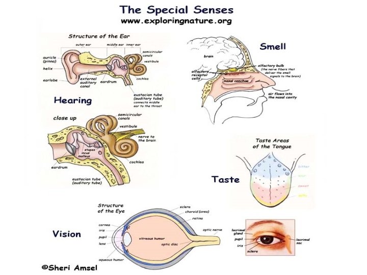
Special Senses What are the 5 special senses? Smell (olfaction) Taste (gustation) Sight Hearing Equilibrium Special Sense Receptors are either large, complex sensory organs (eyes and ears) or localized clusters of receptors (taste buds and olfactory epithelium)
All senses work the same way: 1. Receptors collect information (environmental changes). 2. Stimulate neurons (trigger nerve impulses). 3. Information is sent to the brain (via sensory pathways). 4. The cerebral cortex processes and integrates the information with that from other senses. 5. A perception (a person’s particular view of the stimulus) is formed.
Sensory Pathway
• A sensation or perception occurs when the brain interprets the incoming nerve impulses. • All impulses coming into the brain are alike. The sensation depends on which part of the brain is stimulated.
There are 2 types of sensory receptors: • 1. Somatic senses: touch, pressure, temperature, and pain • 2. Special senses: smell, taste, hearing, equilibrium, and vision
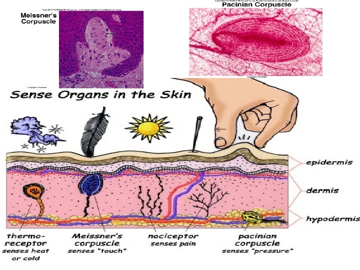
Receptor Types 1. Pain receptors or nociceptors – respond to tissue damage. 2. Thermoreceptors – respond to temperature change (hot or cold)
3. Mechanoreceptors – respond to mechanical forces, such as pressure or fluid movement. 4. Proprioceptors – sense changes in the tension of muscles and tendons. 5. Baroreceptors – in blood vessels – detect changes in blood pressure
6. Stretch receptors – in lungs – sense degree of inflation or stretch. 7. Photoreceptors--respond to light – as little as one photon. (Eyes) 8. Chemoreceptors – sensitive to chemical concentration of various substances. (taste and smell)
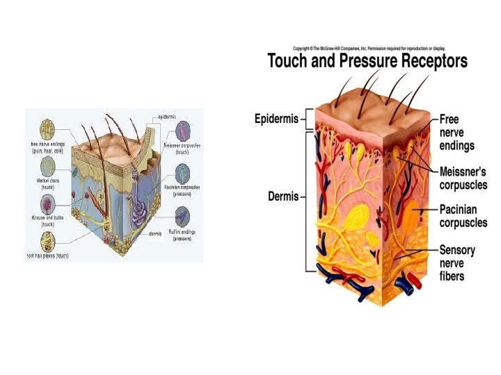
Sense of Touch Cells • 1. Sensory Nerve Fibers--Common in epithelial tissues and are associated with touch and pressure. • 2. Meissner’s Corpuscles--Small masses of connective tissue located in hairless portions of the skin (lips, fingertips, palms, soles). Interpret light touch. —detects exactly what part of the body is touched. • 3. Parcinian Corpuscles--Large structures of connective tissue located in deep tissue layers like ligaments and tendons. Respond to heavy pressure. Layered like an onion surrounding a dendrite. • 4. Root Hair Plexus—dendrites arranged in a network around hair follicles detect movement.
• Special Senses
Chemical Senses: Taste and Smell • Classified as chemoreceptors because they respond to chemicals in solution. Molecules must be released from the substance for you to taste or smell it. • 5 types of taste receptors have been identified, but the olfactory receptors are believed to be sensitive to a much wider range of chemicals. • The receptors for smell and taste complement each other and respond to many of the same stimuli. • Of all the senses, only smell and taste have fibers that run to both cortical areas and the limbic system.
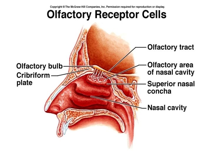
Olfactory Receptors and the Sense of Smell Humans have around 350 olfactory receptors that occupy a postage sized area in the roof of each nasal cavity. They work in various combinations to allow us to smell around 10, 000 odors.
• The olfactory receptor cells are neurons covered in olfactory hairs, long cilia that protrude from the nasal epithelium and are covered in a layer of mucus secreted by underlying glands. When these receptors are stimulated by chemicals dissolved in the mucus, they transmit impulses along olfactory filaments, which are bundled axons of olfactory neurons that together make up the olfactory nerve (cranial nerve #1). The olfactory nerve conducts the impulse to the olfactory cortex of the brain. There the odor is interpreted, and an odor “snapshot” is made.
Olfactory Receptors
• The olfactory receptors are very sensitive— only a few molecules activate them. • They are also adaptive. We tend to be unable to smell our own perfume, but can quickly smell a new scent on someone else….
Olfactory… • organs--Contain the olfactory receptors which are masses that cover the upper parts of the nasal cavity • tracts--Located inside the olfactory bulbs and interpret the nerve impulses • receptors--A chemoreceptor is a sensory receptor that transduces a chemical signal into an action potential. It detects certain chemical stimuli in the environment. • receptor cells--Covered in cilia which have receptor proteins that the odor chemicals bind to • bulbs--Receive the nerve impulses from the receptor cells (located in the brain)
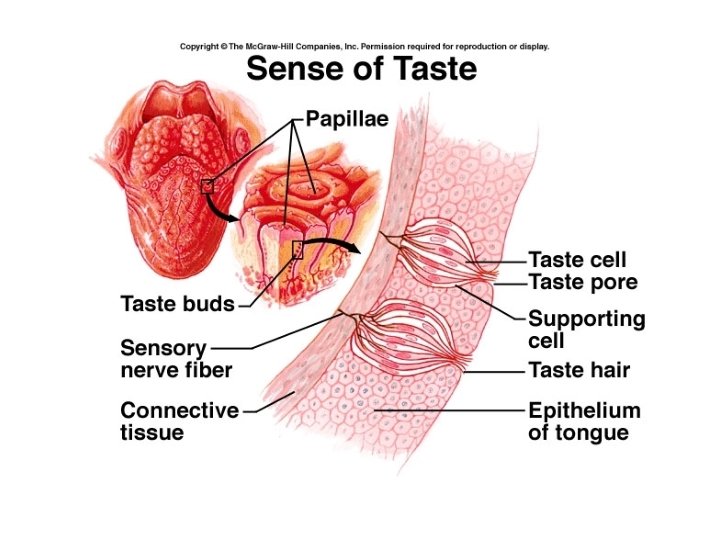
Taste Buds and Sense of Taste • The taste buds, specific receptors for the sense of taste, are widely scattered over the oral cavity. • Of the 10, 000 taste buds, most are in the tongue. A few scattered on the soft palate and inner surfaces of cheeks.
Hot and Spicy Foods • Although there is not a Chemesthetic System per se, the trigeminal nerve (shown in light brown) conveys a great deal of information about the presence of irritating and painful stimuli (like the burn from chili peppers) to the brain. Chemesthetic sensations are perceived by free nerve endings in the tissue and then transmitted to the brain, often through the trigeminal. Different branches of this nerve go the corneas, the nose, the tongue and the teeth, and each relays information about stinging, prickling, burning, and pain in these locations. The trigeminal nerve also conveys information about temperature, like the cooling sensations that arise from the menthol in mouthwash.
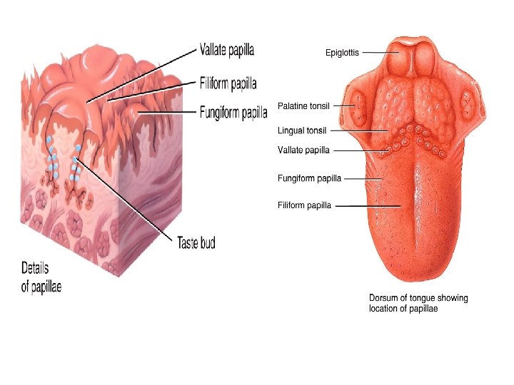
• Tongue surface covered with papillae. • There are 4 types of papillae: filiform, fungiform, circumvallate and foliate. The latter 3 are gustatory papillae, the specific cells that respond to chemicals dissolved in the saliva.
• Circumvallate - taste bud-containing papillae toward the very back of the tongue; they are placed in an inverted “V. ” It can be very hard to see your own, but it is fairly easy to see these in another person, especially if you use a flashlight. • Foliate - taste bud-containing papillae located very far back on the sides of the tongue; they look like a series of folds or lines and can be very difficult to see. • Fungiform—mushroom shaped along tip and sides— sweet and sour tastes • Filiform - papillae that do not contain taste buds. They cover the surface of the tongue in great abundance and are largely responsible for the texture of the tongue. The only purpose it serves in tasting is that it can help to hold taste compounds on the tongue, increasing the chance that the taste compound will interact with a taste receptor cell.
4 types of papillae Fungiform- at surface Circumvallate and Filiform Circumvallate—deep in trenches Filiform—small, pink cone shapes Foliate-deep in folds
The Human Tongue Foliate—a series of folds along the sides of the tongue in the back. Circumvallate—inverted V at the back of the tongue Fungiform—scattered across top, front, and middle Filiform—across top of tongue
How do we taste? • Gustatory cells have gustatory hairs that protrude through the taste pore. When they are stimulated, they depolarize and impulses are transmitted to the brain. • 3 cranial nerves (7, 9, 10) carry taste impulses from the various taste buds to the gustatory cortex. • Taste buds are replaced every 7 -10 days by basal cells (stem cells) found in the deeper regions of taste buds. • Smell accounts for 90% of taste.
Stimulation of taste receptors • Sequence of events – Tastant dissolves in saliva – Enters taste pore contacts gustatory hair – Electrical signal produced – Causes gustatory cell to release neurotransmitter that activates dendrites of first-order neurons • Adaptation occurs within minutes • Different tastes arise from activation of different groups of taste neurons
Gustatory Pathway • Cranial nerves transmit impulses – Facial (CN VII) from anterior of tongue – Glossopharyngeal (CN IX) from posterior – Vagus (CN X) from pharynx, epiglottis • to medulla oblongata thalamus primary gustatory area of cerebral cortex limbic system or hypothalamus
5 basic taste sensations Taste Sensation Sweet Sugars, saccharines, some amino acids, some lead salts Sour H+ (H ions) or acidity Bitter Alkaloids (bases) Salty Metal ions in solution Umami /savory (“delicious”) Glutamate amino acid; responsible for “beef taste” of steak and the flavor of monosodium glutamate, a food additive.
Eating and Taste • Taste is often homeostatic, in that we often crave what our body needs. • Depends strongly on stimulation of olfactory receptors by aromas. • Enhanced by temperature and texture. • “Hot” foods such as peppers stimulate pain receptors in the mouth.
Vision
The Eye and Vision • 70% of the sensory receptors in our body are in the eye. • It is easy to “fool” the eyes Ex: optical illusions, etc. • Optic tracts are huge…over a million nerve fibers. • The adult eye is a sphere and measures about an inch in diameter. • Only the anterior 1/6 can be seen. The rest is protected by a cushion of fat and the orbital socket of the skull, eyebrows, eyelids, and eye lashes.
Accessory Structures – Extrinsic eye muscles – Eyelids—protect eyes • Space between eyelids is palpebral fissure – Conjuctiva--thin membrane that lines the eyelids and covers part of the eye, secretes mucous to lubricate eye – Lacrimal apparatus—cleans and protects the eye as it moistens and lubricates it. § lacrimal glands release dilute salt solutions (tears) onto eyeball. They flush across eyeball into lacrimal canaliculi then into the lacrimal sac, finally into the nasolacrimal duct. § Contain antibodies and lysozyme, an enzyme that destroys bacteria.
Lacrimal System
Review
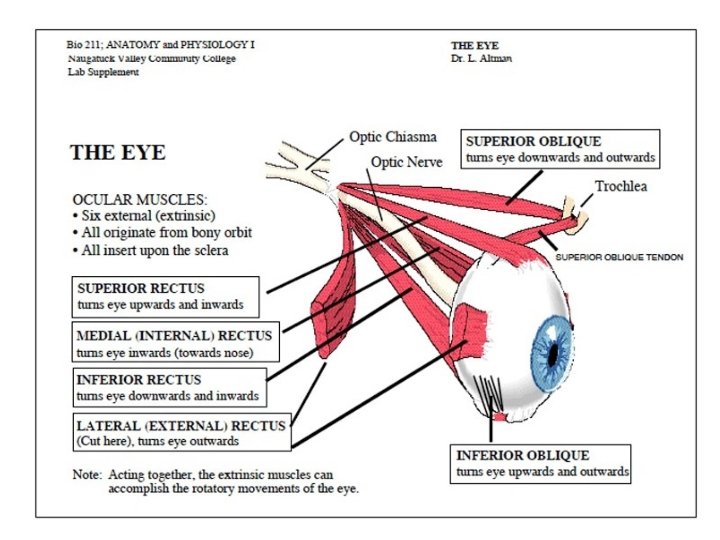
External Eye Muscles Muscle name Action Controlling Cranial Nerve Lateral rectus Moves eye laterally 6 -aducens Medial rectus Moves eye medially 3 -oculomotor Superior rectus Elevates eye and turns it medially 3 -oculomotor Inferior rectus Depresses eye and turns it medially 3 -oculomotor Inferior oblique Elevates eye and turns it laterally 3 -oculomotor Superior oblique Depresses eye and turns it laterally 4 -trochlear
Six Eye Muscles
Internal Structures: The Eyeball • The eyeball is a hollow sphere. • Its wall is composed of 3 layers. • Its interior is filled with fluids called humors that help to maintain its shape. • The lens, the main focusing apparatus, is supported upright within the eye cavity, dividing it into 2 chambers.
Layers forming the wall of the eyeball • 1. Fibrous Layer (outer)— A. Sclera— • Outer layer; White of the eye • Tough coating, helps maintain shape of eye and protects what’s inside • Muscles responsible for moving the eye are attached to the sclera – called EXTRINSIC MUSCLES B. Cornea—The crystal clear “window” through which light enters the eye; many nerve endings/pain fibers. • Front of sclera – clear part (no blood vessels) • Transparent so light rays can pass through • Gets O 2 and nutrients through lymph Aqueous humor —watery substance between cornea and lens.
2. Vascular Layer (middle)— A. Choroid Coat— sheet of cells next to the retina with a black pigment to absorb extra light, and blood vessels to nourish the retina. B. Ciliary body –attaches the ciliary ligament to the eyeball; produces aqueous and vitreous humor. C. Iris--Colored, muscular layer surrounding pupil is IRIS INTRINSIC MUSCLES – change size of iris to control amount of light entering through the pupil—opening in front D. Lens • Crystalline structure located behind iris and pupil • Elastic, disc-shaped, biconvex • Situated between the anterior and posterior chambers • ACCOMMODATION – change in the shape of the lens to allow for near and distant vision • ANTERIOR CHAMBER filled with AQUEOUS HUMOR, a watery fluid. • POSTERIOR CHAMBER filled with transparent, jellylike substance – VITREOUS HUMOR— fills cavity behind the lens • Ciliary Ligament —attached to lens, contracts or relaxes to adjust the lens.
• 3. Sensory Layer (inner)— A. 2 -layered retina • Light rays focus an image on the retina • The image travels to the cerebral cortex via the OPTIC NERVE • If light rays doesn’t focus properly on the retina, corrective lenses can bend the light rays as required. • Retina contains specialized cells – rods and cones • RODS – sensitive to dim light • CONES – sensitive to bright light and color • Fovea—a small area of the retina which is directly in line with the center of the cornea and the lens, concentrated with cones. B. OPTIC DISC – on the retina, known as the blind spot – nerve fibers gather here to form the optic nerve, no rods or cones
Pathway of Vision • Image Cornea Aqueous Humor Pupil Lens(Where light rays are refracted) Vitreous Humor Retina Rods and Cones (pick up stimulus) Optic Nerve Brain • Once the rods and cones are stimulated, a sensory impulse is carried on the: 1. optic nerve (CN 2), which crosses at the 2. optic chiasma forming optic tracts that carry the impulse to the 3. thalamus for direction to the 4. primary visual cortex (occipital lobe) for interpretation.
Photoreceptors 1. Rods—rhodopsin (night vision—silhouettes) • Contains protein (opsin) attached to pigment (retinal). • Light causes retinal to change shape releasing it from opsin. • Chain reaction of events results in closing Na+ channels. • Results in hyperpolarization slowing the tonic firing of AP’s. DARK ADAPTED—all opsin and retinal together, rods very sensitive, vision possible, even in dark. LIGHT ADAPTED—opsin and retinal decompose, cones take over 2. Cones (color vision, sharp images)— 6 million cones • Contains protein iodpsins • A. red—erythrolabe • B. blue—chlorolabe • C. green—cyanolabe • D. different colors—combination of stimulation
• The eye is a slightly asymmetrical globe, about an inch in diameter. The front part of the eye (the part you see in the mirror) includes: • • The iris (the pigmented part) • The cornea (a clear dome over the iris) • The pupil (the black circular opening in the iris that lets light in) • The sclera (the white part) • The conjunctiva (a thin layer of tissue covering the front of the eye, except the cornea) • Just behind the iris and pupil lies the lens, which helps to focus light on the back of the eye. Most of the eye is filled with a clear gel called the vitreous. Light projects through the pupil and the lens to the back of the eye. The inside lining of the eye is covered by special light-sensing cells that are collectively called the retina. The retina converts light into electrical impulses. Behind the eye, the optic nerve carries these impulses to the brain. The center of the retina contains the fovea, a small depression or pit that gives the clearest vision. • Eye color is created by the amount and type of pigment in the iris. Multiple genes inherited from each parent determine a person’s eye color.
Physiology of Vision— 3 steps
Vision • Focus of Lens —When looking at distant objects, the lens is long and thin in shape. When looking at close objects, the lens is short and wide. • Binocular Vision —Two eyes are important in judging distance and depth (3 dimensional) • Pupil Size —In bright light, the iris muscle relaxes and the pupil decreases in size so that less light enters the eye. In dim light, the iris muscle contracts and the pupil increase in size to allow more light to enter.
Vision Problems • Short (near) sightedness (Myopia) – This is a condition where the person can see close objects well, but not distant objects. Light focuses in front of the retina. It is corrected with concave lenses. • Long (far) sightedness (Hyperopia) – This is a condition where the person can see distant objects well, but not close object. Light focuses behind the retina. It is corrected with convex lenses. • Astigmatism – This is a condition where the cornea is curved unevenly, so that different light rays focus in different places. Corrected with glasses.
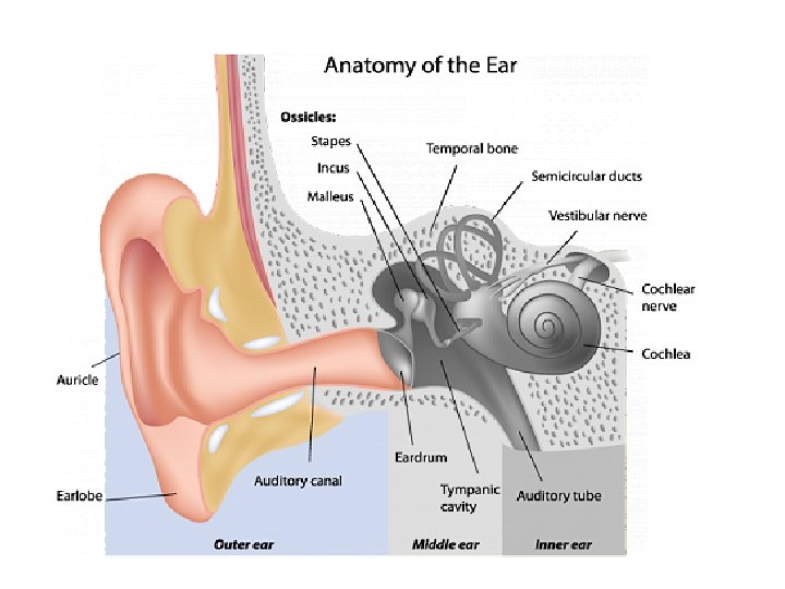
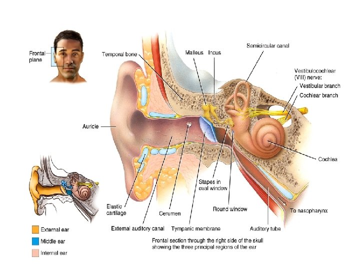
The Ear • Hearing and equilibrium • 3 parts: Outer, middle and inner ear
Hearing and Equilibrium: Ear Structure • Outer ear: auricle, external auditory canal, and tympanic membrane (ear drum) – Canal contains hairs and ceruminous glands • Middle ear: auditory tube (eustachian tube) and ossicles (bones) – Ossicles (malleus, incus, stapes: attached to oval window) • Inner ear: bony labyrinth + membranous labyrinth filled with endolymph – Cochlea: sense organ of hearing , – Vestibule and semicircular canals: organs of balance Copyright 2010, John Wiley & Sons, Inc.
Outer Ear • PINNA (AURICLE) – outer ear, collects sound waves • EXTERNAL AUDITORY CANAL – ear canal • CERUMEN – earwax, protects the ear • TYMPANIC MEMBRANE – ear drum, separates outer and middle ear
Middle Ear • Cavity in temporal bone • Connects with pharynx by EUSTACHIAN TUBE – which equalizes pressure in the middle ear with outside atmosphere • Bones in middle ear that transmit sound waves from ear drum to inner ear 1. MALLEUS (hammer) 2. INCUS (anvil) 3. STAPES (stirrup)
Inner Ear • Contains spiral shaped organ of hearing – the COCHLEA • The cochlea contains a membranous tube, the cochlear duct – which is filled with fluid that vibrates when sound waves are transmitted by the stapes • ORGAN OF CORTI – delicate hair-like cells that pick up vibrations of fluid and transmit them as a sensory impulse along the auditory nerve to the brain • SEMICIRCULAR CANALS – three structures in the inner ear, contain liquid that is set in motion by head and body movements – impulses sent to cerebellum to help maintain body balance(equilibrium).
Inner Ear Structure: 3 Regions • Vestibule includes – Two sacs: utricle and saccule • Semicircular canals: at right angles – Contain membranous semicircular ducts – Each ends in a swelling known as ampulla • Cochlea: 3 levels – Cochlear duct: membranous, has endolymph • Contains spiral organ (sensory organ for hearing) – Above: scala vestibuli: ends at oval window – Below: scala tympani: ends at round window
Inner Ear Structure
Inner Ear Structure
Physiology of Hearing • Sound waves in air auditory canal • Tympanic membrane ossicle movement stapes strikes oval window • Pressure waves in perilymph – Conveyed from scala vestibuli scala tympani • Pressure waves in endolymph cause – Hair cells bend against tectorial membrane – Neurotransmitter released to sensory neurons • Pitch (wavelength): location in cochlea • Volume (loudness): intensity of waves
Pathway of Sound • • • Sound Waves Pinna—outer ear External Auditory Canal Tympanic Membrane/Eardrum Ossicles (malleus, incus & stapes)—middle ear – Hammer, anvil, stirrup • • Oval Window Cochlea—inner ear Auditory nerve (cranial nerve 8) Brain: Thalamus to auditory cortex in temporal lobe.
How do we Hear? • The brain and auditory system work together to control how we hear and how we balance ourselves. The human ear is a complex organ and many scientists consider hearing to be the most complex of the human senses. • Sound can be detected whether a person is on land, underwater or in the air. Hearing is our ability to perceive sound by detecting vibrations that travel through our ears. The main purpose of the ear is to turn sound waves from the air into electrical signals that are interpreted by the brain.
Sound: Rapid Air Waves through the Ear • Sound travels through the auricle and the auditory canal, a short tube that ends at the eardrum. Sound entering the outer ear travels through the middle ear and causes the eardrum and ossicles in the middle ear to vibrate. As it travels, it amplifies (gets louder) and changes from air to liquid. When the stapes moves, it pushes the oval window, which then moves the cochlea. The cochlea takes the fluid vibration of sounds from the surrounding semicircular ducts, translates them into signals sent to the brain by nerves like the vestibular nerve and cochlear nerve. The brain translates the information into recognizable sound patterns. It is a complex process but it occurs in a split-second of time.
Malleus Incus Stapes vibrating. Helicotrema in oval window Cochlea Sound waves Perilymph 3 7 4 2 External auditory canal Scala tympani Scala vestibuli 5 1 8 6 9 Basilar membrane 8 Spiral organ (organ of Corti) Tectorial membrane Vestibular membrane Cochlear duct (contains endolymph) Tympanic membrane Secondary tympanic membrane vibrating in round window Middle ear Auditory tube
Equilibrium • The ears are also responsible for equilibrium. A person with equilibrium problems may complain with nausea, dizziness, and balance problems.
Physiology of Equilibrium • Static equilibrium: senses position relative to gravity – As when head is tilted or a car is speeding up or slowing down • Dynamic equilibrium: senses position in response to head movement – As in spinning movements
Equilibrium • Sensed in maculae of utricle and saccule • Mechanism – Gravity pulls on otoliths in otolithic membrane – Bends hair cells in otolithic membrane – Triggers nerve impulses in vestibular branch of vestibulochochlear nerve
Static Equilibrium
Static Equilibrium
Dynamic Equilibrium • Semicircular canals (3) – At right angles to each other • Cristae in each ampulla contain – Hair cells embedded in jellylike cupula – Supporting cells • Mechanism – When head turns, hair cells move – Endolymph lags and bends hair cells – Nerve impulses in vestibular branch
Dynamic Equilibrium
Dynamic Equilibrium
Equilibrium Pathways • • Axons from vestibular branch medulla or cerebellum Medulla motor control: eye, head, neck Spinal cord tracts for adjusting muscle tone and postural muscles