SENSES Somatic senses Chapter 13 and special senses


 A. Unencapsulated receptors – Free nerve endings – pain and")


 TASTE (TASTE BUDS) VISION (EYE) HEARING")


 Figure 15. 25 b")


 9. Cochlea 10. cochlear nerve 11. semicircular canals")


 start to “ripple” to vestibular membrane to endolymph inside")


,")


- Slides: 41
SENSES Somatic senses -Chapter 13 and special senses- Chapter 15
I. Sensory Receptors A. Receptor – any specialized structure used to detect a stimulus (anything that causes a response. ) 1. Free nerve ending sense organs 2. Major function – changing chemical, thermo, visual, or mechanical sensory information into a nerve impulse.
II. Classification of receptors A. Action – Chemoreceptors – respond to chemicals – smells, taste. – Thermoreceptors – in skin, respond to temperature changes. – Nociceptors – respond to painful stimuli. – Mechanoreceptors – respond to physical change. – Photoreceptors – respond to light – Osmoreceptors-respond to osmotic pressure of body fluids
II. B. Structural classification – General or somatic senses • Free/unencapsulated • Encapsulated – Complex/special sense • Tongue, ear, eye, and nose
II. C. Origin/location of stimulus – Visceroceptors – internal receptors – pickup changes in internal organs. – Externoceptors – external areas – Proprioceptors – position changes – skeletal muscle, joints, and tendons.
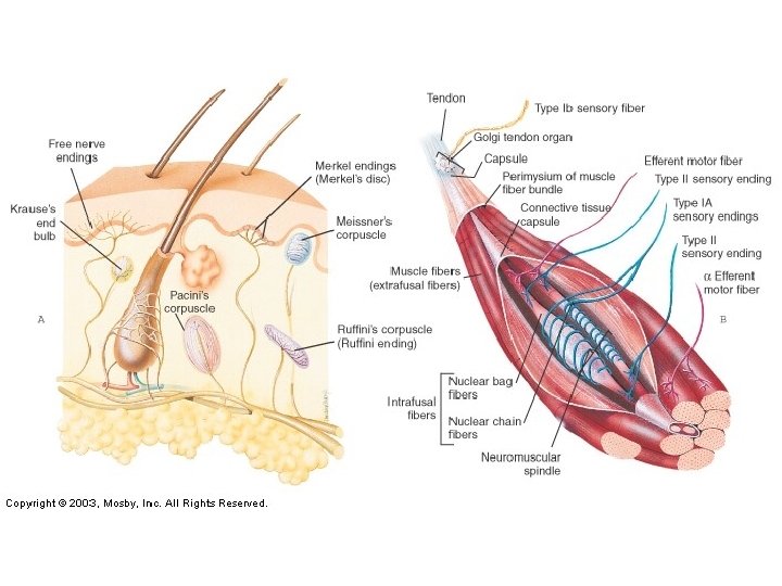
III. Somatic senses (general) A. Unencapsulated receptors – Free nerve endings – pain and temperature. – Merkle cells/discs – tactile – light pressure – Root hair plexus – for hair movement – found around hair bulb, also called hair follicle receptors – Nocioceptors – irritating touch/temperature stimulus – like an itch.
III. B. Encapsulated – Messiner’s corpuscle – light touch – in papillary layer of dermis – Pacinian corpuscle – deep pressure and vibrations – in reticular layer of dermis. – Krause’s end bulb – light touch and temperature – may be responsible for cold mechanoreception – Ruffini corpuscle – touch and temperature – may be responsible for hot temperature.
III. C. Stretch receptors – special type for tendons and muscles – protective feature - Muscle spindles – mechanoreceptors – deals with skeletal muscles. How long the length of a muscle fiber can stretch. - Golgi tendon receptors – tendon, contraction of tendon and skeletal muscle – deals with muscle tension. - Joint kinesthetic receptors- proprioceptors that monitor stretch in articular capsules
IV. SPECIAL SENSES • • OLFACTORY (SMELLING, NOSE) TASTE (TASTE BUDS) VISION (EYE) HEARING AND EQUILIBRIUM (EAR)
IV. ORGAN RECEPTOR TYPE SENSE EYE RODS-MONOCHROME CONES-COLOR PHOTORECEPTORS VISION EAR ORGAN OF CORTI CRISTAE AMPULLARIS MECHANORECEPTORS HEARING EQUILIBRIUM NOSE OLFACTORY CELLS CHEMORECEPTORS SMELL TASTE BUDS GUSTATORY CELLS CHEMORECEPTORS TASTE
Sense of Smell Figure 15. 3
C. Olfactory Pathway 1. If odor dissolved in mucus surrounding olfactory cilia reaches threshold level, AP (action potential) will be generated 2. Impulse passed on to olfactory nerves (I) to olfactory bulb. 3. Passes through olfactory tract to thalamic and olfactory centers for interpretation and memory (temporal lobe)
Taste Buds Figure 15. 1
Taste Sensations • There are five basic taste sensations – Sweet – sugars, saccharin, alcohol, and some amino acids, found at tip – Salt – metal ions, found on anterior sides – Sour – hydrogen ions found on posterior sides – Bitter – alkaloids such as quinine and nicotine, found at posterior – Umami – elicited by the amino acid glutamate
Taste Pathway 1. AP is created in taste cell 2. AP generated to brain by facial nerve (VII), glossopharyngeal (IX), vagus (X) to medulla oblongata to thalamus to gustatory cortex of parietal lobe. – Hypothalamus and limbic system (appreciation of taste)
VII. EAR A. Ear – two functions hearing and equilibrium/balance B. Both senses are because of mechanoreceptors. All are because of hair cell receptors. C. Structures of the ear- 3 major divisionsexternal, middle, and inner ear
The Ear: Hearing and Balance Figure 15. 25 a
Middle Ear (Tympanic Cavity) Figure 15. 25 b
Ear Ossicles Figure 15. 26
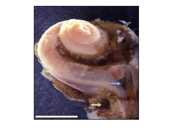
Ear picture- know these on picture 1. 2. 3. 4. 5. 6. 7. Temporal bone External auditory meatus Auricle (pinna) Tympanic membrane (eardrum) Malleus Incus Stapes
8. Auditory tube (Eustachian tube) 9. Cochlea 10. cochlear nerve 11. semicircular canals
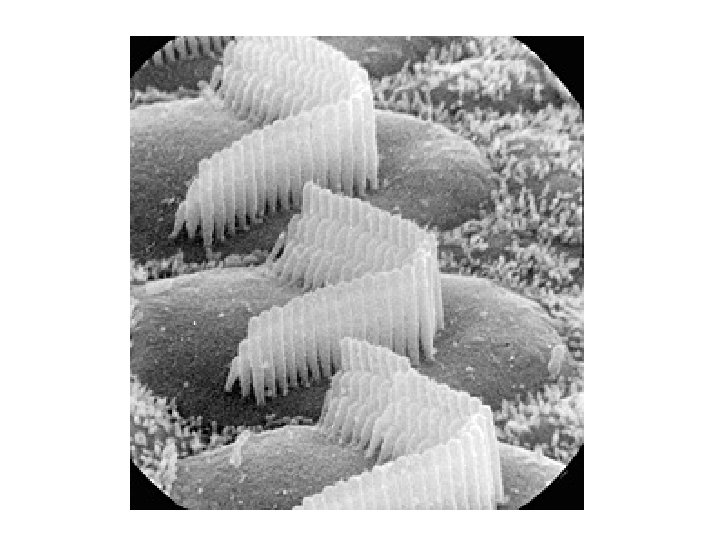
HEARING • Hearing occurs when the auditory area of the temporal lobe is stimulated • Sound waves must be propagated through the air, membranes, bones, and fluids to reach and stimulate the receptor cells in the organ of Corti • Cochlear hair cells are deflected (bent) that causes AP to occur
Transmission of Sound to the Inner Ear Figure 15. 31
Excitation of Hair Cells in the Organ of Corti Figure 15. 28 c
G. Hearing Pathway 1. Sound waves in the air enter external auditory meatus with help from the pinna. 2. Sound waves strike against tympanic membrane moving sound waves to auditory ossicles. 3. The vibrations move malleus incus stapes to oval window 4. Fluid conduction of sound waves begin
5. Perilymph (similar to CSF) start to “ripple” to vestibular membrane to endolymph inside cochlear duct to organ of Corti 6. Hair cells bend starting AP from release of ions and neurotransmitters used transmit nerve impulses to cochlear nerve to brain stem 7. Pass through relay areas in medulla, pons, midbrain, thalamus to auditory centers in temporal lobe
I. Equilbrium – Equilibrium – sense organs for balance – found in the inner ear – vestibule and semicircular canals (called the vestibular apparatus) Equilibrium controlled by vestibular nerve – branch of vestibulocochlear nerve, cranial nerve VIII.
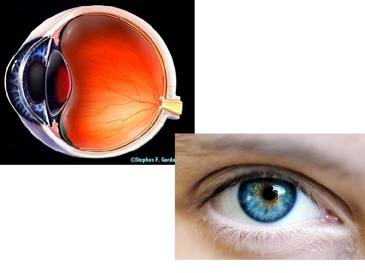
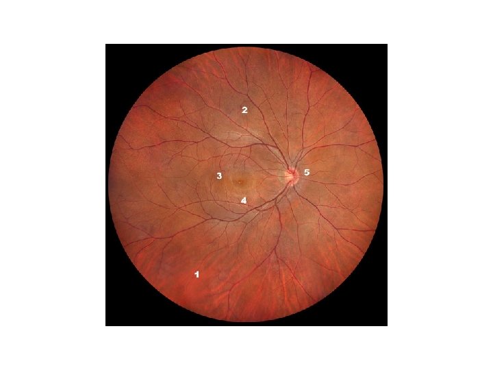
Eyeball Picture- know these on picture 1. 2. 3. 4. 5. 6. 7. Conjunctiva Iris Lens Ciliary muscle Retina Choroid layer sclera
Know these on picture 8. Central retinal artery and vein 9. Optic nerve 10. optic disc (blind spot) 11. anterior cavity 12. pupil 13. cornea
H. Visual Pathway • How we see – light enters cornea anterior cavity aqueous humor lens posterior cavity vitreous humor retina photoreceptors produces on rods and cones formation of image optic nerve visual cortex occipital lobe. • Image must be formed on retina to stimulate rods and cones by refraction of light, accommodation of lens, constriction of pupil, convergence of eye
• Light reaching a photoreceptor causes the breakdown of the chemical rhodopsin, which in turn causes a membrane potential that is transmitted to an action potential. The action potential transfers to synapsed neurons that connect to the optic nerve. The optic nerve connects to the occipital lobe of the brain
I. Vision • Distant vision– eyes are adapted for distant vision (normal 20 ft), eye muscles are relaxed and lens is flat • Close vision- requires active adjustments in the eye to restore focus – Closest point of clear focus is called the near point – This changes as you age (~50 yearspresbyopia)
Focusing for Distant Vision • Light from a distance needs little adjustment for proper focusing • Far point of vision – the distance beyond which the lens does not need to change shape to focus (20 ft. ) Figure 15. 17 a
Focusing for Close Vision • Close vision requires: – Accommodation – changing the lens shape by ciliary muscles to increase refractory power – Constriction – the pupillary reflex constricts the pupils to prevent divergent light rays from entering the eye – Convergence – medial rotation of the eyeballs toward the object being viewed
Focusing for Close Vision Figure 15. 7 b
J. Vision Disorders • Myopia- “near sightedness” distant objects are focused in front of the retina, eyeball may be too long • Hyperopia- “far sightedness” distant objects focused behind the retina, eyeball may be too short