Anatomy Ch 8 SPECIAL SENSES The Eye and


 Choroid �Blood rich �Contains a")
 Iris �Colored region of the eye �Opening in the center is called the")
 Pigmented layer �Outer")
 Neural layer �Inner layer �Contains photoreceptors called rods and cones �Signals pass through")


 ear �Auricle")

 �Area of the ear enclosed by the eardrum and")
 �Incus (anvil) �Stapes (stirrup) �Sound waves vibrate these bones")
 �Maze of bony chambers called the bony labyrinth �The 3")


- Slides: 32
Anatomy Ch. 8 SPECIAL SENSES
The Eye and Vision Of all the sensory receptors in the body 70% are in the eyes. Accessory Features Extrinsic eye muscles � 6 muscles attached to the outer surface of the eye that function in moving the eye Eyelids �Function in protection �Have eyelashes which function in protection �Glands associated with the eyelids called tarsal glands produce an oily secretion that lubricates the eye.
Conjunctiva �Membrane that secretes mucus that lubricates and moistens the eyeball Lacrimal apparatus �Produces tears that clean the eyeball �Secretions contain lysozyme which destroys bacteria �Secretions empty into the nasal cavity
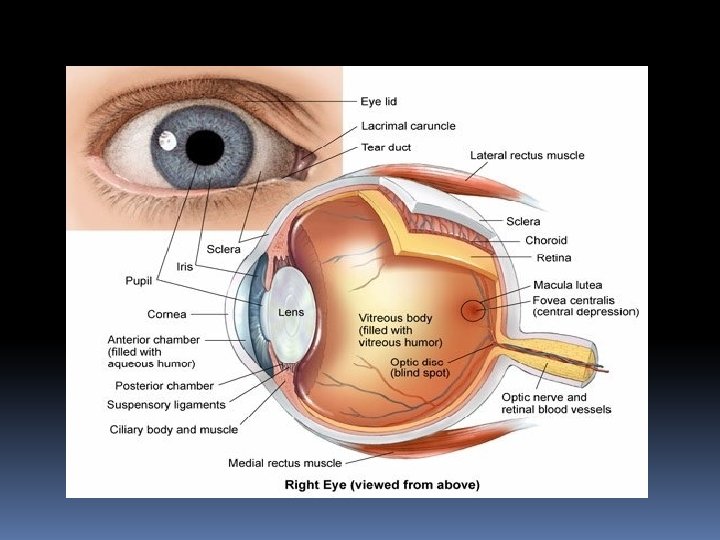
Internal structure of the eyeball The eyeball is a hollow sphere The wall of the eyeball is composed of 3 layers (tunics) The interior of the eyeball is filled with fluids called humors that help the eye maintain shape The lens is the main focusing apparatus of the eye The eye contains 2 chambers
Layers of the Eyeball Wall Fibrous Layer �Outermost layer �Contains the protective sclera and the transparent cornea �The sclera is the white of the eye �The cornea is the area where light enters the eye �Well supplied with nerves �Most exposed part of the eye �Can be damaged easily but can repair itself �Can be transplanted with no possibility of rejection due to lack of blood vessels
Vascular layer �Middle layer � 3 regions 1) Choroid �Blood rich �Contains a dark pigment that prevents light from scattering in the eye 2) Ciliary body �Smooth muscle �Attaches to the lens
3) Iris �Colored region of the eye �Opening in the center is called the pupil that allows light to pass through �The iris is composed of smooth muscle which regulates the amount of light that enters the eye by changing the size of the pupil �Bright light or close vision: pupil constricts �Dim light or distant vision: pupils dilate
Sensory layer �Inner most �Contains the 2 layered retina 1) Pigmented layer �Outer layer �Absorbs light and prevents scattering �Has cells that act as phagocytes
2) Neural layer �Inner layer �Contains photoreceptors called rods and cones �Signals pass through the optic nerve to the occipital lobe
�Photoreceptors are found all over the retina except where the optic nerve leaves the eyeball. �This area is called the optic disk or blind spot.
Rods and Cones �Rods �The majority of rods are found at the edge of the retina �The number decreases as you move to the center �Allow us to see in gray tones in dim light �Provide peripheral vision
�Cones �The majority of cones are found in the center of the retina �The number decreases as you move to the edges �Allow us to see in detail in color in bright light conditions �The fovea centralis is located next to the blind spot. It is loaded with cones and is the area where we have the sharpest vision
�There are 3 types of cones: �Responds to blue light �Responds to green light �Range that includes green and red �Different colors are caused by simultaneous impulses that are mixed and interpreted in the brain.
Lens Biconvex shape Focuses light on the retina Held in position by a ligament that attaches to the ciliary body Divides the eye into 2 chambers
�The anterior segment is filled with a watery fluid called aqueous humor �The posterior segment is filled with a gel like substance called vitreous humor. Vitreous humor helps prevent the eye from collapsing �Both humors help maintain the pressure inside the eye
Physiology of Vision Light rays are refracted or bent as they encounter the cornea, aqueous humor, lens, and vitreous humor The refractive power of the cornea and humors does not change but the refractive power of the lens can change by changing shape The more the lens bulges the more it bends light and vice versa. In order to make close vision possible the lens must bulge more. This process is called accommodation.
The image formed on the retina is called a real image. A real image is reversed from left to right, upside down, and smaller The image is placed in the correct orientation by the occipital lobe.
Visual Pathway to the Brain Axons from the eye bundle together to form the optic nerve At a point in the brain called the optic chiasma fibers from the medial (inner) side of each eye cross over to the opposite side of the brain. Fibers from the lateral (outer) side of each eye stay on the same side of the brain. Fibers on each side of the brain join together and are called optic tracts.
Each tract contains fibers from both eyes The optic tracts pass through the thalamus and enter the occipital lobe where seeing occurs Each side of the brain receives input from both eyes. This causes binocular vision.
Eye Reflexes Intrinsic eye muscles �Autonomic �Affect lens curvature �Affect muscles of the iris which controls pupil size Extrinsic eye muscles �Controls eye movement and allows the eyes to follow moving objects �Allows for convergence which allows us to view close objects �Controlled by cranial nerves III, IV, and VI
Photopupillary reflex �Constricting of the pupils when exposed to bright light Accommodation pupillary reflex �Constricting of the pupils when we view close objects allowing for more acute vision
The Ear and Hearing and Balance Structure of the Ear External (outer) ear �Auricle (Pinna) �Outer visible portion of the ear �External acoustic meatus (auditory canal) �Short chamber that enters the temporal bone �Contains ceruminous glands that secrete earwax
�The tympanic membrane or eardrum is found at the end of the canal �It separates the outer and middle ear �Sound waves cause the eardrum to vibrate
Middle Ear (tympanic cavity) �Area of the ear enclosed by the eardrum and oval window �The auditory tube connects the middle ear to the throat �This tube is normally flat but swallowing or yawning can open it to equalize pressure �The middle ear contains the 3 smallest bones in the body called ossicles which transmits vibrations from the eardrum to the fluid of the inner ear.
�The 3 ossicles �Malleus (hammer) �Incus (anvil) �Stapes (stirrup) �Sound waves vibrate these bones in order with the last vibrating the oval window which vibrates fluid in the inner ear.
Internal (Inner ear) �Maze of bony chambers called the bony labyrinth �The 3 regions of the labyrinth are the cochlea, vestibule, and semicircular canals �The bony labyrinth is filled with a fluid called perilymph �Suspended in the perilymph is a membranous labyrinth �The membranous labyrinth contains a fluid called endolymph
Equilibrium The equilibrium sense responds to movements of the head The equilibrium receptors are found in the inner ear There are 2 types of equilibrium: static and dynamic
�Static equilibrium �Occurs in the vestibule of the bony labyrinth �Receptors called maculae report on changes in position of the head when the body is not moving �The maculae help us keep our head erect �Movements of the maculae send messages to the vestibular nerve which sends a message to the cerebellum.
�Dynamic Equilibrium �Occurs in the semicircular canals of the bony labyrinth �Responds to angular or rotation of the head rather than straight line movements �Receptor regions are called crista ampullaris �Messages are sent to the vestibular nerve which sends the message to the cerebellum
Hearing Within the cochlea is the spiral Organ of Corti which contains hair cells Sound waves reach the cochlea through vibrations of the eardrum, ossicles, and oval window The vibrations set fluids in the cochlea in motion The force of sound waves is increased as it passes through the ossicles
Fluid in the Organ of Corti is vibrated by sound waves and sets fibers in motion Fibers attached to the membrane of this region have different lengths �High pitched sounds affect short fibers �Low pitched sounds affect long fibers These fibers stimulate hair cells in the cochlea The hair cells transmit impulses along the cochlear nerve to the auditory cortex of the temporal lobe where hearing occurs