Ventilation and ED Monitoring for Dummies Todd Ring

- Slides: 79
Ventilation and ED Monitoring for Dummies… Todd Ring July 24/03
Objectives Invasive (IPPV) vs. Non Invasive Positive (NPPV) Pressure Ventilation The different modes of IPPV and typical ventilator settings CPAP vs. BPAP Clinical scenarios requiring mechanical ventilation Pulse oximetry, non-invasive BP monitoring and capnography
Case 1: “Dumb” Motor. Biker 28 yo male, riding in Calgary, no helmet, crashes, unconscious, EMS scoop and run, in ED no spontaneous respirations, bag and mask ventilation. What to do? Indications for Mechanical Ventilation?
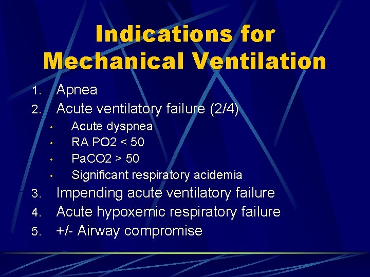
Indications for Mechanical Ventilation Apnea 2. Acute ventilatory failure (2/4) 1. • • Acute dyspnea RA PO 2 < 50 Pa. CO 2 > 50 Significant respiratory acidemia Impending acute ventilatory failure 4. Acute hypoxemic respiratory failure 5. +/- Airway compromise 3.
Case 1: Cont’d The patient was successfully intubated. The RT is now “bagging” the patient manually and asks you to give orders for mechanical ventilation. What mode to choose? Where do you set your ventilator parameters?
Invasive Mechanical Ventilation Volume vs. Pressure control Spontaneous, controlled or combined ventilation
Invasive Mechanical Ventilation Modes used in the ED: Assist Control (A/C) Pressure Support (PSV) Simulated Intermittent Mechanical Ventilation (SIMV) +/- Pressure Control (PCV)
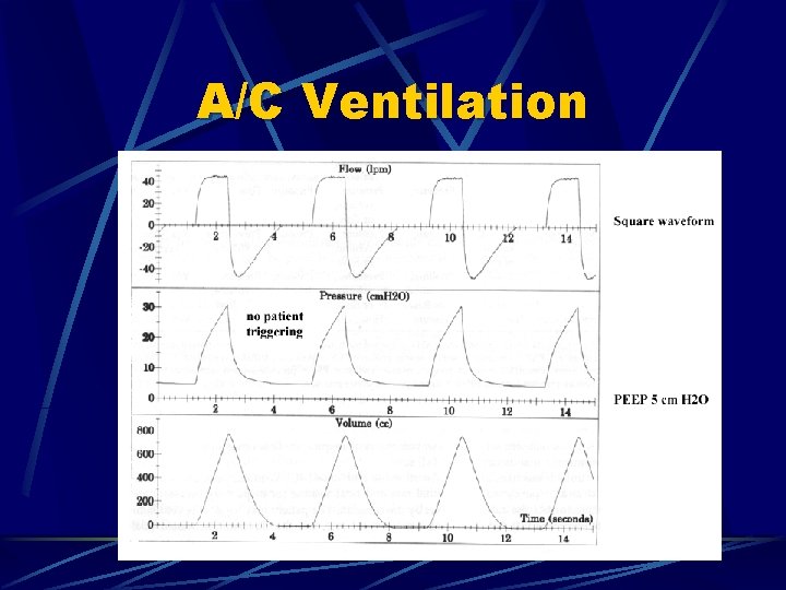
Assist Control Ventilation (Volume Control) In volume control ventilation (VCV) the mechanically ventilated patient receives a preset volume of gas with every breath. VCV is frequently administered in an assist control (A/C) environment l l If the patient is apneic the ventilator will deliver a preset rate If the patient is breathing they are able to initiate or trigger a greater then the preset number of breaths.
A/C Ventilation
Parameters for A/C TV Freq Fi. O 2 PEEP 6 – 8 mls/kg IBW 10 – 18 bpm. 4 –. 6 5 cm H 2 O Other parameters set in A/C are: Flow Trigger Sensitivity
Disadvantages of A/C Patients receive a mandatory breath every time they trigger the ventilator therefore hypocapnia with respiratory alkalemia may occur Uncomfortable for patient l l Receive mandatory breath every time patient triggers ventilator Patient unable to vary volume and flow Results in patient-ventilator asynchrony Heavy sedation required which delays weaning
Observations to make when using A/C Is the patient triggering the ventilator? l l If the objective of ventilation is to rest the patient, frequent triggering may require significant work by the patient component of the inspiratory work in the A/C mode can be significant (30 to 50%) When delivering a preset volume of gas, the pressure in the lungs varies with changes in lung stiffness (compliance) and airway resistance l airway pressures should be closely monitored and when they consistently exceed 35 cm H 2 O a change in ventilating strategy should generally
Case 2: Mrs. Seizure 35 yo female, former Et. OH abuse, known Et. OH seizure x’s 2, seizure free for 5 years, no meds, presents to Olds seizes in ED status epilepticus x’s 1 h, STARS RSI intubation. Arrives @ FMC ED, GCS (E=1, M=5), spontaneous respirations. What mode of MV would you choose?
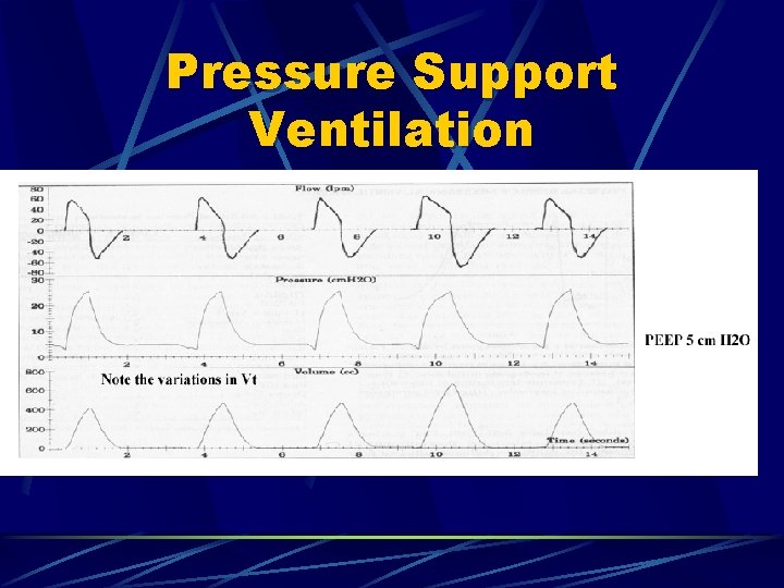
Pressure Support Ventilation In pressure support ventilation (PSV) the patient receives a preset pressure of gas with every breath The patient initiates every breath The preset pressure is rapidly achieved and maintained throughout inspiration Volume, flow and duration of inspiration are variable and change according to patient demand.
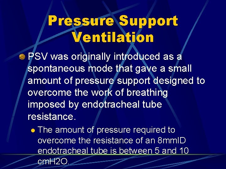
Pressure Support Ventilation PSV was originally introduced as a spontaneous mode that gave a small amount of pressure support designed to overcome the work of breathing imposed by endotracheal tube resistance. l The amount of pressure required to overcome the resistance of an 8 mm. ID endotracheal tube is between 5 and 10 cm. H 2 O.
Pressure Support Ventilation
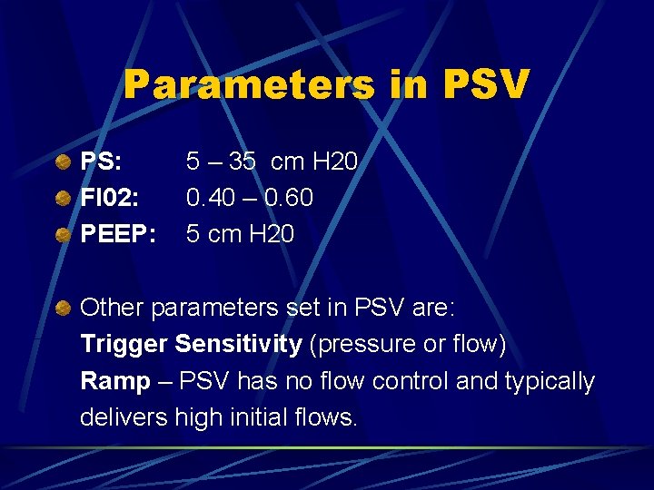
Parameters in PSV PS: FI 02: PEEP: 5 – 35 cm H 20 0. 40 – 0. 60 5 cm H 20 Other parameters set in PSV are: Trigger Sensitivity (pressure or flow) Ramp – PSV has no flow control and typically delivers high initial flows.
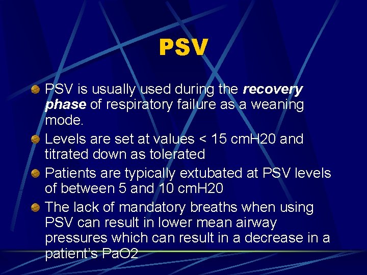
PSV is usually used during the recovery phase of respiratory failure as a weaning mode. Levels are set at values < 15 cm. H 20 and titrated down as tolerated Patients are typically extubated at PSV levels of between 5 and 10 cm. H 20 The lack of mandatory breaths when using PSV can result in lower mean airway pressures which can result in a decrease in a patient’s Pa. O 2
Disadvantages of PSV Potential for increased work of breathing at lower levels of PSV Reduction in mean airway pressure with decreased patient oxygenation
Observations Patient tidal volume should be above 250 – 350 mls (5 mls/kg) as a minimum. l When the level of PSV is inadequate the VT will drop below this range. Patient rate (f) l When low-level PSV is not being tolerated the VT falls and the f rises. Rates greater than 35 bpm are generally undesirable and signal the need for higher levels of PSV or a switch to a mandatory mode (VCV, A/C or PCV).
Case 1: The sequale… The RT from case one comes up to you annoyed and states “we need more sedation/paralysis for the motor-biker who hit his head. He is starting to fight the ventilator. ” Do you tell the nurse to give more sedation/paralytic or is there something else you can do?
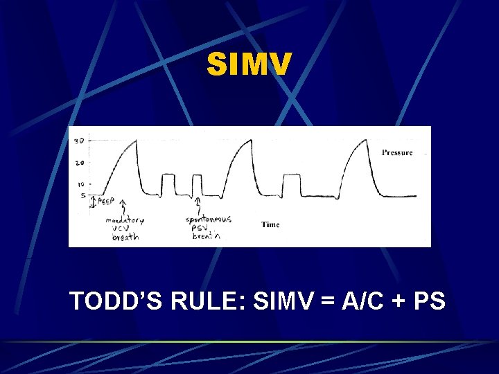
SIMV The patient gets a preset number of mandatory breaths (A/C) volume-controlled and they are synchronized with the patient’s breathing l Synchronization means that the mandatory breath will be delivered when triggered by the patient. The patient will only get the set number of mandatory breaths per minute. If the patient’s rate exceeds the set rate the additional breaths will be spontaneous breaths l Usually pressure supported
SIMV TODD’S RULE: SIMV = A/C + PS
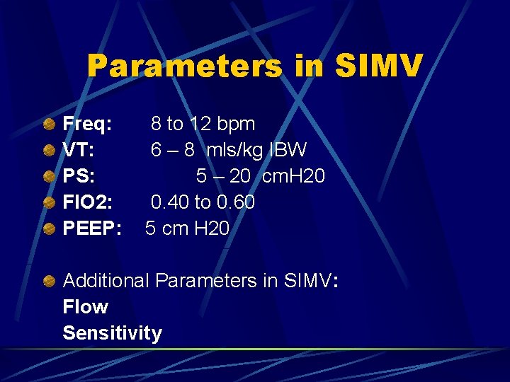
Parameters in SIMV Freq: VT: PS: FIO 2: PEEP: 8 to 12 bpm 6 – 8 mls/kg IBW 5 – 20 cm. H 20 0. 40 to 0. 60 5 cm H 20 Additional Parameters in SIMV: Flow Sensitivity
Disadvantages of SIMV Mandatory support can be set inappropriately low when SIMV is used as the vehicle for VCV or PCV.
Observations in SIMV Patients total rate Tidal volume during spontaneous breaths Airway pressures during VCV mandatory breaths Careful assessment of adequate mandatory support
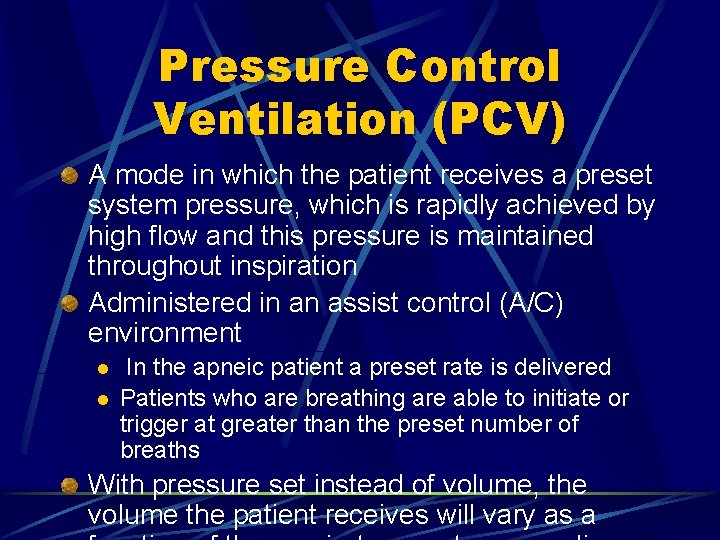
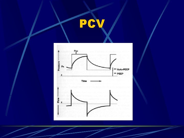
Pressure Control Ventilation (PCV) A mode in which the patient receives a preset system pressure, which is rapidly achieved by high flow and this pressure is maintained throughout inspiration Administered in an assist control (A/C) environment l l In the apneic patient a preset rate is delivered Patients who are breathing are able to initiate or trigger at greater than the preset number of breaths With pressure set instead of volume, the volume the patient receives will vary as a
PCV
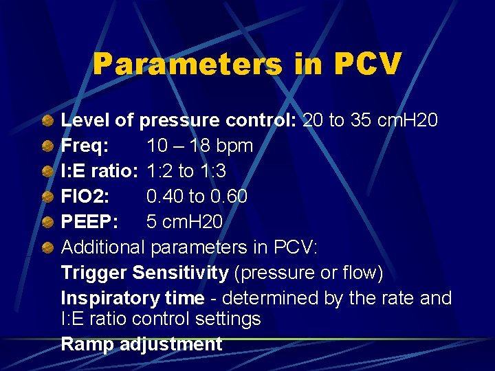
Parameters in PCV Level of pressure control: 20 to 35 cm. H 20 Freq: 10 – 18 bpm I: E ratio: 1: 2 to 1: 3 FIO 2: 0. 40 to 0. 60 PEEP: 5 cm. H 20 Additional parameters in PCV: Trigger Sensitivity (pressure or flow) Inspiratory time - determined by the rate and I: E ratio control settings Ramp adjustment
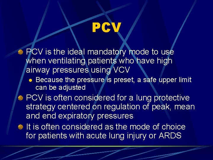
PCV is the ideal mandatory mode to use when ventilating patients who have high airway pressures using VCV l Because the pressure is preset, a safe upper limit can be adjusted PCV is often considered for a lung protective strategy centered on regulation of peak, mean and expiratory pressures It is often considered as the mode of choice for patients with acute lung injury or ARDS
Disadvantages of PCV Inability to maintain a constant tidal volume and Pa. CO 2 When compliance decreases and resistance increases, tidal volume falls and Pa. CO 2 may rise (hypercapnia) The set I: E ratios are not maintained when the patient rate exceeds set rate (when in A/C mode) resulting in shortening expiratory time which leads to increasing Auto PEEP, decreasing tidal volumes and increasing asynchrony.
Observations in PCV Careful monitoring of tidal volume Actual patient rate l When patient rate significantly exceeds set rate I: E ratios will inverse.
Potential Adverse Effects of IPPV Increased mean intrathoracic pressure Decreased venous return and cardiac output Increased ventilation/perfusion ratio Decreased renal blood flow and glomerular filtration rate with fluid retention Air trapping and intrinsic positive end-expiratory pressure (i. PEEP, auto-PEEP) Barotrauma Nosocomial infections of the lungs and sinuses Respiratory alkalosis Agitation and increased respiratory distress Increased work of breathing
Ventilator Settings Rule of 10’s VT 10 ml/kg IBW l Freq 10 bpm l Fi. O 2 100% l Fi. O 2: start high and wean down VT: Between 5 – 10 ml/kg; be aware of lung compliance
More Ventilator Settings… Respiratory Rate: wide range; increase or decrease according to ventilatory parameters Inspiratory Pressure: < 35 cm H 2 O; generally 10 – 30 Pressure Support: 5 – 30 cm H 2 O; when weaning pressures should be < 10 cm Trigger Sensitivity: amount of negative pressure that the patient must establish for the machine to sense patient effort and thus deliver a breath; generally − 1 to − 2 cm H 2 O
Even More Ventilator Settings… PEEP: the level of positive pressure that is maintained in the airways at the end of expiration; range from 5– 20 mm H 2 O I: E ratio: generally set at 1: 2; may increase to facilitate complete expiration in obstructive lung disease
PEEP Preprogrammed level of positive pressure maintained at the end of exhalation Improvement in oxygenation with increased PEEP The advantages of PEEP are balanced by its potential detrimental effects l A worsening of gas exchange is possible if an increase in dead space ventilation occurs
Auto-PEEP A phenomenon that is seen most often in patients with airflow limitation or with high respiratory rates combined with a shortened expiratory time. In this situation, expiration to functional residual capacity is not accomplished before the next inspiratory cycle begins, resulting in dynamic hyperinflation.
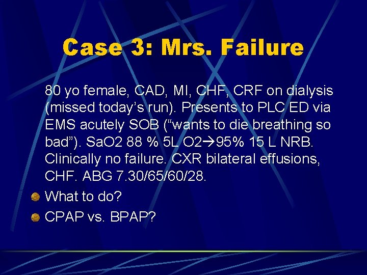
Case 3: Mrs. Failure 80 yo female, CAD, MI, CHF, CRF on dialysis (missed today’s run). Presents to PLC ED via EMS acutely SOB (“wants to die breathing so bad”). Sa. O 2 88 % 5 L O 2 95% 15 L NRB. Clinically no failure. CXR bilateral effusions, CHF. ABG 7. 30/65/60/28. What to do? CPAP vs. BPAP?
Non-Invasive Positive Pressure Ventilation (NPPV) Positive pressure is delivered by way of a tightly fitting mask In 1938 continuous positive airway pressure (CPAP) shown to be effective for the treatment of acute pulmonary edema Nasal CPAP mask for the treatment of obstructive sleep apnea in the 1980 s In the 1990’s an alternative to endotracheal intubation in acute respiratory failure
Advantages of NPPV Avoids complications that are related to endotracheal intubation and mechanical ventilation and the loss of airway defenses caused by endotracheal intubation NPPV is not as invasive as endotracheal intubation; it offers a means of temporarily supporting some patients who do not wish to have aggressive or prolonged ventilatory means used in their care.
Evidence? A case-controlled study by Girou et al found the use of NPPV in critically ill patients with acute COPD and CHF exacerbations was associated with a lower risk for pulmonary infections, lower antibiotic use, shorter length of stay, and lower mortality Girou E, Schortgen F, Delclaux C, et al. Association of noninvasive ventilation with nosocomial infections and survival in critically ill patients. JAMA 2000; 284: 2361
CPAP Delivers a constant level of positive pressure throughout the respiratory cycle Main use is for hypoxemic respiratory failure Improve oxygenation by: l increasing the mean airway pressure, increasing functional residual capacity, and opening underventilated and collapsed alveoli, enhancing gas exchange and oxygenation
CPAP Initiated between 0 – 15 cm H 2 O Level is set low initially and slowly increased to allow adequate oxygenation with as low an FIO 2 as possible Important to check the mask for leaks
BPAP Two different pressure levels are cycled between inspiration and expiration Inspiratory pressure (IPAP) > expiratory pressure (EPAP) The primary benefit of this mode over CPAP is in patients with ventilatory fatigue or failure l increases airway pressure in expiration and decreases WOB by aiding inspiration Inspiratory pressure set at 8 – 20 cm H 2 O and expiratory pressure is set at 0 – 15 cm H 2 O
Complications of NPPV Often minor: l nasal congestion, eye irritation, discomfort, and pressure necrosis caused by the tight mask fit Airway protection must be addressed in patients with altered sensorium l especially with tight fitting mouth mask Gastric distension is uncommon unless pressures exceed 20– 25 cm H 2 O
NPPV The most common reason for failure of NPPV is patient intolerance Key to success is adequate patient preparation and acclimatization to the device Choice of mask depends on familiarity and availability l l Nasal masks allow patients to communicate verbally, but they suffer from air leaks unless the patient keeps his or her mouth closed. Full-face masks offer the tightest seal but can be hazardous in patients who cannot completely protect their airway or who are at risk for vomiting
CPAP vs. BPAP In patients with hypoventilatory respiratory failure, BPAP should be the preferred mode because of its theoretic advantage in providing ventilatory assistance and therefore decreasing the work of breathing. With hypoxemic ventilatory failure, CPAP and BPAP should be similarly efficacious given that both improve oxygenation l Though BPAP has theoretic advantages over CPAP, the choice of modes should be based on familiarity with and patient tolerance of a given mode
Case 4: Mr. Puff 76 yo male, known COPD, seen at PLC ED 3 times in prior week for SOB. Dx: COPD exacerbation. Presents SOB Sa. O 2 90 % RA, 95 % 10 L O 2. Working ++ hard to breath. Initial ABG 7. 30/65/70/30 What ventilation strategy would you employ?
COPD 1 st line = consideration of NPPV Good evidence that NPPV decreases incidence of the need for intubation and invasive ventilation l l Bott J, Carroll MP, Conway JH, et al. Randomised controlled trial of nasal ventilation in acute ventilatory failure due to chronic obstructive airways disease. Lancet 1993; 341: 1555 Celikel T, Sungur M, Ceyhan B, et al. Comparison of noninvasive positive pressure ventilation with standard medical therapy in hypercapnic acute respiratory failure. Chest 1998; 114: 1636
Case 4: Mr. Puff Breath’s on… You initiate BPAP. One hour later Mr. Puff is beginning to tire despite continuous ventolin and atrovent, and 4 mg IV Mg. SO 4. You repeat the ABG: 7. 15/60/138/30 What is your next step?
COPD: Goals of IPPV Gradually correct respiratory acidosis (over hours) Normalization of lung volume Decrease auto-PEEP by: l l l higher flow rates (eg, 100 L/min) during inspiration increasing I: E ratio (1: 3 to 1: 4) occasionally disconnecting the patient from the ventilator to allowing complete exhalation use of bronchodilators and steroids adding extrinsic PEEP (no more than auto-PEEP) to decrease work required to maintain auto-PEEP
Asthmatics with impending respiratory failure demonstrate some of the most difficult management issues with regard to mechanical ventilation Asthma is predominantly a problem with expiration with increased airway resistance resulting in pulmonary hyperinflation
Asthma Mechanical ventilation can help decrease the work of breathing BUT intubation exaggerates expiratory obstruction by the fixed diameter of the endotracheal tube Potential for significant auto-PEEP and decreased venous return Therefore, all attempts should be made to avoid the initiation of mechanical ventilation in asthmatic patients except as a last resort
NPPV in Asthmatics NPPV has not been studied prospectively Meduri described the use of BPAP in 17 patients with severe asthma and impending respiratory failure. Intubation was averted in all but two patients and no complications were observed. Meduri G, Cook T, Turner R, et al. Noninvasive positive pressure ventilation in status asthmaticus. Chest 1996; 110: 767
Mechanical Ventilation and Asthma Limit the negative effects of positive pressure Allow maximal expiratory time to minimize the chances of auto-PEEP and resultant dynamic pulmonary hyperinflation When pulmonary pressures remain elevated, deep sedation and neuromuscular blockade should be considered in an effort to minimize ventilator asynchrony In severe cases controlled hypoventilation with permissive hypercapnea should be considered
Case 3: Mrs. Failure continues to fail… Despite BPAP for 45 min Mrs. Failure shows no signs of improvement. Her O 2 sat has slowly been decreasing (now 85%) on 15 L and she is starting to become obtunded. You repeat her ABG 7. 25/55/65/30 What is your next step?
Cardiogenic Pulmonary Edema A systematic review of CPAP in CHF revealed a pooled decrease in need for intubation of 26% and a trend toward decreased mortality Pang D, Keenan SP, Cook DJ, et al. The effect of positive pressure airway support on mortality and the need for intubation in cardiogenic pulmonary edema: a systematic review. Chest 1998; 114: 1185 One randomized trial compared CPAP with BPAP in a total of 27 patients and found a more rapid improvement in ventilatory parameters (Pa. CO 2, p. H) and vital signs with BPAP than CPAP in acute pulmonary edema Mehta S, Jay GD, Woolard RH, et al. Randomized, prospective trial of bilevel versus continuous positive airway pressure in acute pulmonary edema. Crit Care Med 1997; 25: 620
Mechanical Ventilation in CHF In mechanically ventilated patients PEEP can help to reduce extravascular lung water and thus can result in improved oxygenation However, PEEP can have an adverse effect in patients with CHF who are in cardiogenic shock by decreasing pulmonary venous return Compromise is to use only enough PEEP to decrease Fi. O 2 to less than 60 %
Acute Lung Injury (ALI) and Acute Respiratory Distress Syndrome (ARDS) One small study looked at the use of NPPV in 10 hemodynamically stable patients with ALI/ARDS and averted intubation in 6 of the 10 patients Rocker GM, Mackenzie MG, Williams B, et al. Noninvasive positive pressure ventilation: successful outcome in patients with acute lung injury/ARDS. Chest 1999; 115: 173 Most cases of ALI and ARDS require invasive mechanical ventilation
ALI/ARDS Indication for PCV Use small tidal volumes (6 m. L/kg) Keep plateau pressure ≤ 30 cm H 2 O. Increase PEEP and RR to help decrease tidal volumes Watch for significant amounts of auto. PEEP with the increase in respiratory rate
Physiologic Changes in Pregnancy Mother has reduced oxygen reserve (decreased FRC) and increased oxygen consumption (15 – 20 %) The fetus is very vulnerable to any reduction in oxygen delivery l 30% decrease in uterine blood flow with maternal hypoxia Higher risk of aspiration in pregnancy Pregnant patient = difficult airway Physiologic respiratory alkalosis (Pa. CO 2 of 30 mm Hg) in the last stage of pregnancy
Mechanical Ventilation in Pregnancy Rapid sequence induction is recommended for intubation Increase tidal volumes when ventilating Hyperventilate to maintain physiologic respiratory alkalosis
ED Monitoring Clinical observation Routine vital sign measurement Electrocardiographic monitoring Pulse oximetry End-tidal carbon dioxide (CO 2 ) measurement Noninvasive blood pressure (BP) measurement
Non-Invasive Blood Pressure Monitoring Use a detection system based on auscultatory, oscillometric, or Doppler principles Automatic oscillometric devices determine BP by electronically determining the pulse amplitude l l Cuff inflated at predetermined intervals to a preset level Cuff deflated sensing the amplitude of oscillations Abrupt increase in the magnitude of the oscillation is the systolic pressure. The point where there is no longer an alteration in the magnitude of the oscillation is the diastolic pressure. The mean arterial pressure (MAP) is the cuff pressure at the point of largest oscillation
Noninvasive BP Monitoring More accurate, precise, and reliable than auscultation in patients with very low or high BP Limitations common to regular BP measurement l obese arms, uncooperative moving patients, and those with very high or very low BP Cycle length of the inflation-deflation sequence in the older machines was exceedingly long and led to frequent failure; resolved in newer machines
Indications for Invasive Monitoring Exceedingly high (>250 mm Hg systolic) or low (<80 mm Hg systolic) pressures 2. Patients who are rapidly going into shock 1. l best chance to insert an arterial line may be in the ED while the arterial pulse is still palpable Anatomic indications in critically ill patients with either has no limb or no suitable limb (e. g. , too obese) to undertake conventional measurement 4. Frequent arterial sampling is required 3.
Pulse Oximetry Noninvasive and continuous means of rapidly determining arterial oxygen saturation and its changes Based on differences in the optical transmission spectrum of oxygenated and deoxygenated hemoglobin Uses Beer Lambert Law = relates the concentration of a solute to the intensity of light transmitted through a solution Light absorption is divided into a pulsatile (AC) component (arterial blood) and a nonpulsatile (venous and capillary)
Limitations of Pulse Oximetry Severe vasoconstriction (e. g. , shock, hypothermia) Excessive movement Synthetic fingernails and nail polish Severe anemia Presence of abnormal hemoglobins l Carboxy and Methemoglobin
COHgb and Met. Hgb Pulse oximeter senses COHgb as oxyhemoglobin and therefore gives an erroneously high Sa. O 2 Met. Hgb absorbs light at both red and IR wavelengths therefore the net effect is a reading of 85 %
Caution with Pulse Oximetry Does not give any information with regards to adequate ventilation
Capnography/ Capnogram The measurement and display of CO 2 concentrations on a visual display
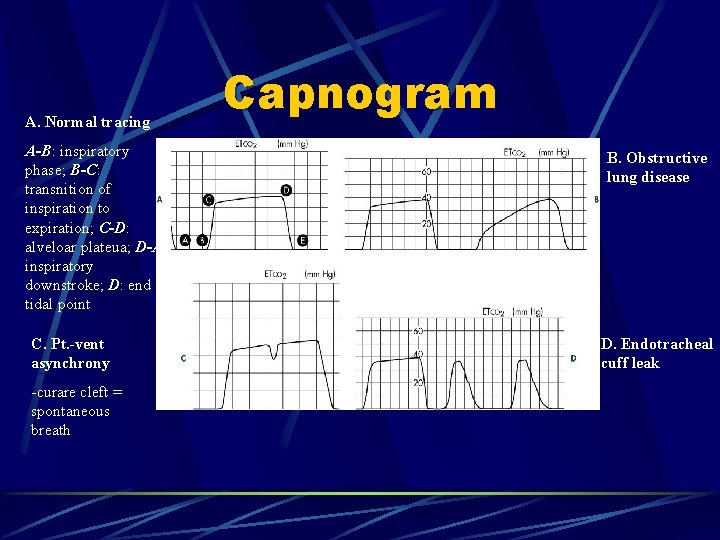
A. Normal tracing A-B: inspiratory phase; B-C: transnition of inspiration to expiration; C-D: alveloar plateua; D-A: inspiratory downstroke; D: end tidal point C. Pt. -vent asynchrony -curare cleft = spontaneous breath Capnogram B. Obstructive lung disease D. Endotracheal cuff leak
Capnometers Either sidestream (ED) or mainstream in design Sidestream capnometers aspire a sample of gas through a small catheter into a measuring chamber l l lightweight and can be used in intubated and nonintubated patients disadvantages include plugging by secretions, 2 -or 3 -second delays in response time, and air leaks, which can dilute the sample
Capnometers Colorimetric capnometers use p. H-sensitive filter paper impregnated with metacresol purple, which changes color from purple (<4 mm Hg CO 2 ) to tan (4 to 15 mm Hg CO 2 ) to yellow (>20 mm Hg CO 2 ) depending on the concentration of CO 2 l l Yellow = yes (in the right place) Purple = poor (wrong tube) The indicator is inserted between the endotracheal tube and the ventilator bag l detects changes on a breath-by-breath basis
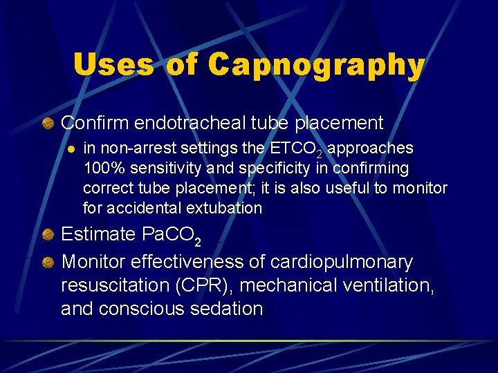
Uses of Capnography Confirm endotracheal tube placement l in non-arrest settings the ETCO 2 approaches 100% sensitivity and specificity in confirming correct tube placement; it is also useful to monitor for accidental extubation Estimate Pa. CO 2 Monitor effectiveness of cardiopulmonary resuscitation (CPR), mechanical ventilation, and conscious sedation
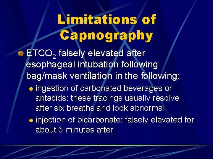
Limitations of Capnography ETCO 2 falsely elevated after esophageal intubation following bag/mask ventilation in the following: ingestion of carbonated beverages or antacids: these tracings usually resolve after six breaths and look abnormal l injection of bicarbonate: falsely elevated for about 5 minutes after l
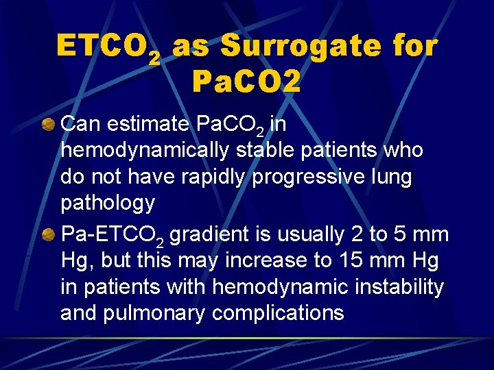
ETCO 2 as Surrogate for Pa. CO 2 Can estimate Pa. CO 2 in hemodynamically stable patients who do not have rapidly progressive lung pathology Pa-ETCO 2 gradient is usually 2 to 5 mm Hg, but this may increase to 15 mm Hg in patients with hemodynamic instability and pulmonary complications
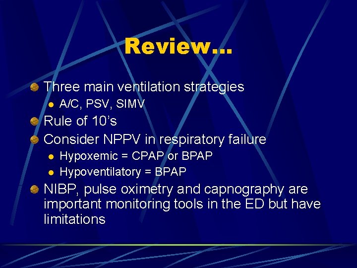
Review… Three main ventilation strategies l A/C, PSV, SIMV Rule of 10’s Consider NPPV in respiratory failure l l Hypoxemic = CPAP or BPAP Hypoventilatory = BPAP NIBP, pulse oximetry and capnography are important monitoring tools in the ED but have limitations