Dr Tabrez Inguinal Canal Introduction This is an

Dr. Tabrez

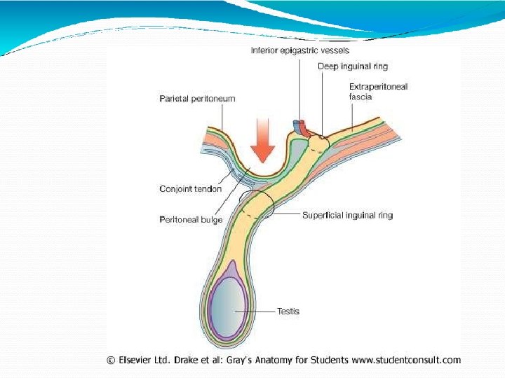
Inguinal Canal Introduction §This is an oblique intermuscular passage in the lower part of the anterior abdominal wall , Situated just above the medial half of the inguinal ligament

Inguinal Canal Location Inferior part of the anterolateral abdominal wall
 long, and is")
Length & direction • It is about 4 cm(1. 5 inches) long, and is directed downwards, forwards and medially.

• The inguinal canal extends from the deep inguinal ring to the superficial inguinal ring.

A Box? Lateral Floor Imagine the right side inguinal canal viewed from the front as a box with anterior & posterior walls, a roof & floor. The arrow indicates that structures can run through it from lateral to medial – e. g. in males it transmits the spermatic cord, and in females, the round ligament of the uterus. Medial

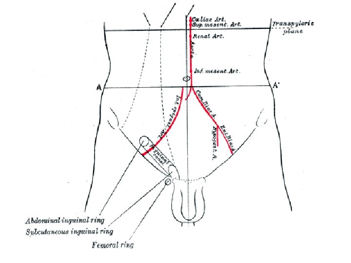
Deep inguinal ring An oval opening in the fascia transversalis situated 1. 2 cm above the midinguinal point, and immediately lateral to the stem of the inferior epigastric artery

Deep inguinal ring Inguinal canal Lateral Medial Here are the posterior wall, which has the DEEP inguinal ring situated laterally, and the floor. (Roof and anterior wall removed). Floor

Structures passing through the deep inguinal ring

Superficial inguinal ring • Is a triangular gap in the external oblique aponeurosis. • It is shaped like an obtuse angled triangle. The base of the triangle is formed by the pubic crest, the two sides of the triangle from the lateral or lower and the medial or upper margins of the opening. • It is 2. 5 cm long and 1. 2 cm broad at the base these margins are referred as crura. • At and beyond the apex of the triangle 2 crura are united by intercrural fibers

Inguinal canal Lateral Medial Superficial inguinal ring Here are the anterior wall (which has the SUPERFICIAL inguinal ring situated medially), and the roof. Floor

Structures passing through the superficial inguinal ring

BOUNDARIES OF INGUINAL CANAL THE ANTERIOR WALL 1. In its whole extent a. Skin b. Superficial fascia c. External oblique aponeurosis 2. In its lateral one-third The fleshy fibres of the internal oblique muscle.

Inguinal canal The anterior wall is made up of the external oblique muscle throughout, and is reinforced by the internal oblique m. laterally. The transversus abdominus m. lies even more laterally as part of the anterior abdominal wall. Lateral Medial Superficial inguinal ring 13

THE POSTERIOR WALL 1. In its whole extent a. The fascia transversalis b. The extra peritoneal tissue c. The parietal peritoneum. 2. In its medial two-thirds a. The conjoint tendon b. At its medial end by the reflected part of the inguinal ligament.

Posterior wall of the inguinal canal Deep inguinal ring Posterior wall Conjoint tendon medially Lateral Medial The posterior wall is formed by transversalis fascia (orange) throughout and the conjoint tendon (red) medially. The wall is particularly weak over the deep inguinal ring 15

Inguinal canal Conjoint tendon The conjoint tendon attaches to the pubic crest, reinforces the posterior canal wall medially and also forms the ROOF of the canal Lateral Medial The transversus abdominis and internal oblique mm. combine to form the CONJOINT tendon that arches over the contents of the inguinal canal Floor Spermatic cord 16

ROOF OF THE INGUINAL CANAL It is formed by the arched fibres of the internal oblique and transverse abdominis muscles.

Roof and anterior wall of the inguinal canal Superficial inguinal ring Lateral Medial The anterior wall of the canal is formed by external oblique muscle (orange) throughout and by internal oblique muscles (red/black/white) laterally. This wall is weak medially because of the “hole” in the external oblique muscle (= superficial inguinal ring). 18

FLOOR It is formed by the grooved upper surface of the inguinal ligament; and at the medial end by the lacunae ligament

Floor of the inguinal canal Lateral Medial The floor is formed by an incurving of the inguinal ligament, which is part of the external oblique muscle, forming a gutter. (Medially it forms the lacunar ligament which is not illustrated). Floor 20

SEX DIFFERENCE The inguinal canal is larger in males than in females.

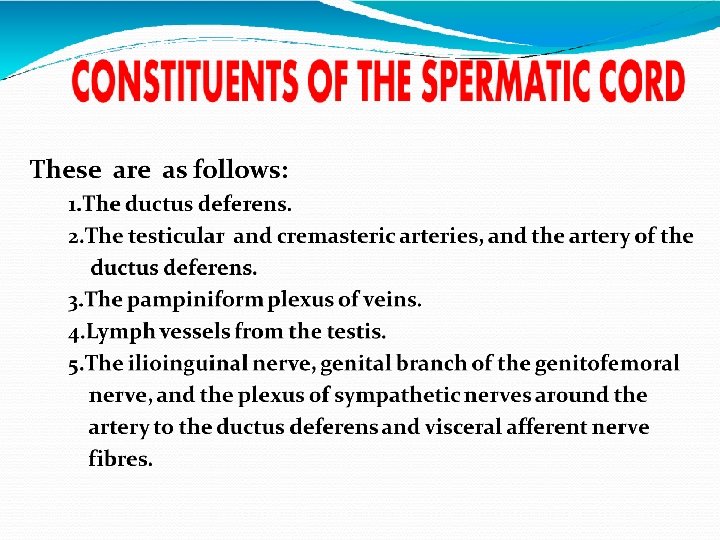
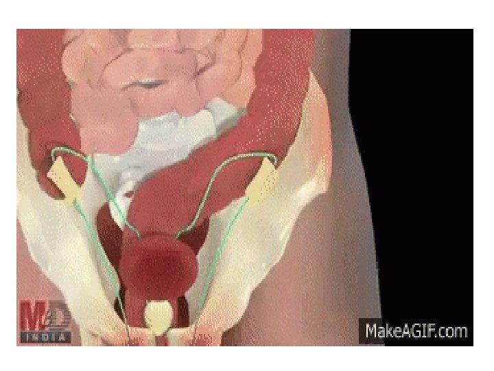
STRUCTURES PASSING THROUGH THE CANAL 1. The spermatic cord in males, or the round ligament of the uterus in females, enters the inguinal canal through the deep inguinal ring and passes out through the superficial inguinal ring. 2. The ilioinguinal nerve enters the canal through the interval between the external and internal oblique muscles and passes out through the superficial inguinal ring.

Deep inguinal ring Inguinal canal Spermatic cord enters the inguinal canal through the deep inguinal ring Medial Lateral Superficial inguinal ring Spermatic cord exits through the superficial inguinal ring Floor

Inguinal canals – why have them? �Allow contents of the scrotum to communicate with intra-abdominal contents �Prevent mobile intra-abdominal contents (e. g. intestine) from entering the scrotum and possibly becoming damaged, while at the same time permitting blood vessels, nerves, lymphatics, vas deferens etc. to supply the scrotal contents

MECHANISM OF INGUINAL CANAL The presence of the inguinal canal is the cause of weakness in the lower part of the anterior abdominal wall. This weakness is compensated by the following factors

Obliquity of the inguinal canal The two inguinal rings do not lie opposite to each other. Therefore, when the intraabdominal pressure rises the anterior and posterior walls of the canal are approximated, thus obliterating the passage. This is known as the flap valve mechanism.

The superficial inguinal ring is guarded from behind by the conjoint tendon and by the reflected part of the inguinal canal.

The deep inguinal ring is guarded from the front by the fleshy fibres of the internal oblique.

Shutter mechanism of the internal oblique This muscle has a triple relation to the inguinal canal. It forms the anterior wall, the roof, and the posterior wall of the canal. When it contracts the roof is approximated to the floor, like a shutter.

Ball valve mechanism Contraction of the cremaster helps the spermatic cord to plug the superficial inguinal ring

Slit valve mechanism Contraction of the external oblique results in approximation of the two crura of the superficial inguinal ring. The integrity of the superficial inguinal ring is greatly increased by the intercrural fibres.

Hormones may play a role in maintaining the tone of the inguinal musculature

Whenever, there is a rise in intra abdominal pressure as in coughing , sneezing, lifting heavy weights all these mechanisms come to play, so that the inguinal canal is obliterated, its openings are closed, and herniation of abdominal viscera is prevented.
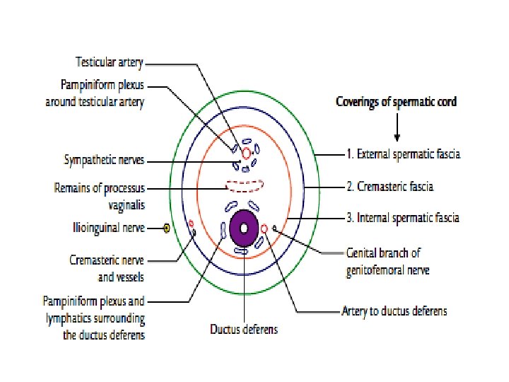

From within outwards, these are as follows: 1. The internal spermatic fascia , derived from the fascia transversalis; it covers the cord in its whole extent. 2. The cremasteric fascia is made up of the muscle loops costituting the cremaster muscle, and the intervening areolar tissue. It is derived from the internal oblique and transversus abdominis muscles.

Round ligament of uterus The round ligaments are two fibro muscular flat bands , 10 to 12 cm long, which lie between the two layers of broad ligament , begins at the lateral angle of the uterus, passes through the deep inguinal ring , traverses the inguinal canal and merges with the areolar tissue of the labium majus
 Triangle is an important structure as it is the")
HESSELBACH’S TRIANGLE � Hesselbach’s (Inguinal) Triangle is an important structure as it is the site for direct hernias. The triangle has the following borders: 1) Medial border of rectus abdominus(medially) 2) Inguinal ligament (inferiorly) 3) Inferior epigastric vessels(laterally)

A Brief Mention of Hernias are abnormal outpouchings of the abdominal contents (such as the small intestine) from the cavity in which they belong. There are two main types of hernias that occur at the inguinal region. Direct hernia and indirect hernia. .

�The posterior wall of the canal is particularly weak laterally because of the deep inguinal ring �The anterior wall opposite the deep ring is reinforced laterally by the internal oblique muscles. �A hernia (e. g. of small bowel) that comes through the deep inguinal ring will have to travel along the inguinal canal as it cannot push into the reinforced layers of muscle in the anterior wall of the canal directly opposite the deep inguinal ring

�The anterior wall of the canal is weak medially where the superficial inguinal ring is situated �The posterior wall, opposite the superficial ring, is reinforced medially by the conjoint tendon that is formed by fibres of the internal oblique and transversus abdominis muscles �Abdominal contents cannot normally force themselves through the superficial ring directly because of the reinforced posterior wall medially

Indirect or oblique hernia These are the most common inguinal hernias, in this the contents of the abdomen enter the deep inguinal ring and traverse the whole length of the inguinal canal to come out through the superficial inguinal ring
 �Cremaster muscle &")
Coverings of indirect hernias �Peritoneum �Internal spermatic fascia (from transversalis fascia) �Cremaster muscle & fascia (from transversus abdominis and internal oblique mm. ) �External spermatic fascia (from external oblique m. ) �Superficial fascia �Skin 44

Direct Hernias Direct hernias occurs lateral to the epigastric vessels. They do not protrude through any ring, but through an area of weakness in the posterior wall of the inguinal canal; this area is likely to be Hesselbach’s Triangle. The hernia is often parallel to the spermatic cord, but almost never enters the scrotum

Coverings of direct hernias �Peritoneum �Transversalis fascia �Conjoint tendon �External oblique aponeurosis �Superficial fascia �Skin 46
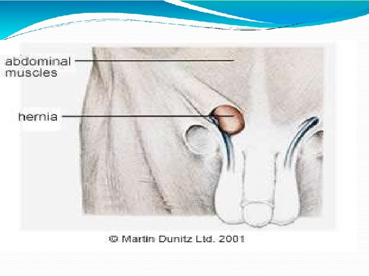

Inguinal hernia results because pressure finds weak spot at inguinal canal

- Slides: 57