Heart Structure Heart located in mediastinum within the


 Node = pacemaker")


 – ventricles contract faster than 350 bpm •")


- Slides: 66
Heart Structure
Heart – located in mediastinum within the thoracic cavity
The Pericardium – membrane surrounding heart
Serous Pericardium = 1. Parietal - outer layer 2. Visceral - inner layer (part of epicardium)
Layers of Heart Wall • Epicardium – outermost layer – synonymous with visceral portion of serous membrane • Myocardium – middle layer – Cardiac muscle – Pumping chamber • Endocardium – innermost layer – Smooth surface to reduce friction
Layers of Heart Wall
Layers of Heart Wall
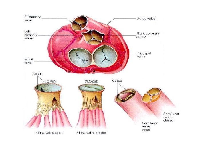
Chambers of Heart
Heart Chambers and Valves
Heart Valves
AV Valves - structure
Heart Chambers, Valves and Vessels
Heart Chambers, Valves, and Vessels
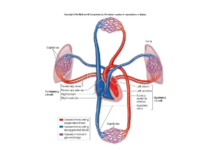
Pulmonary vs Systemic Circulation
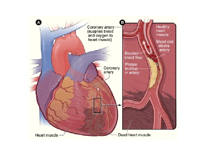
Coronary Circulation
Homeostatic Imbalance: Mycardial Infarctions
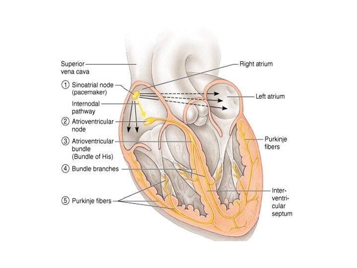
Intrinsic Conduction System of the Heart
Intrinsic Conduction System • Also called nodal system • Sinoatrial (SA) Node = pacemaker Atria Contract Atrioventricular (AV) Node • Brief Delay Atrioventricular bundle (bundle of His) Bundle branches Purkinje fibers Contraction of ventricle from bottom up Blood pushed out of ventricles
Homeostatic Imbalances • Heart block = Damage to AV node which allows ventricles to pump at own rate, which is typically slower than normal • Fibrillation = rapid uncoordinated shuddering of heart muscle, makes heart useless, major cause of death from heart attacks • Tachycardia = rapid heart rate +100 bpm • Bradycardia = slow heart rate < 60 bpm
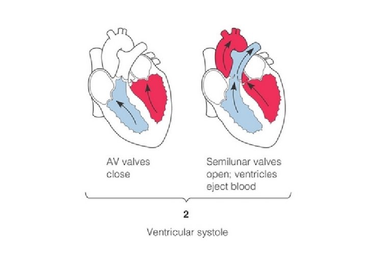
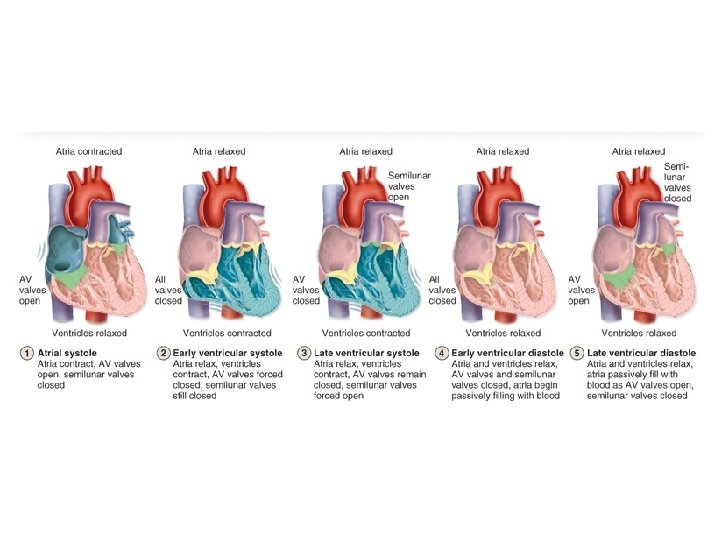
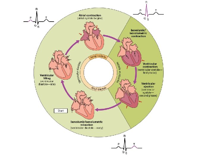
Cardiac Cycle =. 8 seconds • Sequence of Events in one heartbeat – Contraction of both atria – Contraction of both ventricles • • Systole = contraction – of ventricle Diastole = relaxation – of ventricle Lub = closing of AV valves Dub = closing of semilunar valves
Cardiac Cycle • Systole: – Ventricles contract – AV Close = “Lub” – Semi-lunar open • Diastole: – Ventricle relax – AV valve open – Semi-lunar closed= “Dub”
Cardiac Cycle
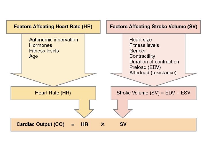
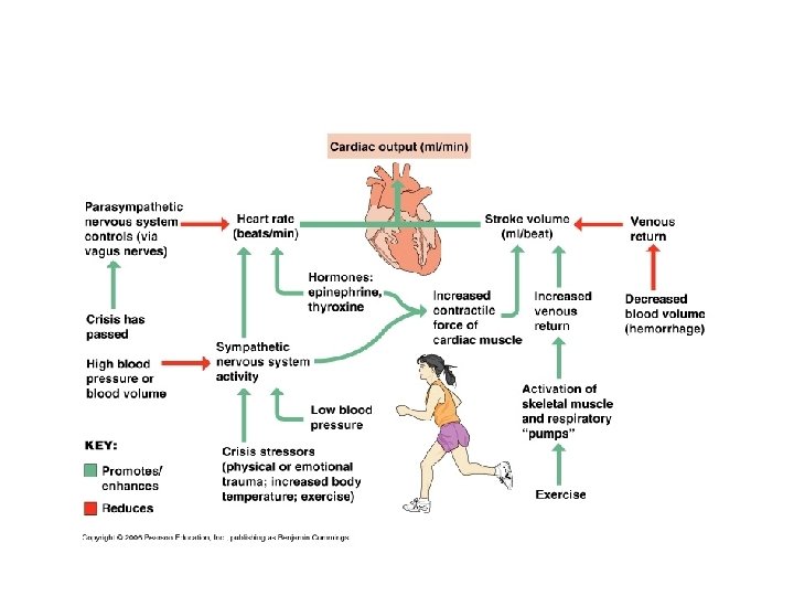
Cardiac Output • Cardiac Output = amount of blood pumped out by EACH side of the heart in 1 minute • CO = HR X SV • HR = heart rate or beats per minute • SV = stroke volume = amount of blood pumped out be each ventricle with each heartbeat • If either HR or SV varies, the other tries to compensate to keep the cardiac output stable
Factors Affecting Heartrate • Autonomic NS – most important external influence on heart – Sympathetic – increase heart rate – Parasympathetic – slow and steady heartrate (vagus nerve) • Hormones – Epinephrine and thyroxine – increase heartrate • Physical factors – Age, gender, exercise, body temperature
Factors Affecting Stroke Volume • How much cardiac muscle stretched before contraction • More stretch = more contraction • Venous return determines amount of stretch • Greater venous return = Greater SV • Increase SV – Slow Heartrate – Exercise • Decrease SV – Severe blood loss – Very Rapid heartrate
Regulation of Heart Rate = most important external influence on heart rate
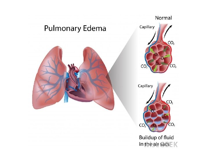
Homeostatic Imbalance: Congestive Heart Failure • When the pumping efficiency of the heart is depressed so that circulation is inadequate to meet tissue needs • Usually progressive condition that reflects weakening of the heart by atherosclerosis, persistent high blood pressure, or multiple myocardial infarctions
Congestive Heart Failure Pulmonary Congestions Peripheral Congestion • Left side failure • Right side pumps to lungs • Left side cannot pump to body • Blood pressure inside lungs increases and fluid leaks into lung tissue • Pulmonary edema • Person can suffocate • Right side failure • Blood backs up in systemic circulation • Edema in most distal parts of body
Review Questions – True of False The ventricles contract in diastole The AV valves are open during systole The SA node is the pacemaker of the heart Increased venous return will decrease stroke volume • Parasympathetic NS keeps heartrate slow and steady • •
Electrocardiogram • ECG or EKG – electrical activity of the heart – depolarize–contract – repolarize–relax
Electrocardiogram = ECG • P wave – Depolarization of atria – Atria contract • QRS complex – Depolarization of ventricles – Repolarization of atria – Ventricles contract (atria relax) • T wave – Repolarization of ventricles – Ventricles relax
Normal ECG
Cardiac Arrhythmias • bradycardia – slow heart beat • tachycardia – fast heart beat • premature atrial contraction (PACs) – atria contracts before SA node
Cardiac Arrhythmias • atrial fibrillation – atria contract faster than 350 bpm • premature ventricular contractions (PVCs) – ventricles contract too soon • ventricular tachycardia (VT) – ventricles, rather than SA node, cause beat
Cardiac Arrhythmias • ventricular fibrillation (VF) – ventricles contract faster than 350 bpm • heart block – impulse from SA node to AV node • first–impulse delayed • second–intermittently blocked • third–completely blocked
Blood Vessels and Circulation • • • blood vessels: the transport network circulation: moving blood around the body taking vital signs know your numbers Closed system – blood always in a vessel or heart
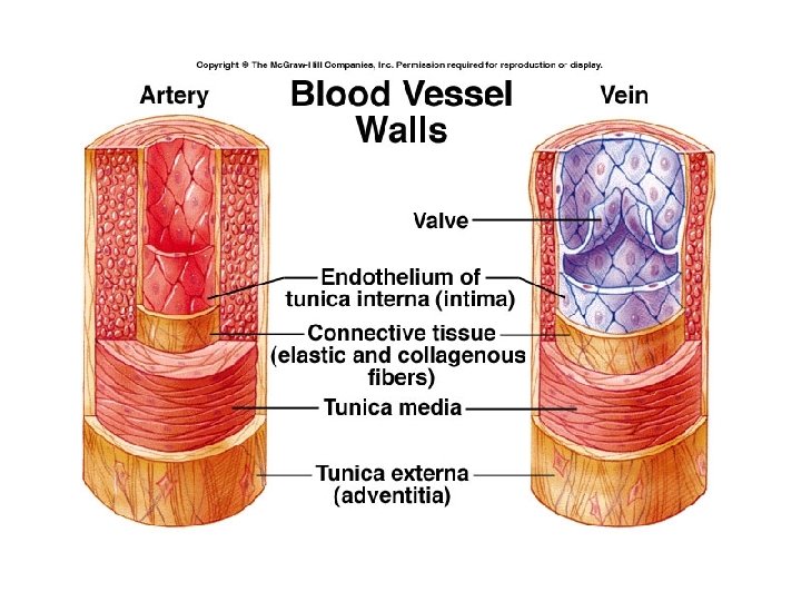
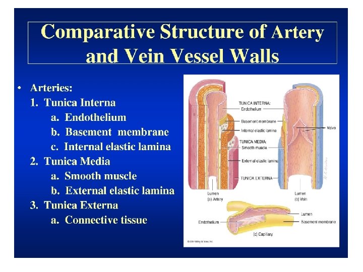
The Three Layers of Blood Vessels • tunica intima – innermost layer – Epithelial tissue • tunica media – middle layer – Smooth muscle • tunica externa – outermost layer – Connective tissue
Blood Pathway Heart Arteries Arterioles Capillaries Venules Veins Heart
Arteries • Carry blood AWAY from heart • Blood usually oxygenated – exception is pulmonary artery • Thick walls, narrow lumen • Walls elastic – expand contract with pulse • Arterioles – smaller arteries – Dilate and constrict to alter blood flow – Ex. Muscles dilate when running • Two organs blood supply never changes = brain and kidneys
Capillaries Smallest of all blood vessels Only one cell thick Narrow – blood cells flow single file Very large surface area for gas, nutrient, and waste exchange • Blood moves slower through here to allow time for exchanges • •
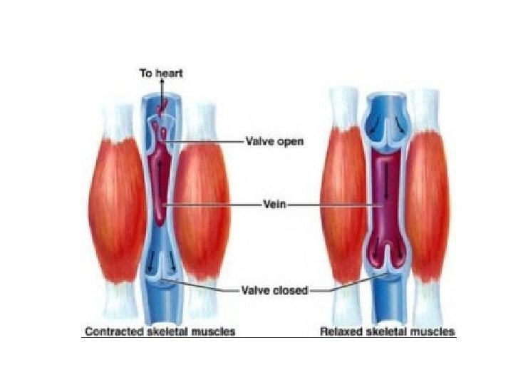
Veins • Carries deoxygenated back to the heart – Exception: Pulmonary vein • Thinner and less elastic, more flexible • Larger veins contain valves – Prevent backflow • Skeletal muscles and breathing help return blood to heart • When inhale, drop in pressure occurs in thorax causing large veins near heart to expand fill
Blood Vessels: The Transport Network • structure and function of vessels
Factors Influencing Venous Return • Lumen of veins larger than arteries • Large veins have valves that prevent backflow • Skeletal muscles help “milk” blood through veins towards heart • When inhale, drop in pressure occurs in thorax causing large veins near heart to expand fill
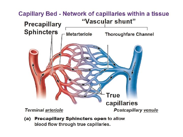
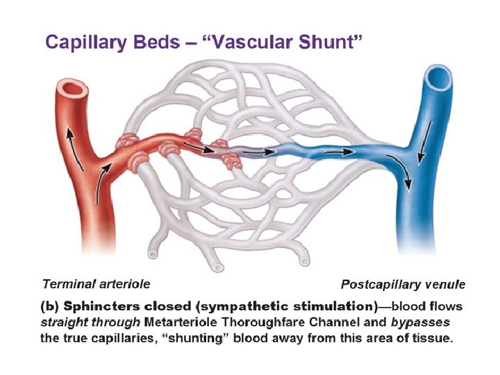
Anatomy of Capillary Bed • Two types of vessels • A. vascular shunt – vessel that directly connect the arteriole and venule at opposite ends of the bed • B. true capillaries – actual exchange vessels – 100 per capillary bed – Precapillary sphincter surrounds each to act as cut -off valve and regulate blood flow
Capillary Exchange • Substances tend to move into and out of body cells according to their concentration gradients. • Oxygen and nutrients: blood tissue cells • CO 2 and waster: tissue cells blood
How Solutes Move In and Out • 1. Diffusion – lipid soluble • 2. Endocytosis and Exocytosis – lipid insoluble • 3. Intercellular clefts – special diffusion through gaps between cell membranes of adjoining cells • 4. Fenestrated capillaries – special capillaries filled with pores. Found in areas where absorption and filtration important
Fluid Movement - due to differences in pressure • Blood pressure highest - arterial end – Fluid moves out of capillaries • Osmotic pressure highest - venule end – Fluid moves into capillaries
Homeostatic Imbalance – Varicose Veins
Thrombophlebitis • Complication of varicose veins • Inflammation of a vein that results when a clot forms in vein with poor circulation • Common Consequence: clot detachment and pulmonary embolism
Special Circulations • Arterial Supply of Brain and Circle of Willis – – Supplied by 2 sets of arteries – These blood supplies are united by special arteries – Result is complete circle of connecting blood vessels called the circle of Willis – Protects the brain because it provides more than one route for blood to reach the brain
Special Circulations • Hepatic Portal Circulation – Veins drain the digestive organs and deliver blood to liver – Liver processes nutrients before make it to systemic circulation • Fetal Circulation – All nutrient, excretory, and gas exchange occur through placenta – Umbilical veins and arteries