PLEURA LUNG By Prof Saeed Abuel Makarem Pleura

PLEURA & LUNG By Prof. Saeed Abuel Makarem

Pleura ﺍﻟﻐﺸﺎﺀ ﺍﻟﺮﺋﻮﻱ • Double-layered serous membrane enclosing the lung. • Has two layers: – Parietal layer, which lines the thoracic walls. – Visceral layer, which covers the surfaces of the lung. • The two layers continue with each other around the root of the lung ﺍﻧﻈﺮ ﺍﻟﺼﻮﺭﻩ , where it forms a loose cuff hanging down called the pulmonary ligament. • The space between the two layers, the pleural cavity, contains a thin film ﺩﻗﻴﻖ ﺟﺪﺍ of pleural fluid ( 5 -10 ml. ). Root of the lung: ﻣﻨﻄﻘﻪ ﺩﺧﻮﻝ ﺍﻟﺸﺮﺍﻳﻴﻦ ﻭﺍﻻﻭﻋﻴﻪ ﻭﺍﻟﻘﺼﺒﺎﺕ ﻟﻠﺮﺋﻪ ﻭﻓﻲ ﻫﺬﻩ ﺍﻟﻤﻨﻄﻘﻪ ﻳﺘﻢ ﺍﻟﺘﺤﻢ ﻃﺒﻘﺘﻲ ﺍﻟﻐﺸﺎﺀ ﺍﻟﺮﺋﻮﻱ ﺑﻄﺒﻘﻪ ﻭﺍﺣﺪﻩ Pulmonary ligament

Parietal Pleura • Divided according to the region in which it lies and the surfaces it covers, into: 1 - Cervical ﻋﻨﻘﻲ 2 - Costal 3 - Mediastinal 4 - Diaphragmatic ﺗﺸﺮﺡ ﻣﻔﺼﻼ ﺑﺎﻟﺸﺮﺍﺋﺢ ﺍﻟﻘﺎﺩﻣﻪ

Parietal Pleura • Cervical Pleura: Pleura • Projects up ﺗﺒﺮﺯ into the neck about 1 -1. 5 inches above the medial 1/3 rd of clavicle. • It lines the under surface of the suprapleural membrane ﻏﺸﺎﺀ ﻓﻮﻕ ﺍﻟﻐﺸﺎﺀ ﺍﻟﺮﺋﻮﻱ. • Costal pleura: • lines, the back of the: • 1 - sternum, • 2 - Ribs & costal cartilages, • 3 - Intercostal spaces & • 4 - Sides of vertebral bodies

Parietal Pleura • Mediastinal pleura: pleura covers the mediastinum ﺍﻧﻈﺮ ﺍﻟﺼﻮﺭﻩ. • At the hilum ﻣﻨﻄﻘﻪ ﺩﺧﻮﻝ ( ﺍﻻﻭﻋﻴﻪ ) ﺳﺒﻖ ﺷﺮﺣﻬﺎ , it is reflected on to the vessels and bronchi, to become continuous with the visceral pleura • Diaphragmatic pleura: covers the thoracic (upper) surface of the diaphragm

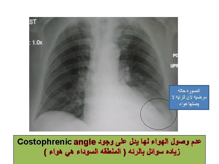
Pleural Recesses ﺗﺠﻮﻳﻔﺎﺕ : • 1 - Costodiaphragmatic: • Slit ﺗﺠﻮﻳﻒ ﺩﻗﻴﻖ ﻋﻤﻴﻖ like space between costal and diaphragmatic pleurae. The inferior border ﻳﺎﺗﻲ ﻻﺣﻘﺎ of the lung descends ﻳﻨﺰﻝ into it during deep inspiration • 2 - Costomediastinal: • Slit like space between costal and mediastinal pleurae, is filled by the anterior border of the lung during deep inspiration

Pleura: Nerve Supply • Parietal pleura: • is sensitive to pain, temperature, touch & pressure and is supplied as follows: follows v Costal pleura segmentally supplied by the intercostal nerves. v Mediastinal pleura supplied by phrenic nerves. v Diaphragmatic pleura supplied over the domes by phrenic nerves, around the periphery by lower six intercostal nerves. Visceral pleura sensitive to • stretch only and is supplied by the autonomic fibers from the pulmonary plexus

Lungs

Lungs • Located in the thoracic cavity, one on each side of the mediastinum • Each lung is: Ø Conical in shape. Ø Covered by the visceral pleura. Ø Suspended ﻣﻌﻠﻘﻪ free in its own pleural cavity. Ø Attached to the mediastinum only by its root.

Borders ﻣﻌﻨﺎﻫﺎ ﺣﺪﻭﺩ ﺍﻟﺮﺋﻪ ﻳﻌﻨﻲ ﺍﻃﺮﺍﻓﻬﺎ • Each lung has: v 1 - A thin anterior ﺍﻣﺎﻣﻲ border that overlaps the heart. v The left lung shows cardiac notch along this border. v 2 - A thick posterior ﺧﻠﻔﻲ border that lies beside the vertebral column. v 3 - A thin inferior ﺳﻔﻠﻲ border, that is related to diaphragm cardiac notch

Surfaces ﺳﻄﻮﺡ ﺍﻟﺮﺋﻪ ﻳﻌﻨﻲ ﺍﻻﺟﺰﺍﺀ ﺍﻟﺒﺎﻗﻴﻪ ﻏﻴﺮ ﺍﻟﺤﺪﻭﺩ Each lung has: • An apex ﻗﻤﻪ , ﻗﻤﻪ which projects upward into the neck for about 1 inch ﺗﺬﻛﺮ ﺍﻟﻐﻼﻑ ﻳﺘﻤﺪ ﻻﻧﺶ ﻭﻧﺼﻒ above the clavicle. • A concave base, base which rests on the diaphragm. • A convex costal surface, which corresponds to the concave chest wall.
 ﻣﺜﻞ ﺍﻟﺼﻮﺭﻩ ﻭﺍﻟﻠﻲ")
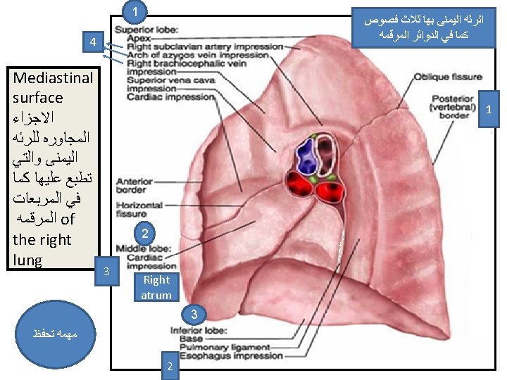
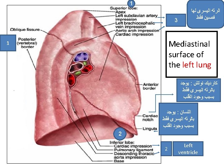
• A mediastinal surface, which is molded ﺗﻄﺒﻊ ﻋﻠﻴﻬﺎ ) ﻣﺜﻞ ﺍﻟﺼﻮﺭﻩ ﻭﺍﻟﻠﻲ ( ﻭﺭﻭﻧﺎ ﺑﺎﻟﻌﻤﻠﻲ to the mediastinal structures. • At bout ﻧﻮﺑﻪ - ﻣﺮﺽ the middle of this surface is a depression ﺗﻨﻀﻐﻂ , the hilum, where the structures enter the lung: • (bronchi, bronchial & pulmonary arteries) or leave (bronchial & pulmonary veins, nerves & lymphatics)

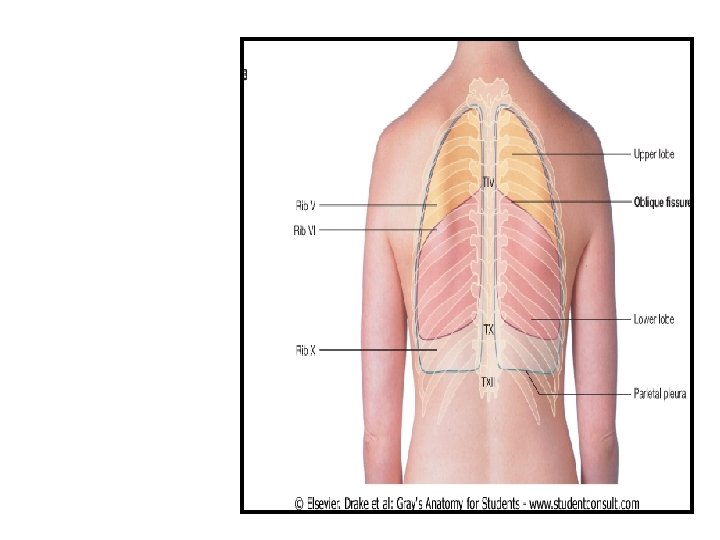
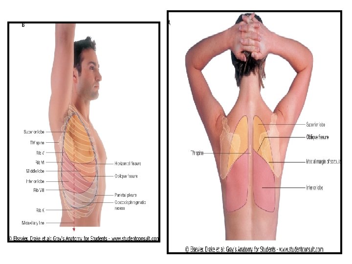
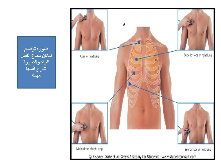
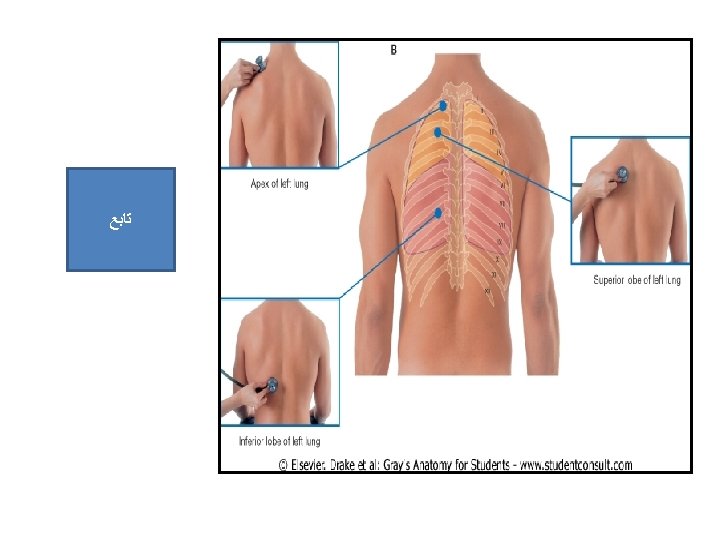
Fissures & ﺍﻟﺨﻂ ﺍﻟﻔﺎﺻﻞ Lobes ﺍﻟﻔﺼﻮﺹ • Right Lung: • Divided by two fissures, the oblique & horizontal, into: • 1 - Superior, • 2 -Middle and • 3 - Inferior • Left lung: • Divided by only one oblique fissure into: • 1 - Superior and • 2 - Inferior

Fissures • Oblique fissure: • Runs from the inferior border upward and backward across the medial and costal surfaces until cuts the posterior border about 2½ inches below the apex • Horizontal fissure: runs horizontally across the costal surface at the level of right 4 th costal cartilage to meet the oblique fissure in the midaxillary ﺍﻟﺨﻂ ﺍﻟﻔﺎﺻﻞ ﺍﻻﺑﻄﻲ line

Blood Supply ﻟﻠﺮﺋﻪ • Arterial supply: • By bronchial arteries, branches of the descending thoracic aorta. • Venous Drainage ﺍﻻﻭﺭﺩﻩ : • By bronchial veins, which drain into azygos & hemiazygos veins.

Nerve Supply • Through pulmonary plexuses located at the root of each lung, and composed of: – Sympathetic fibers from the sympathetic trunk – Parasympathetic fibers from the vagus nerve

Root of the Lung • Formed by the structures entering or leaving the lung: bronchi, pulmonary vessels, lymphatics, bronchial vessels and nerves. • Surrounded by a tubular sheath of pleura which hangs down to form pulmonary ligament. • The pulmonary ligament provides a potential space for the movement of pulmonary vessels and large bronchi


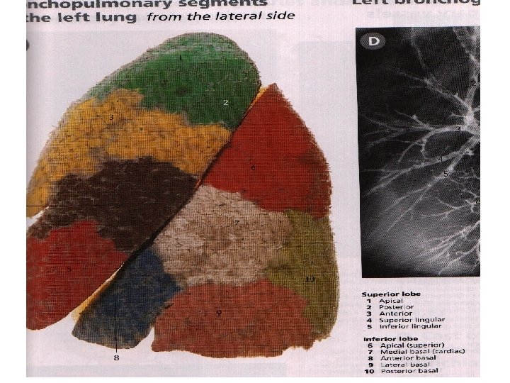
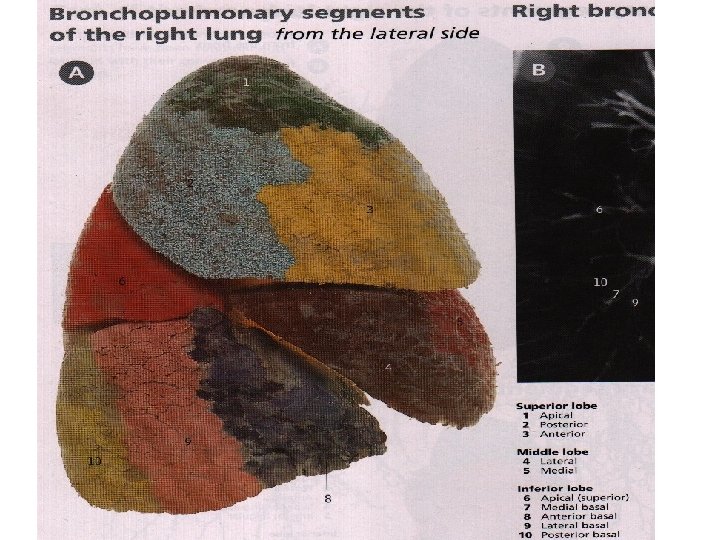
Bronchopulmonary segments: ﻋﻨﺪﻣﺎ ﺗﺪﺧﻞ ﺍﻟﺸﻌﺐ ﻟﻠﺮﺋﻪ ﺗﺘﻔﺮﻉ ﺣﺘﻰ ﺗﺮﺗﺒﻂ ﺑﺎﻟﺮﺋﻪ ﻋﻨﺪﻣﺎ ﺗﺮﺗﺒﻂ ﺑﺎﻟﺮﺋﻪ ﺗﻜﻮﻥ ﻫﺬﻩ ﺍﻟﺴﻴﻘﻤﻴﻨﺖ ﻓﺎﺋﺪﺗﻬﺎ ﺍﻥ ﻟﻜﻞ ﺳﻴﻘﻤﻨﺖ ﺷﺮﻳﺎﻥ ﻭﻻ ﺗﺆﺜﺮ ﻋﻠﻰ ﻋﻤﻞ ﺍﻟﺮﺋﻪ ﻭﻋﺼﺐ ﺧﺎﺹ ﺑﻪ ﻓﻌﻨﺪ ﺣﺪﻭﺙ ﻣﺸﻜﻠﻪ ﺑﻪ ﻳﻤﻜﻦ ﺍﺯﺍﻟﺘﻬﺎ • Definition: Are the smallest anatomic, surgical, and functional units of the lung. Each segment is pyramidal in shape with its apex directed medially toward the lung root, and its base toward the lung surface. Each segment receives segmental bronchus, branch of pulmonary artery, its own lymphatic vessels, and autonomic nerve. The branch of pulmonary vein lie in the connective tissue between the segment.

● ● ● Pleural effusion Pleuritis, pleural rub, pleural adhesions Pneumothorax Empyema ﻗﺎﻝ ﺍﻟﺪﻛﺘﻮﺭ ﺑﺎﺷﺮﺣﻬﺎ ﺍﻟﺪﺭﺱ ﺍﻟﺠﺎﻱ Clinical Notes

- Slides: 30