Coronary Artery Disease Introduction Coronary heart disease CHD


- Slides: 120

Coronary Artery Disease

Introduction • Coronary heart disease (CHD) is the most common form of heart disease • Approximately 62 million people in India have CAD, with 23 million of these below 40 years of age

Risk factors for cardiovascular disease

Risk factors definition • Concept of “risk factors” for CAD evolved from prospective epidemiological studies in US and Europe • Consistent association among characteristics observed at one point of time in apparently healthy individuals and subsequent incidence of CAD in these patients. • But, presence of a risk factor does not necessarily imply a direct causal relationship.

ATP III classifies Risk factors for CVD into three categories: • Underlying • Major (traditional) • Emerging

Underlying risk factors include: • Obesity • Disinclination to exercise • Atherogenic diet

Major (traditional risk factors): -Age -Male gender -Dyslipidemia High LDL cholesterol Low HDL cholesterol -DM -HTN -Smoking -Family history of premature CAD in first degree relative

Emerging risk factors: -Metabolic syndrome -Triglyceride -Lp(a) -Lp-PLA 2 -Fibrinogen -Homocysteine -Urine microalbuminuria/creatinine ratio -Hs CRP -Impaired fasting glucose (100 -125 mg/dl per ADA) -Markers of subclinical ASCVD ABI Exercise testing EBCT/MRI Carotid IMT

Dyslipidemia Better term than hyperlipidemia as it includes the risk of having low HDL Serum total cholesterol (TC) is a composite of: LDL cholesterol- directly related to CVD HDL cholesterol- inversely related to CVD VLDL cholesterol- related to CVD in patients with DM and low HDL Best single predictor for CVD risk is TC/HDL ratio. Ideal ratio is <3, intermediate 3 -5, high risk >5 This ratio is also the best predictor of treatment benefits


Hypertension • Potent risk factor for all CVD and dominant risk factor for stroke • Graded relationship between level of BP and outcomes. • SBP rises with age, whereas DBP plateaus in the late middle life and decreases somewhat then. • Trials for isolated systolic hypertension have shown benefits for both stroke and CHD

• Systolic and diastolic hypertension increase the RR for CVD by 1. 6 times • For combined Systolic and diastolic HTN the RR is 2. 0 • The risk for CVD is increased even in individuals with “high normal BP” (130 -39/85 -89 mm Hg)

Smoking • This habit increases the risk of vascular outcomes by 2 fold. • Both, regular and filter cigarettes have same adverse effects. • Low tar/low nicotine products have not been shown to reducethe risk • Unlike other modifiable risk factors, cigarette smoking can be eliminated entirely • Benefits of quitting smoking are dramatic. Risk in ex-smokers falls to near non-smoking levels in 2 yrs.

Obesity • It contributes independently to CVD risk and also aggravates known CVD risk factors. • Measures of obesity include: -BMI -Waist: hip ratio.


Synergy of risk factors: • The CHD death risk in men who smoke, have DBP>90 mm Hg, TC>250 mg/dl, the actual risk is 82/1000 v/s 43/1000 if all the three risk factors are added • Thus there is multiplicative effect of multiple risk factors acting in concert. • Also control of one risk factor provides substantial benefit in persons with multiple risk factors


Diabetes Mellitus • Patients with either type I or type II diabetes have increased risk for CVD • Risk of CHD is increased 2 -fold in young men and 3 -fold in young women with type 2 diabetes • Type II diabetics have one or more metabolic abnormalities (hypertriglyceridemia, low HDL, hypertension) • They may also have normal LDL levels but LDL particles are dense and small thus being more atherogenic

(Circulation 1998; 97: 1837)

Metabolic syndrome: 1. Abdominal obesity: waist circumference -Men >40 inches; Women >35 inches 2. Triglycerides >150 mg/dl 3. HDL: Men <40 mg/dl; Women <50 mg/dl 4. BP >130/85 mm Hg 5. Fasting glucose >100 mg/dl (presence of 3 or more criteria constitutes metabolic syndrome)

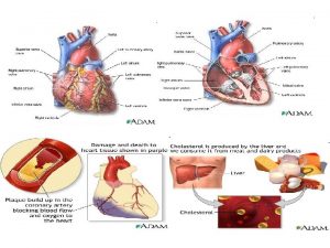
Pathophysiology • Disease of the coronary arteries is almost always due to atheroma and its complications • particularly thrombosis

Myocardial Ischemia • Results when there is an imbalance between myocardial oxygen supply and demand • Most occurs because of atherosclerotic plaque with in one or more coronary arteries • Limits normal rise in coronary blood flow in response to increase in myocardial oxygen demand

Oxygen Carrying Capacity • The oxygen carrying capacity relates to the content of hemoglobin and systemic oxygenation • When atherosclerotic disease is present, the artery lumen is narrowed • Coronary blood flow cannot increase in the face of increased demands and ischemia may result


Ischaemic Heart Disease § Angina § Stable § Unstable § Prinzmetal’s § Myocardial Infarction § NSTEMI § STEMI

Clinic pathological correlation Clinical Problem Pathology Stable angina Ischaemia due to fixed atheromatous stenosis of one or more coronary arteries Unstable angina Ischaemia caused by dynamic obstruction of a coronary artery due to plaque rupture with superimposed thrombosis and spasm Myocardial infarction Myocardial necrosis caused by acute occlusion of a coronary artery due to plaque rupture and thrombosis Heart failure Myocardial dysfunction due to infarction or ischaemia Arrhythmia Altered conduction due to ischaemia or infarction Sudden death Ventricular arrhythmia, asystole or massive myocardial infarction


Evaluation of Chest pain

Acute Coronary Syndromes Stable Angina Acute Coronary Syndromes Unstable Angina STEMI NSTEMI Rest pain Relievers Responds to GTN No GTN effect Enzymes Normal Elevated Often normal Often ST depression ST segment elevation No ST segment elevation Character of pain Exertional pain ECG

Angina • When ischemia results it is frequently accompanied by chest discomfort: Angina Pectoris • In the majority of patients with angina, development of myocardial ischemia results from a combination of fixed and vasospastic stenosis

Chronic Stable Angina • May develop sudden increase in frequency and duration of ischemic episodes occurring at lower workloads than previously or even at rest • Known as unstable angina: up to 70% patients sustain MI over the ensuing 3 months

Angina: cont • Patients with mild obstruction coronary lesions can also experience unstable angina • >90% of Acute MI result from an acute thrombus obstructing a coronary artery with resultant prolonged ischemia and tissue necrosis

Quality • Tightness, squeezing, heaviness, pressure, burning, indigestion or aching sensation • It is rarely “PAIN” • Never: sharp, stabbing, prickly, spasmodic, or pleuritic • Lasts a few seconds < 10 minutes • Relieved by NTG s/l • Levine Sign: clench fist to sternum

Signs & Symptoms accompany Angina • Dyspnea, nausea, diaphoresis resolve quickly after cessation of angina • Angina is a diffuse sensation rather than discrete

Ischemic Heart Disease • Imbalance between Myocardial oxygen supply and demand = Myocardial hypoxia and accumulation of waste metabolites due to atherosclerotic disease of coronary arteries

Stable Angina • Stable Angina: chronic pattern of transient angina pectoris precipitated by physical activity or emotional upset, relieved by rest with in few minutes • Temporary depression of ST segment with no permanent myocardial damage

Angina Pectoris • Angina Pectoris: uncomfortable sensation in the chest or neighboring anatomic structures produced by myocardial ischemia

Variant Angina • Typical anginal discomfort usually at rest • Develops due to coronary artery spasm rather than increase myocardial oxygen demand • Transient shifts of ST segment – ST elevation

Unstable Angina • Increased frequency and duration of Angina episodes, produced by less exertion or at rest = high frequency of myocardial infarction if not treated

Silent Ischemia • Asymptomatic episodes of myocardial ischemia • Detected by electrocardiogram and laboratory studies

Myocardial Infarction • Region of myocardial necrosis due to prolonged cessation of blood supply • Results from acute thrombus at side of coronary atherosclerotic stenosis • May be first clinical manifestation of ischemic heart disease or history of Angina Pectoris

Precipitants • Exertion: walking, climbing stairs, vigorous work using arms, sexual activity • Vasoconstriction: extremities, increased systemic vascular resistance, increased in myocardial wall tension and oxygen requirements • Myocardial Ischemia displays a circadian rhythm threshold for Angina it is lower in morning hours.

Physical Examination • Arcus senilis, xanthomas, funduscopic exam: AV nicking, exudates • Signs and symptoms: hyperthyroidism with increased myocardial oxygen demand, hypertension, palpitations • Auscultate carotid and peripheral arteries and abdomen: aortic aneurysm • Cardiac: S 4 common in CAD, increased heart rate, increased blood pressure

Physical Examination • Myocardial ischemia may result in papillary muscle regurgitation • Ischemic induced left ventricular wall motion abnormalities may be detected as an abnormal precordial bulge on chest palpation • A transient S 3 gallop and pulmonary rales = ischemic induced left ventricular dysfunction

Diagnostic Tests • Blood tests include serum lipids, fasting blood sugar, Hematocrit, thyroid (anemias and hyperthyroidism can exacerbate myocardial ischemia • Resting Electrocardiogram: CAD patients have normal baseline ECGs • pathologic Q waves = previous infarction • minor ST and T waves abnormalities not specific for CAD

Electrocardiogram • Electrocardiogram: is useful in diagnosis during cc: chest pain • When ischemia results in transient horizontal or downsloping ST segments or T wave inversions which normalize after pain resolution • ST elevation suggest severe transmural ischemia or coronary artery spasm which is less often

Exercise Stress Test • Used to confirm diagnosis of angina • Terminate if hypotension, high grade ventricular disrrhythmias, 3 mm ST segment depression develop • (+): reproduction of chest pain, ST depression • Severe: chest pain, ST changes in 1 st 3 minutes, >3 mm ST depression, persistent > 5 minutes after exercise stopped • Low systolic BP, multifocal ventricular ectopy or V- tach, ST changes, poor duration of exercise (<2 minutes) due to cardiopulmonary limitations

Other Diagnostic Tests • Radionuclide studies • Myocardial perfusion scintigraphy • Exercise radionuclide ventriculography • Echocardiography • Ambulatory ECG monitoring • Coronary arteriography

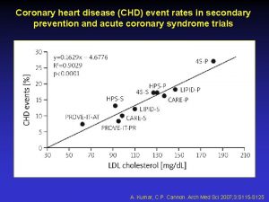
Management Goals to reduce Anginal Symptoms • Prevent complications – myocardial infarction, and to prolong life • No smoking, lower weight, control hypertension and diabetes • Patients with CAD – LDL cholesterol should achieve lower levels (<100) • HMG-COA reductase inhibitors are effective


Pharmacologic Therapy • Therapy is aimed in restoring balance between myocardial oxygen supply and demand • Useful Agents: nitrates, beta-blockers and calcium channel blockers

Nitrates • Reduce myocardial oxygen demand • Relax vascular smooth muscle • Reduces venous return to heart • Arteriolar dilators decrease resistance against- which left ventricle contracts and reduces wall tension and oxygen demand

Nitrates: cont • Dilate coronary arteries with augmentation of coronary blood flow • Side effects: generalized warmth, transient throbbing headache, or lightheadedness, hypotension • ER if no relief after X 2 nitro's: unstable angina or MI

Problems with Nitrates • Drug tolerance • Continued administration of drug will decrease effectiveness • Prevented by allowing 8 – 10 hours nitrate free interval each day. • Elderly/inactive patients: long acting nitrates for chronic antianginal therapy is recommended • Physical active patients: additional drugs are required

Beta Blockers • Prevent effort induced angina • Decrease mortality after myocardial infarction • Reduce Myocardial oxygen demand by slowing heart rate, force of ventricular contraction and decrease blood pressure

Beta -1 • Block myocardial receptors with less effect on bronchial and vascular smooth muscle- patients with asthma, intermittent claudication

Beta-Agonist blockers • With partial B-agonist activity: • Intrinsic sympathomimetic activity (ISA) have mild direct stimulation of the beta receptor while blocking receptor against circulating catecholamines • Agents with ISA are less desirable in patients with angina because higher heart rates during their use may exacerbate angina • not reduce mortality after AMI

Beta-blockers • Duration of beta-blockers depends on lipid solubility • Accounts for different dosage schedules

Contraindications • Symptomatic CHF, history of bronchospasm, bradycardia or AV block, peripheral vascular disease with s/s of claudication

Side Effects • Bronchospasm (RAD), CHF, depression, sexual dysfunction, AV block, exacerbation of claudication, potential masking of hypoglycemia in IDDM patients

Abrupt Cessation • Tachycardia, angina or MI • Inhibit vasodilatory beta 2 receptors • Should be avoided in patients with predominant coronary artery vasospasm

Beta-Blockers: Long Term effects • Serum lipids: decrease of HDL cholesterol and increased triglycerides • Effects do not occur with beta-blockers with B-agonist activity or alpha-blocking properties

Calcium Channel Blockers • Anti-anginal agents prevent angina • Helpful: episodes of coronary vasospasm • Decreases myocardial oxygen requirements and increase myocardial oxygen supply • Potent arterial vasodilators: decrease systemic vascular resistance, blood pressure, left ventricular wall stress with decrease myocardial oxygen consumption

Nifedipine and other dihydropyridine CCB • Fall in blood pressure, trigger increase heart rate • Undesirable effect associated with increased frequency of myocardial infarction and mortality

Calcium Channel Blockers • Secondary agents in management of stable angina • Are prescribed only after beta blockers and nitrate therapy has been considered • Potential to adversely decrease left ventricular contractility • Used cautiously in patients with left ventricular dysfunction

Amlodipine and Felodipine • Are newer CCB • Decrease (-) inotropic effects • Amlodipine is tolerated in patients with advanced heart failure without causing increase mortality when added with ace inhibitor, diuretic, and digoxin

Drug Selection • Chronic Stable Angina: beta blocker and long acting nitrate or calcium channel blocker (not verapamil: bradycardia) or both. • If contraindication to BB a CCB is recommended (bronchospasm, IDDM, or claudication) any of CCB approved for angina are appropriate.

Drugs • Verapamil and Cardizem is preferred because of effect on slowing heart rate • Patients with resting bradycardia or AV block, a dihydropyridine calcium blocker is better choice • Patients with CHF: nitrates preferred amlodipine should be added if additional therapy is needed

Drugs • Primary coronary vasospasm: no treatment with beta blockers, it could increase coronary constriction • Nitrates and CCB are preferred • Concomitant hypertension: BB or CCB are useful in treatment • Ischemic Heart Disease & Atrial Fibrillation: treatment with BB, verapamil or Cardizem can slow ventricular rate

Combination Therapy • If patients do not respond to initial antianginal therapy – a drug dosage increase is recommended unless side effects occur. • Combination therapy: successful use of lower dosages of each agent while minimizing individual drug side effects

Combination Therapy Include: • Nitrate and beta blocker • Nitrate and verapamil or cardizem for similar reasons • Long acting dihydropyridine calcium channel blocker and beta blocker • A dihydropyridine and nitrate is often not tolerated without concomitant beta blockade because of marked vasodilatation with resultant head ache and increased heart rate

Combinations • Beta blockers should be combined only very cautiously with verapamil or cardizem because of potential of excessive bradycardia or CHF in patients with left ventricular dysfunction

Other methods • Patients with 1 – 2 vessel disease with normal left ventricular function are referred for catheter based procedures • Patients with 2 and 3 vessel disease with widespread ischemia, left ventricular dysfunction or DM and those with lesions are not amendable to catherization based procedures and are referred for CABG

PCI vs CABG

Metal stents

PCI

PCI

Pre & post PCI

Angiography of normal coronaries

Acute Coronary Syndromes • Unstable Angina • Non-ST-Segment Elevation MI (NSTEMI) • ST-Segment Elevation MI (STEMI) Similar pathophysiology Similar presentation and early management rules STEMI requires evaluation for acute reperfusion intervention

Diagnosis of Acute MI STEMI / NSTEMI • At least 2 of the following • Ischemic symptoms • Diagnostic ECG changes • Serum cardiac marker elevations

Diagnosis of Angina • Typical angina—All three of the following • Substernal chest discomfort • Onset with exertion or emotional stress • Relief with rest or nitroglycerin • Atypical angina • 2 of the above criteria • Noncardiac chest pain • 1 of the above

Diagnosis of Unstable Angina • Patients with typical angina - An episode of angina • Increased in severity or duration • Has onset at rest or at a low level of exertion • Unrelieved by the amount of nitroglycerin or rest that had previously relieved the pain • Patients not known to have typical angina • First episode with usual activity or at rest within the previous two weeks • Prolonged pain at rest

Unstable Angina Non occlusive thrombus Non specific ECG Normal cardiac enzymes NSTEMI Occluding thrombus sufficient to cause tissue damage & mild myocardial necrosis ST depression +/T wave inversion on ECG Elevated cardiac enzymes STEMI Complete thrombus occlusion ST elevations on ECG or new LBBB Elevated cardiac enzymes More severe symptoms

Acute Management • Initial evaluation & stabilization • Efficient risk stratification • Focused cardiac care

Evaluation • Efficient & direct history • Initiate stabilization interventions Plan for moving rapidly to cardiac care Occurs simultaneously indicated

Chest pain suggestive of ischemia Immediate assessment within 10 Minutes Initial labs and tests • 12 lead ECG • Obtain initial cardiac enzymes • electrolytes, cbc lipids, bun/cr, glucose, coags • CXR Emergent care n IV access n Cardiac monitoring n Oxygen n Aspirin n Nitrates History & Physical n Establish diagnosis n Read ECG n Identify complication s n Assess for reperfusion

Focused History • Aid in diagnosis and rule out other causes • Palliative/Provocative factors • Quality of discomfort • Radiation • Symptoms associated with discomfort • Cardiac risk factors • Past medical history especially cardiac • Reperfusion questions • Timing of presentation • ECG c/w STEMI • Contraindication to fibrinolysis • Degree of STEMI risk

Targeted Physical Examination • Vitals • Cardiovascular system • Respiratory system • Abdomen • Neurological status • Recognize factors that increase risk • Hypotension • Tachycardia • Pulmonary rales, JVD, pulmonary edema, • New murmurs/heart sounds • Diminished peripheral pulses • Signs of stroke

ECG assessment ST Elevation or new LBBB STEMI ST Depression or dynamic T wave inversions NSTEMI Non-specific ECG Unstable Angina

ECG changes with MI

Normal or non-diagnostic ECG

ST Depression or Dynamic T wave Inversions

ST-Segment Elevation MI

New LBBB QRS > 0. 12 sec L Axis deviation Prominent S wave V 1 -V 3 Prominent R wave 1, a. VL, V 5 -V 6 with t-wave inversion

Cardiac markers • Troponin ( T, I) • Very specific and more sensitive than CK • Rises 4 -8 hours after injury • May remain elevated for up to two weeks • Can provide prognostic information • Troponin T may be elevated with renal dz, poly/dermatomyositis • CK-MB isoenzyme • Rises 4 -6 hours after injury and peaks at 24 hours • Remains elevated 36 -48 hours • Positive if CK/MB > 5% of total CK and 2 times normal • Elevation can be predictive of mortality • False positives with exercise, trauma, muscle dz, DM, PE

Cardiac markers

Risk Stratification YES Based on initial Evaluation, ECG, and Cardiac markers STEMI Patient? - Assess for reperfusion - Select & implement reperfusion therapy - Directed medical therapy NO UA or NSTEMI - Evaluate for Invasive vs. conservative treatment - Directed medical therapy

Cardiac Care Goals • Decrease amount of myocardial necrosis • Preserve LV function • Prevent major adverse cardiac events • Treat life threatening complications

STEMI cardiac care • STEP 1: Assessment • Time since onset of symptoms • 90 min for PCI / 12 hours for fibrinolysis • Is this high risk STEMI? • KILLIP classification • If higher risk may manage with more invasive rx • Determine if fibrinolysis candidate • Meets criteria with no contraindications • Determine if PCI candidate • Based on availability and time to balloon rx

Fibrinolysis indications • ST segment elevation >1 mm in two contiguous leads • New LBBB • Symptoms consistent with ischemia • Symptom onset less than 12 hrs prior to presentation

Absolute contraindications for fibrinolysis therapy in patients with acute STEMI • Any prior ICH • Known structural cerebral vascular lesion (e. g. , AVM) • Known malignant intracranial neoplasm (primary or metastatic) • Ischemic stroke within 3 months EXCEPT acute ischemic stroke within 3 hours • Suspected aortic dissection • Active bleeding or bleeding diathesis (excluding menses) • Significant closed-head or facial trauma within 3 months

STEMI cardiac care • STEP 2: Determine preferred reperfusion strategy Fibrinolysis preferred if: n n <3 hours from onset PCI not available/delayed n door to balloon > 90 min n door to balloon minus door to needle > 1 hr Door to needle goal <30 min No contraindications PCI preferred if: n n n PCI available Door to balloon < 90 min Door to balloon minus door to needle < 1 hr Fibrinolysis contraindications Late Presentation > 3 hr High risk STEMI n n Killup 3 or higher STEMI dx in doubt

Medical Therapy MONA + BAH • Morphine (class I, level C) • Analgesia • Reduce pain/anxiety—decrease sympathetic tone, systemic vascular resistance and oxygen demand • Careful with hypotension, hypovolemia, respiratory depression • Oxygen (2 -4 liters/minute) (class I, level C) • Up to 70% of ACS patient demonstrate hypoxemia • May limit ischemic myocardial damage by increasing oxygen delivery/reduce ST elevation

• Nitroglycerin (class I, level B) • • Analgesia—titrate infusion to keep patient pain free Dilates coronary vessels—increase blood flow Reduces systemic vascular resistance and preload Careful with recent ED meds, hypotension, bradycardia, tachycardia, RV infarction • Aspirin (160 -325 mg chewed & swallowed) (class I, level A) • • Irreversible inhibition of platelet aggregation Stabilize plaque and arrest thrombus Reduce mortality in patients with STEMI Careful with active PUD, hypersensitivity, bleeding disorders

• Beta-Blockers (class I, level A) • 14% reduction in mortality risk at 7 days at 23% long term mortality reduction in STEMI • Approximate 13% reduction in risk of progression to MI in patients with threatening or evolving MI symptoms • Be aware of contraindications (CHF, Heart block, Hypotension) • Reassess for therapy as contraindications resolve • ACE-Inhibitors / ARB (class I, level A) • Start in patients with anterior MI, pulmonary congestion, LVEF < 40% in absence of contraindication/hypotension • Start in first 24 hours • ARB as substitute for patients unable to use ACE-I

• Heparin (class I, level C to class IIa, level C) • LMWH or UFH (max 4000 u bolus, 1000 u/hr) • Indirect inhibitor of thrombin • less supporting evidence of benefit in era of reperfusion • Adjunct to surgical revascularization and thrombolytic / PCI reperfusion • 24 -48 hours of treatment • Coordinate with PCI team (UFH preferred) • Used in combo with aspirin and/or other platelet inhibitors • Changing from one to the other not recommended

Additional medication therapy • Clopidodrel (class I, level B) • Irreversible inhibition of platelet aggregation • Used in support of cath / PCI intervention or if unable to take aspirin • 3 to 12 month duration depending on scenario • Glycoprotein IIb/IIIa inhibitors (class IIa, level B) • Inhibition of platelet aggregation at final common pathway • In support of PCI intervention as early as possible prior to PCI

Additional medication therapy • Aldosterone blockers (class I, level A) • Post-STEMI patients • • no significant renal failure (cr < 2. 5 men or 2. 0 for women) No hyperkalemis > 5. 0 LVEF < 40% Symptomatic CHF or DM

STEMI care CCU • Monitor for complications: • recurrent ischemia, cardiogenic shock, ICH, arrhythmias • Review guidelines for specific management of complications & other specific clinical scenarios • PCI after fibrinolysis, emergent CABG, etc… • Decision making for risk stratification at hospital discharge and/or need for CABG

Unstable angina/NSTEMI cardiac care • Evaluate for conservative vs. invasive therapy based upon: • Risk of actual ACS • TIMI risk score • ACS risk categories per AHA guidelines Low Intermediate High

Risk Stratification to Determine the Likelihood of Acute Coronary Syndrome Assessment Findings indicating HIGH likelihood of ACS Findings indicating History Chest or left arm pain or discomfort as chief symptom Reproduction of previous documented angina Known history of coronary artery disease, including myocardial infarction Chest or left arm pain or discomfort as chief symptom Age > 50 years Probable ischemic symptoms Recent cocaine use Physical examination New transient mitral regurgitation, hypotension, diaphoresis, pulmonary edema or rales Extracardiac vascular disease Chest discomfort reproduced by palpation ECG New or presumably new transient ST-segment deviation (> 0. 05 m. V) or T-wave inversion (> 0. 2 m. V) with symptoms Fixed Q waves Abnormal ST segments or T waves not documented to be new T-wave flattening or inversion of T waves in leads with dominant R waves Normal ECG Serum cardiac markers Elevated cardiac troponin T or I, or elevated CK-MB Normal INTERMEDIATE likelihood of ACS in absence of highlikelihood findings Findings indicating LOW likelihood of ACS in absence of high- or intermediate-likelihood findings

TIMI Risk Score Predicts risk of death, new/recurrent MI, need for urgent revascularization within 14 days

Low Intermediate High risk Chest Pain center Conservative therapy Invasive therapy

Invasive therapy option UA/NSTEMI • Coronary angiography and revascularization within 12 to 48 hours after presentation to ED • For high risk ACS (class I, level A) • MONA + BAH (UFH) • Clopidogrel • 20% reduction death/MI/Stroke – CURE trial • 1 month minimum duration and possibly up to 9 months • Glycoprotein IIb/IIIa inhibitors

Conservative Therapy for UA/NSTEMI • Early revascularization or PCI not planned • MONA + BAH (LMW or UFH) • Clopidogrel • Glycoprotein IIb/IIIa inhibitors • Only in certain circumstances (planning PCI, elevated Tn. I/T) • Surveillence in hospital • Serial ECGs • Serial Markers

Secondary Prevention • Disease • HTN, DM, HLP • Behavioral • smoking, diet, physical activity, weight • Cognitive • Education, cardiac rehab program

Secondary Prevention disease management • Blood Pressure • Goals < 140/90 or <130/80 in DM /CKD • Maximize use of beta-blockers & ACE-I • Lipids • LDL < 100 (70) ; TG < 200 • Maximize use of statins; consider fibrates/niacin first line for TG>500; consider omega-3 fatty acids • Diabetes • A 1 c < 7%

Secondary prevention behavioral intervention • Smoking cessation • Cessation-class, meds, counseling • Physical Activity • Goal 30 - 60 minutes daily • Risk assessment prior to initiation • Diet • DASH diet, fiber, omega-3 fatty acids • <7% total calories from saturated fats

Thank You
 Coronary artery disease
 Coronary artery disease pathophysiology
 Coronary heart disease
 Good morning blood
 Where is the pulmonary semilunar valve located
 Chd meridian healthcare
 Certification study guide: certified hvac designer (chd)
 Cjanoza
 Chd pulmonary hypertension
 Mesenteric artery origin
 Av
Av Blood vessels crash course
 Coronary circulation of heart
 Bharathi viswanathan
 Heart disease symptoms
 Randi sokol
 Cyanotic congenital heart disease
 Charyties
 Murmur in asd
 Juxtaductal position
 Heart disease cat
 Heart disease data
 Ie
Ie Mitral regurgitation symptoms
 Site:slidetodoc.com
 Terrible t's cyanotic heart disease
 Causes of valvular heart disease
 Pathophysiology of ischemic heart disease
 Dopamine uses
 Pathophysiology of valvular heart disease
 Nursing assessment for congestive heart failure
 Pathophysiology of valvular heart disease
 Causes of valvular heart disease
 Beau's lines autoimmune disease
 Site:slidetodoc.com
 Farah garmany
 Fish mouth buttonhole stenosis
 Rhuematic
 Ischemic heart disease classification
 Strepsore
 Rheumatic heart disease causes
 Superadded changes in coronary atherosclerosis
 /watch?v=0evgft8hgns
 Nyha
Nyha Cvds
Cvds Vijaya's echo criteria
 Grade 9 circulatory system parts and functions
 Base and apex of the heart
 Coronary personality
 Coronary sinus opening
 Ali sepahdari
 Cpr routine
 Atrioventricular groove
 Dipyrimadole
 Annulus of vieussens
 Global registry of acute coronary events
 Pk papyrus covered coronary stent system
 Flow of coronary circulation
 Acute coronary syndrome
 Coronary ligament
 Coronary blood flow
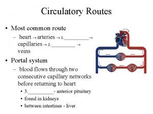
 Coronary circulatory routes
 Mesa coronary calcium score
 Heart cycle animation
 Mesa coronary calcium score
 Thebasian veins
 Coronary calcium score guidelines
 Qfr coronary
 Coronary sinus
 Papyrus stent graft
 Ductus choledochus
 Dr eter
 Percussion of right border of heart
 Sheep heart vs human heart
 Hrt to hrt merrylands
 Introduction to heart
 Heart of darkness historical context
 Introduction of cardiovascular system
 Foot vascular anatomy
 Wrist anastomosis
 Internal maxillary artery branches
 A thyroidea ima
 Kidneys location and structure figure 15-2
 Upper arm
 Scarpa triangle
 Cervical plexus
 Knee joint classification
 Right marginal artery
 M
M Apex of axilla
 Structure of an artery
 Artery kidney
 Anterior stomach
 Plantar artery
 Pterion middle meningeal artery
 Subclavian artery
 Persistent stapedial artery
 Porto systemic anastomosis
 Teochanter
 Pleura
 Pelvic arteries
 Internal maxillary artery branches
 Sinusoidal capillaries
 Superior rectal artery is a branch of
 Weber syndrome
 Tunica intima
 Mastoid process
 Typical intercostal nerve diagram
 Spermatic cord components
 Bursa coracobrachialis
 A. poplitea palpation
 Femoral nerve root value
 Facial artery branches
 Pa systolic pressure range
 External carotid artery terminal branches
 Termination of femoral artery
 Superior epigastric artery
 øit
øit The common hepatic artery branches into the
 Blood supply of the pons
 Saclike formation in the wall of an artery
