Three basic components 1 Heart 2 Blood vessels

Three basic components 1. Heart 2. Blood vessels 3. Blood Cardiovascular system function: 1. 2. 3. 4. Transport oxygen Nutrients Cell wastes Hormones via the blood

ØCone shape ØMuscular pump Ø 250 – 300 g ØSize of a person’s fist

Location – Mediastinum – Superior surface of diaphragm – Anterior to the vertebral column – Posterior to the sternum – Apex is directed toward the left, rests on the diaphragm, at about the fifth intercostals space

Pericardiu m

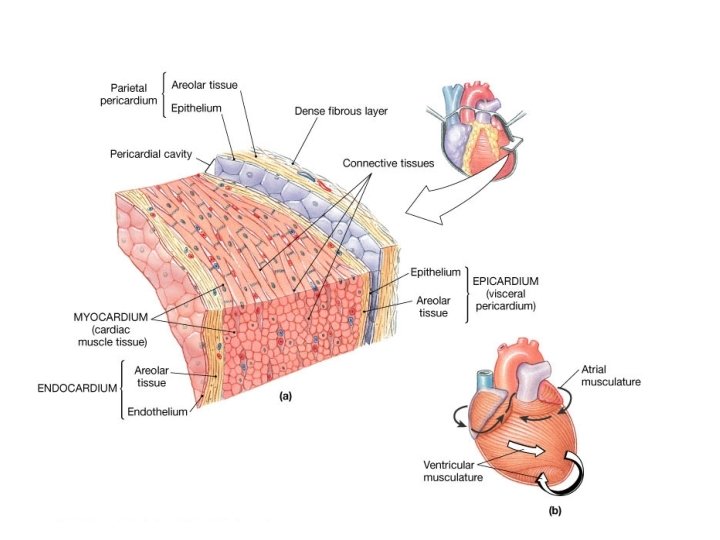
Pericardium : • • A superficial fibrous pericardium A deep two-layer serous pericardium 1. The parietal layer 2. The visceral layer 3. They are separated by the fluid-filled pericardial cavity The pericardium: Protects Prevents overfilling Allows for the heart to work in a relatively friction-free environment

Heart Wall • Epicardium – visceral layer of the serous pericardium • Myocardium • Endocardium

Anatomical position • surface 1. Sternocostal 2. Diapgragmatic 3. Pulmonary R &L • Borders 1. 2. 3. 4. Left Right Inferior superior • Base • Apex

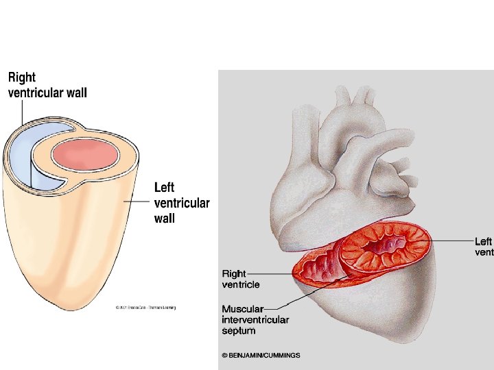
Four hollow chambers: • 2 atria – receiving chambers – divided by the interatrial septum • 2 ventricles – discharging chambers – divided by the interventricular septum

Atria Septum Ventricles

Sternocostal surface Left Atrium Right Atrium Sinus coronary sulcus Anterior Inter ventricular sulcus Right Ventricle Left Ventricle

Diaphragmatic surface Left Ventricle posterior Inter ventricular sulcus Coronary Sulcus

Right atrium – Ineratrial septum – Fossa ovalis – Right auricle – Superior Vena Cava – inferior Vena Cava – Coronary sinus – Right atrioventricular foramen

Pectinate Muscle Fovea Ovalis

Blood Enters The Right Atrium Via Three Veins Superior vena cava 1. Superior vena cava 2. Inferior vena cava 3. Coronary sinus Inferior vena cava

Right ventricle – Papillary muscles – Chorda tendineae Right ventricle: • Papillary muscles Chorda tendineae Tricuspid valves Pulmonary artery – – – Tricuspid valves – Pulmonary artery Trabeculae carneae

Chordae tendoneae Papillary muscles

Left atrium – Left auricle – Left atrioventricular foramen – Aorta foramen

Blood enters the left atrium via four veins Right and left pulmonary veins Left pulmonary veins Right Pulmonary veins

Left ventricle • – Papillary muscles – Chorda tendineae – Bicuspid valves – Aorta Papillary muscles – Chorda tendineae – bicuspid valves – Aorta –

Radiographic Landmarks

Major Vessels of the Heart Vessels returning blood to the heart include: • • • Superior venae cavae inferior venae cavae Right and left pulmonary veins Vessels conveying blood away from the heart include: • Pulmonary trunk • Ascending aorta
 • Pulmonary")
Valves of the Heart Atrioventricular valves Semilunar valves • Right AV (Tricuspid) • Pulmonary valve • Left AV (Bicuspid) • Aortic valve
 valve Chordai tendineae Papillary muscle Pulmonary semilunar valve Aortic semilunar valve")
Right AV (tricuspid) valve Chordai tendineae Papillary muscle Pulmonary semilunar valve Aortic semilunar valve Left AV (bicuspid) valve
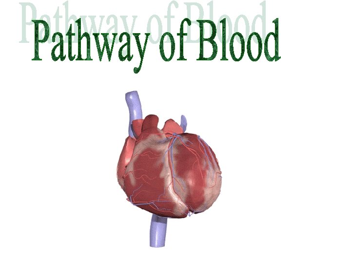

Circulatory System • Pulmonary circulation • Systemic circulation

Pathway of Blood Through the Heart and Lungs • RA tricuspid valve RV • RV pulmonary semilunar valve pulmonary arteries lungs • Lungs pulmonary veins LA • LA bicuspid valve LV • LV aortic semilunar valve aorta • Aorta systemic circulation

Pulmonary Circulation

Systemic Circulation
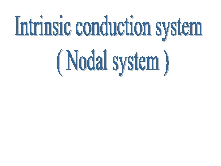

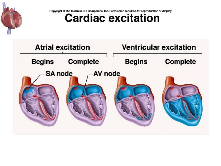
Two systems act to regulate heart activity: 1. Extrinsic conduction system 2. Intrinsic conduction system = or nodal system Setting the basic rhythm 1. Sinoatrial (SA) node : • • 2. located in right atrium pacemaker of the heart Atrioventricular (AV) node : • at junction of atria and ventricles 3. Atrioventricular (AV) Bundle (Bundle of his) 4. Bundle branches 5. Purkinje fibers
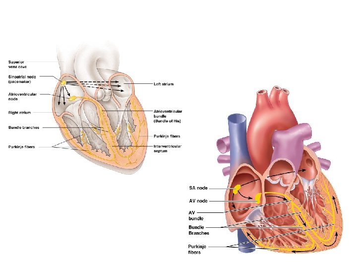

SA Node Bundle of His Internodal Pathways Bundle Branches AV Node Purkinje Fibers

Regulation of Heart Rate Increased heart rate · · · Sympathetic nervous system thoracic N – 1 - 4 Low blood pressure Hormones 1. Epinephrine 2. Thyroxine Exercise Decreased blood volume Decreased heart rate · Parasympathetic vagus N · High blood pressure or blood volume

Heart sounds – Lub dup – Lub – closing of AV valves ( systole ) – Dup – closing of semilunar valves (diastole ) • Murmurs = abnormal heart sounds

Cardiac Cycle Systole : lup • Rising ventricular pressure results in closing of AV valves • Atria relax Diastole : dup • relaxation of heart muscle

Capillary Beds Figure 19. 4 a
 • Force wall of a blood vessel by its contained blood")
Blood Pressure (BP) • Force wall of a blood vessel by its contained blood – Expressed in millimeters of mercury (mm Hg) – in large arteries near the heart Systemic pressure: – Is highest in the aorta – Declines throughout the length of the pathway – Is 0 mm Hg in the right atrium

Measuring Blood Pressure first sound heard = systolic pressure Second sound heard = diastolic pressure Systole = contraction Diastole = relaxation

Blood Pressure: Effects of Factors · Normal 140– 110 mm Hg systolic 80– 75 mm Hg diastolic · Hypotension Low systolic (below 110 mm HG) · Hypertension High systolic (above 140 mm HG) · Neural factors (sympathetic division) · Renal factors ( Renin ) · Temperature Ø Heat has a vasodilation effect Ø Cold has a vasoconstricting effect · Chemicals Ø Various substances can cause increases or decreases · Diet
 • Coronary heart disease (CHD) ---Myocardial")
Types Of Cardiovascular Disease • Atherosclerosis ----(Plaque ) • Coronary heart disease (CHD) ---Myocardial infarction (MI) or heart attack • Chest pain (angina pectoris) – Ischemia Irregular heartbeat (arrhythmia) » Tachycardia = more than 100 beats/min » Bradycardia = less than 60 beats/min • • Congestive heart failure (CHF) – heart muscle is unable to keep blood circulating normally • Congenital heart disease • Stroke--- Myocardial infarction (MI) • Embolism: blockage of blood vessels

Common Blood Vessel Disorders

Angioplasty Versus Bypass Surgery Coronary bypass surgery

Pericarditis Inflammation of pericardial sac • Clinical Manifestations of Pericarditis 1. 2. 3. 4. Chest pain Dyspnea Chills Fever

Clinical Manifestations of Mitral Stenosis • • Cough Frequent respiratory infections Weakness, fatigue Diastolic murmur

Varicose Veins
- Slides: 51