FOR DEPT OF PRACTICE OF MEDICINE PULSE AN


- Slides: 38

FOR DEPT. OF PRACTICE OF MEDICINE PULSE - AN OUT LOOK & EXAMINATION. . DR. Arun nair

DEFINITION PULSE is a transmitted pressure wave along with the arterial wall that is felt by the palpating finger and it is produced by cardiac systole which transverses the arterial tree in a peripheral direction at a rate faster than that of blood column. 2 Caused by pressure changes in aorta. systolic ejection of bl. From LV, aorta expands & then recoils- setting up pressure wave or pulse wave. Pulse wave travels faster than blood with velocity of 5 -8 m/sec, (bl. flows at velocity of 0. 5/sec) Dr. A. R. NAIR 4/22/2015

What do u understand by term PULSE? The alternate expansion and recoil of elastic arteries after each systole of the left ventricle creating a traveling pressure wave that is called the PULSE. 3 Dr. A. R. NAIR 4/22/2015

The normal pulse rate is 60 -100 bpm. �It is under control of ANS �HR increases with incr. sympathetic activity �Decreases with increased parasympathetic activity �A HR more than 100 - Tachycardia �HR less than 60 -Bradycardia. �Normally higher in children and low in elderly. �HR is higher in inspiration and lower in expiration.

5 Dr. A. R. NAIR 4/22/2015

INTERPRETATION(1/2) �Anacrotic limb is the ascending limb, percusssion wave –expansion of artery due to ventricle systole, corresponds to max. ejection phase. �Catacrotic limb is the descending limb.

�INTERPRETATION(2/2) Dichrotic notch- notch on downstroke, similar to incisura of of aortic pressure curve-due to reflux of blood back into aorta, closure of aortic valve. negative wave. Dicrotic wave-positive wave, wave below dicrotic notch, blood rebounds from closed aortic valve

Abnormal character of the arterial pulse wave 8 Dr. A. R. NAIR 4/22/2015

Anacrotic pulse� 2 upbeats, seen in aortic stenosis, �Results in slow ejection of blood from LV �Pulse is typically small volume, slow rising �Peak is delayed compared with normal pulse.

Slow rising or Pulsus Parvus �(Parvus=small), small, weak pulse rises slowly and has a late systolic phase. �Weak upstroke is due to decreased stroke volume. �Seen in conditions diminished LV stroke volume and narrow pulse pressure-Aortic, Mitral stenosis, LVF and hypovolemia

Collapsing or water hammer pulse �Corrigan pulse �This is a sign of aortic regurgitation, although it is sometimes also seen in patients with a hyperdynamic circulation and with a rigid arterial system. A stiff arterial system leads to an accentuated systolic peak in the peripheral pulses. The pulse has an early peak and then quickly falls away, giving it a tapping quality. The preferred method is to palpate the brachial pulse with the whole palm applied to the flexor aspect of the wrist

COLLAPSING PULSE When a collapsing pulse is detected look for the following signs, Duroziez sign: Seen in severe aortic regurgitation. Place the diaphragm of the stethoscope over the femoral artery and press downwards. Initially a systolic murmur will be heard. Gradually increase pressure over the artery- a diastolic murmur will become evident also related to the flow reversal with profound aortic regurgitation. Now tilt the proximal edge of the stethoscope further downwards – if aortic regurgitation is present the systolic murmur is accentuated and the diastolic component is diminished. Now tilt the distal edge of the stethoscope downwards, the diastolic component will now be accentuated and the systolic reduced. This sign has a positive predictive value of close to 100% for aortic regurgitation, and can detect this lesion in some patients in whom it is not possible to hear the characteristic diastolic murmur on auscultation of the heart 12 Dr. A. R. NAIR 4/22/2015

COLLAPSING PULSE Traubes sign: A “pistol shot” sound heard over the femoral artery with the aid of a stethoscope. It is necessary to compress the femoral artery distal to the stethoscope head to produce the characteristic double tone sound. Hills sign: This is a nonspecific sign of aortic regurgitation- it is also seen in other causes of a hyperdynamic circulation, such as thyrotoxicosis, beri-beri, or pregnancy. Check the blood pressures in the upper and lower limbs. If the pressure in the lower limbs exceeds that in the upper limbs by more than 20 mm. Hg then the sign is positive. Quinkes sign: Pulsatile blanching of the nail bed De Musset’s sign: Named after the famous French poet whose head nodded in time with his arterial pulsations due to his syphilis related aortic regurgitation 13 Dr. A. R. NAIR 4/22/2015

Pulsus bisfereins �Combination of above 2. �Slow rising and collapsing pulses. �Seen in combined aortic stenosis and aortic regurgitation.

Pulses paradoxus �Pulse becomes smaller or disappear at end of deep inspiration. (dec. in pulse volume, normal fall is 8 -10 mm. Hg). �It is called paradoxus – as heart sounds still be heard on auscultation over precordium, when no pulse is palbable at radial artery. �Commonly seen in- large pericardial effusion(Cardiac tamponade). �Constrictive pericarditis.

Pulsus Alterans �Arterial pulses typically show alternate large & small amplitude, but regular beats. �It is seen when Lt. ventricle is severely damaged (Myocardial infarction), LVF. �This causes alternate strong and weak contractions, beats. �Seen in arterial hypertension, coronary artery disease.

Pulse delay � Delay in appearance between radial and femoral pulse is useful for diagnosis of – � Coarctation of aorta. � Significance� To asses the state of myocardium, valves. � To asses cardiac arrythmias � To asses condition of vessel wall � Rough guide to estimate the arterial BP.

Dicrotic Pulse �There are 2 palpable waves � 1 in systole other in diastole �Encountered in patients with very low stroke volume. �In those with dilated(congestive) cardiomytopathy, infarction.

Plateau Pulse �Pulse wave rises slowly �Delayed and sustained peak �Pulse fades away slowly �Such pulse is sometimes seen in aortic stenosis.

Apical impulse-apex beat �Lower most, outer most point of cardiac pulsation. �Normally felt in Lt. 5 th intercostal space �Half inch medial to midclavicular line. �This is apical area, corresponds with mitral area. �Impact of ventricles against chest wall.

Describing the pulse The pulse is described by �Rate �Rhythm �Volume �State of the vessel wall �Synchronous with other pulse /EQUALITY. 21 Dr. A. R. NAIR 4/22/2015

1. RATE �The rate of the pulse is recorded in beats per minute. The rate should be counted over a minimum of thirty seconds. �The normal resting pulse rate is 72/min. �Abnormal slow (bradycardia)<60/min �Abnormal fast (tachycardia) >100/min 22 Dr. A. R. NAIR 4/22/2015

2. RHYTHM �Normally pulse beat at – Regular inteverval. �Regular or Irregular, �If irrrgular-Regularly irregular( Extra systole). �Irregularly irregular-( due to atrial fibrillation).

Irregularly irregular pulse �Seen in atrial fibrillation �Irregularity occurs not only in interval between beats. �Also in the volume of beats.

Irregular rhythm is due to�Sinus irregularity, or premature contractions. �Sinus irregularity- S. arrhythmia- pulse rate is greater in inspiration but it becomes marked in sinus arrythmia. �Premature contractions- Extrasystole, occurs due to impulse generation from ectopic focus( ectopic beat).

3. VOLUME �The volume of the pulse is a crude indicator of the stroke volume of the heart (the amount of blood ejected by the heart) �It is increased in exercise (full or bounding) and reduced in states of low blood volume (weak or thready) 26 Dr. A. R. NAIR 4/22/2015

4. STATE OF THE VESSEL WALL �The normal arterial wall is compressible and has an elastic feel �Diseased arteries may feel inelastic and even hard in cases of calcification 27 Dr. A. R. NAIR 4/22/2015

5. EQUALITY ON BOTH SIDES �Compare each of the above features with other sides. �Normally in health all features should be same. �Important note�Other main peripheral arterial pulses; brachial, carotid, femoral, popliteal, post. tibial and dorsalis pedis artery pulse is noted. �Volume is compared with other side.

READING THE PULSE 29 Dr. A. R. NAIR 4/22/2015

While palpating radial pulse�Middle 3 fingers are used �Index finger is used for occlusion or compression. �Middle is used for assessing the volume and rate of pulse �Ring finger to study the condition of vessel wall.

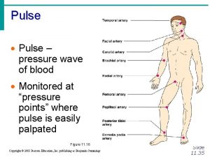
Common pulse sites Radial Pulse Lateral aspect of the lower forearm just proximal to the wrist joint Feel the bony prominence Move fingertips medially Tips of fingers drop into a groove in which lies the artery Examine the pulse by compressing the artery backwards against the bone, using the finger tips 31 Dr. A. R. NAIR 4/22/2015

The brachial pulse Medial aspect of the antecubital fossa at the line of the elbow joint. The artery is felt by compressing backwards with fingers or thumb. Divides just below elbow to form radial and ulnar arteries 32 Dr. A. R. NAIR 4/22/2015

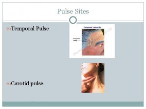
Carotid pulse 1 -1. 5 cm lateral of the midline in the neck at the upper level of the thyroid cartilage Readily palpable at anterior border of sternomastoid muscle May be felt with finger tips or thumb which are used to push posteriorly 33 Dr. A. R. NAIR 4/22/2015

Femoral artery The femoral artery enters the upper leg by passing under the inguinal ligament. It enters the leg at the mid-inguinal point. The femoral artery is usually easily palpated and is an important point of access to the arterial system. 34 Dr. A. R. NAIR 4/22/2015

Popliteal artery The popliteal artery is palpable in the popliteal fossa. The artery passes through the fossa slightly medially to laterally. The popliteal artery can be palpated in about the midline of the fossa at the level of the femoral condlyes. Artery best felt with knee in slight flexion. 35 Dr. A. R. NAIR 4/22/2015

Tibialis posterior artery The tibialis posterior artery is found on the medial aspect of the ankle. It is palpable at a position midway between the prominence of the medial malleolus and the prominence of the calcaneus. 36 Dr. A. R. NAIR 4/22/2015

Dorsalis pedis artery Dorsalis pedis is a continuation of the tibialis anterior. Tibialis anterior is often palpable at the ankle joint in a mid-malleolar position, medial to the extensor hallucis longus tendon. 37 Dr. A. R. NAIR 4/22/2015

. . e r a c e k a t. . s w e l l i t e y b d o o i a g a t e mee dn o o g h n wit w G THANKS FOR SILENT WATCHING & SLEEP. . 38 Dr. Arun R Nair. Dept. of PM SKHMC 4/22/2015
 Dept nmr spectroscopy
 Florida dept of agriculture and consumer services
 Finance dept structure
 Inspectors in worcester
 Dept. name of organization (of affiliation)
 Mn dept of education
 Dept of finance and administration
 Dept. name of organization (of affiliation)
 Ohio dept of dd
 Dept. name of organization (of affiliation)
 Vaginal dept
 Gome dept
 Gome dept
 Gome dept
 Gome dept
 Hoe dept
 Lafd interview score
 Maine department of agriculture conservation and forestry
 Dept of education
 Florida dept of agriculture and consumer services
 Florida dept of agriculture and consumer services
 Dept a
 Central islip fire department
 Rowan county medicaid
 Dept of education
 Tabella chemical shift c13
 Pt dept logistik
 Nys dept of homeland security
 La dept of revenue
 Geaux biz login
 Continuing education library oxford
 Nebraska dept of agriculture
 Iit
Iit Dept ind onegov
 Albany county dss
 Fspos
Fspos Typiska drag för en novell
 Nationell inriktning för artificiell intelligens
 Vad står k.r.å.k.a.n för
