PELVIC ARTERIES Dr Abeera SarfrazDr Muhammad Zubair Several

PELVIC ARTERIES Dr Abeera Sarfraz/Dr Muhammad Zubair

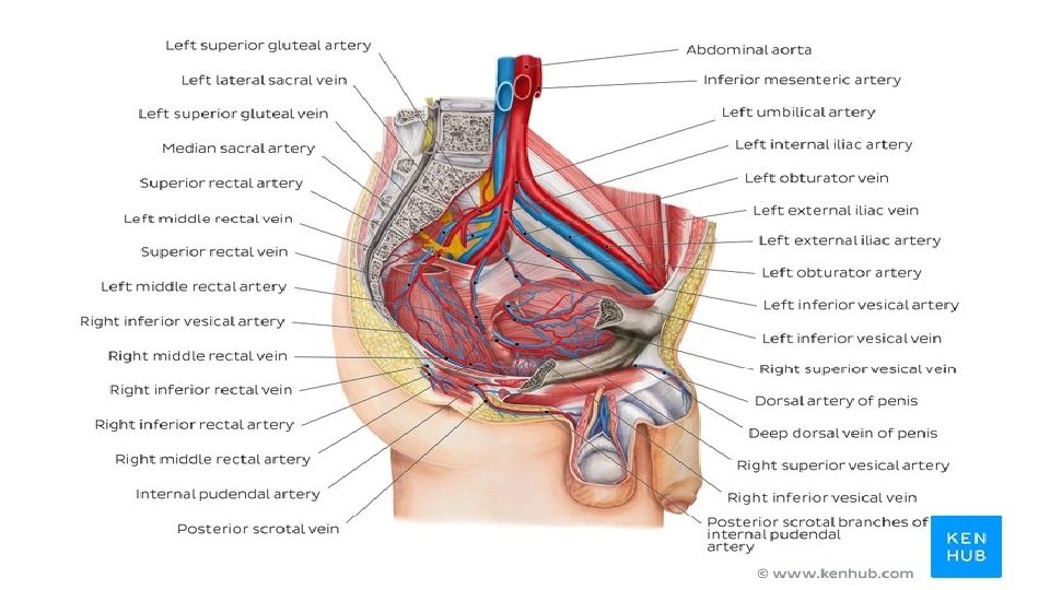
Several arteries stream across the pelvic walls. The common and external iliac arteries are related to the pelvic brim. The internal iliac, superior rectal, ovarian, and median sacral arteries run into the pelvic cavity. The arteries related to the pelvic walls are densely packed in the pelvic cavity and subject to significant variation in their branching patterns of origin. However, these vessels always terminate where they are supposed. Thus, identifying the individual arteries by their destinations is more reliable than by their origins. Common Iliac Artery Each common iliac artery ends at the pelvic inlet in front of the sacroiliac joint by dividing into the external and internal iliac arteries.

The external iliac artery runs along the medial border of the psoas muscle, following the pelvic brim and gives off the inferior epigastric and deep circumflex iliac branches. It leaves the false pelvis by passing under the inguinal ligament to become the femoral artery. EXTERNAL ILIAC ARTERY True Pelvis Arteries The following arteries enter the pelvic cavity: Internal iliac artery Superior rectal artery Ovarian artery Median sacral artery

Key facts about the pelvic arteries Common iliac artery Source: abdominal aorta (at the level of L 4) Branches: branches to ureter and peritoneum Terminal branches: external and internal iliac arteries External iliac artery Source: common iliac artery (at the level of sacroiliac joint) Branches: inferior epigastric artery, deep circumflex artery Terminal branch: femoral artery Supplies: anterior compartment of the thigh (femoral artery), iliac crest bone flap (deep circumflex artery), anterior abdominal wall (inferior epigastric artery) Internal iliac artery Source: common iliac artery (at the level of sacroiliac joint) Anterior branches: obturator, middle rectal, superior vesical, uterine (female), inferior gluteal, inferior vesical (male) or vaginal (female), internal pudendal arteries Posterior branches: iliolumbar, lateral sacral, superior gluteal arteries Mnemonic: PILS Supplies: Psoas major muscle, quadratus lumborum, iliacus muscle, uterus, vagina, bladder, prostate, semicanal vesicle

INTERNAL ILIAC ARTERY The internal iliac artery passes down into the pelvis to the upper margin of the greater sciatic foramen, where it divides into anterior and posterior divisions. The branches of these divisions supply the pelvic viscera, the perineum, the pelvic walls, and the buttocks

Umbilical artery: the superior vesical artery arises from the proximal patent part of the umbilical artery. It supplies the upper portion of the bladder. It also gives off the artery to the vas deferens. Obturator artery: this artery runs forward along the lateral wall of the pelvis with the obturator nerve and leaves the pelvis through the obturator canal. ANTERIOR DIVISION BRANCHES Inferior vesical artery: this artery supplies the base of the bladder and the prostate and seminal vesicles in the male. Middle rectal artery: commonly, this artery arises with the inferior vesical artery. It supplies the muscle of the lower rectum and anastomoses with the superior rectal and inferior rectal arteries. Internal pudendal artery: this artery leaves the pelvis through the greater sciatic foramen and enters the gluteal region below the piriformis muscle. It then curls around the ischial spine (with the pudendal nerve) and enters the perineum by passing through the lesser sciatic foramen. It passes forward in the pudendal canal with the pudendal nerve. Its branches supply the musculature of the anal canal and the skin and muscles of the perineum.

Inferior gluteal artery: this artery leaves the pelvis through the greater sciatic foramen below the piriformis muscle. It passes between the first and second or second and third sacral nerves. Uterine artery: this artery runs medially on the floor of the pelvis and crosses the ureter superiorly. It passes above the lateral fornix of the vagina to reach the uterus. Here, it ascends between the layers of the broad ligament along the lateral margin of the uterus. It ends by following the uterine tube laterally, where it anastomoses with the ovarian artery. The uterine artery gives off a vaginal branch. Vaginal artery: this artery usually takes the place of the inferior vesical artery present in the male. It supplies the vagina and the base of the bladder.

POSTERIOR DIVISION BRANCHES Iliolumbar artery: this artery ascends across the pelvic inlet posterior to the external iliac vessels, psoas, and iliacus muscles. Lateral sacral arteries: these arteries descend in front of the sacral plexus, giving off branches to neighboring structures and entering the anterior sacral foramina. Superior gluteal artery: this artery leaves the pelvis through the greater sciatic foramen above the piriformis muscle. It supplies the gluteal region.

SUPERIOR RECTAL ARTERY The superior rectal artery is a direct continuation of the inferior mesenteric artery. The name changes as the latter artery crosses the common iliac artery. It supplies the mucous membrane of the rectum and the upper half of the anal canal. Median Sacral Artery The median sacral artery is a small artery that arises at the bifurcation of the aorta. It descends over the anterior surface of the sacrum and coccyx.

The ovarian artery arises from the abdominal part of the aorta at OVARIAN ARTERY the level of the first lumbar vertebra. The artery is long and slender and passes downward and laterally behind the peritoneum. It crosses the external iliac artery at the pelvic inlet and enters the suspensory ligament of the ovary. It then passes into the broad ligament and enters the ovary by way of the mesovarium. The testicular artery enters the inguinal canal and does not enter the pelvis

PELVIC VEINS For the greater part, the pelvic veins correspond to the pelvic arteries. The external iliac vein begins behind the inguinal ligament as a continuation of the femoral vein. It runs along the medial side of the corresponding artery and joins the internal iliac vein to form the common iliac vein. It receives the inferior epigastric and deep circumflex iliac veins. The internal iliac vein begins by the joining together of tributaries that correspond to the branches of the internal iliac artery. It passes upward in front of the sacroiliac joint and joins the external iliac vein to form the common iliac vein. The median sacral veins accompany the corresponding artery and end by joining the left common iliac vein.

PELVIC LYMPHATICS The lymph nodes and vessels are arranged in a chain along the main blood vessels. The nodes are named after the blood vessels with which they are associated. Thus, there are external iliac nodes, internal iliac nodes, and common iliac nodes.
- Slides: 13