Digestive tract Liver Pancreas GYTK 2018 03 19


 –")


- Slides: 45
Digestive tract Liver Pancreas GYTK 2018. 03. 19. Dr. Szuák András
The great glands • Liver • Pancreas
Right coronary lig. Left coronary lig. Right lobe Left lobe Falciform lig Gall bladder
Ligamentum venosum Caudate lobe Inferior vena cava Bare area Left lobe Hepatorenal lig. Liver hilum Portal vein Hepatic duct Proper hepatic artery Round lig. Gall bladder Right lobe
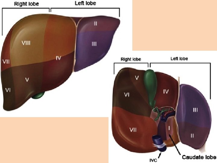
Segments of the liver
Lobes of the liver • The actual boarder is the so called Rex-Cantlie line – Line between • inferior vena cava • gall bladder
Diaphragm Topography of the liver Left coronary lig. Falciform lig. Round lig. Liver Spleen Stomach Gall bladder Hepatoduodenal lig. Lesser omentum Right colic flexure Left colic flexure Transverse mesocolon Transverse colon Descending colon Ascending colon Greater omentum
Hepatoduodenal lig. Hepatogastric lig. Omental bursa Greater omentum L e s s e r o m e n t u m
Bile passages Cystic duct Bile duct Duodenum Hepatic duct Pancreatic duct
Blood supply of the liver
Portal vein
Histology of the liver • Hepatic lobules – hexagonal shape (portal lobule, acinus) – Portal triad – portal vein, artery, bile duct – Hepatic sinusoids between the hepatic chords – Central vein
Hepatic lobule Central vein Hep. sinus Hep. chord Interlobular vein Interlobular artery Portal triad Interlobular duct
Hepatic sinus
Bile canaliculi
Portal triad Central vein
Interlobular duct Interlobular artery Interlobular vein Portal triad
Endothel cell Kupffer-cell Hep. chord Bile canaliculus
Gall bladder • • • Simple columnar epith. Lamina propria – glands, lymphocytes NO Muscularis mucosae layer NO Submucosa layer Muscularis externa – cirlular, longitudinal and oblique layers • Serosa OR Adventitia
Gall bladder
The great glands • Liver • Pancreas
Pancreas
Ducts opening into the duodenum Right hepatic duct Cystic duct Gall bladder Bile duct Minor duodenal papilla Major duodenal papilla Left hepatic duct Common hepatic duct Accessory pancreatic duct Pancreatic duct
Blood supply of the pancreas
Histology of the pancreas • Serous gland • Centroaciner cell • Langerhans islets – endocrine part
Kidney Urinary tract
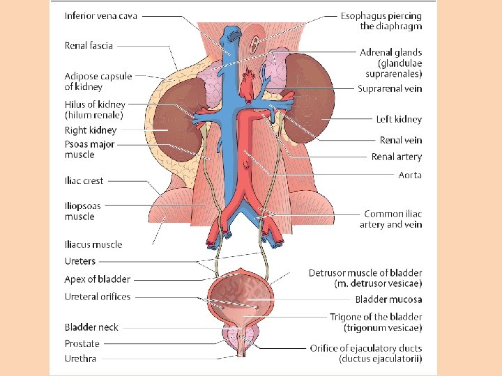
Position of the kidneys
Kidney Adrenal gland Renal artery Renal vein Ureter Fibrous capsule Adipose capsule
cortex medulla
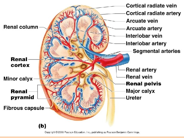
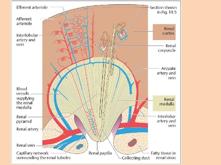
Kidney • Medulla – pyramids – Renal lobes • Cortex – Bertini columns – Convoluted part – Straight part • Renal artery – interlobar aa. –arcuate aa. – interlobular aa.
Nephron
Nephron
Macula densa Malphigian corpuscule Podocytes interdigitating
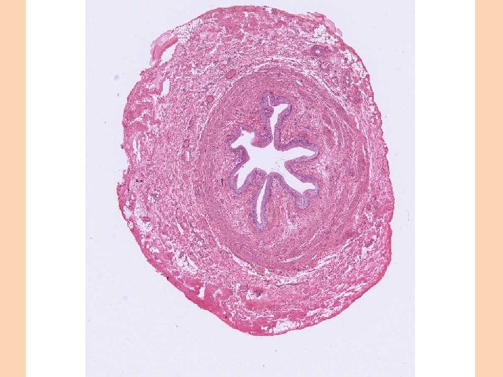
Ureter • Urothelium • Dense fibro-elastic connective tissue in the lamina propria • NO muscularis mucosae&submucosa • Muscularis externa – Inner longitudinal, outer circular layer – Serosa or Adventitia
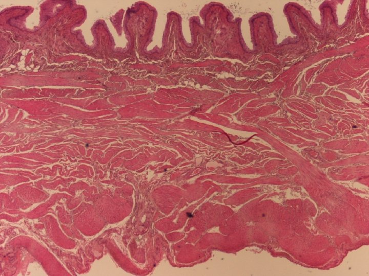
Urinary bladder • Urothelium • Dense fibro-elastic connective tissue in the lamina propria • Muscularis mucosae – Not forming a continous layer, only some islets of smooth muscle • Submucosa – loose connective tissue • Muscularis externa – Inner&outer longitudinal layers, middle circular layer • Serosa or Adventitia
Urothelium, umbrella cells
Male Bladder&Urethra
Female Bladder&Urethra
Thank you for your attention!