Liver Gallbladder Pancreas And Salivary Glands Undergraduate Graduate

Liver, Gallbladder, Pancreas, And Salivary Glands Undergraduate – Graduate Histology Lecture Series Larry Johnson, Professor Veterinary Integrative Biosciences Texas A&M University College Station, TX 77843

Objectives To understand the general organization of the accessory organs of the digestive system and how they contribute to obtaining metabolites necessary for growth and energy for the body. To learn the origin of these glands and how structural features of these glands contribute to their function in digestion and absorption of food stuffs

Origin And Distribution Of Epithelium Ectoderm - epidermis of skin and epithelium of cornea together covers the entire surface of the body; sebaceous and Ectoderm mammary glands, oral cavity Endoderm - alimentary tract, Liver, pancreas, gastric glands, intestinal glands – Endocrine glands - lose connection with surface Mesoderm Endothelium - lining of blood vessels Mesothelium - lining serous cavities Endoderm

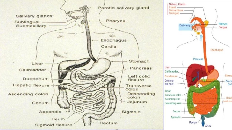
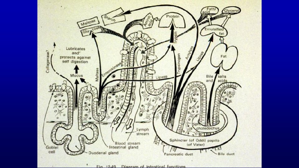
Function of the Digestive System Role of liver, gall bladder, salivary glands, and pancreas Movement of food Salivary glands lubricates Secretion of digestive juices Salivary glands and pancreas secretes digestive juices and liver secretes bile Absorption of digested foods, water, and electrolytes Liver stores nutrients and cleans the blood. Also, the accessory digestive organs contribute antibodies and antibacterial/viral growth substances.

ORIGIN AND DISTRIB UTION OF EPITHELI UM con’d Salivary gland 19758 Liver Histo 67 155 Gallbladder Pancreas 158
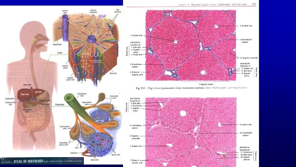

454 Histo 067 pig liver Human liver Classical liver lobules Separated and surrounded with connective tissue in the pig Mesothelium Connective tissue capsule Monkey liver 118

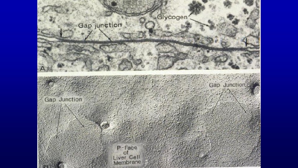
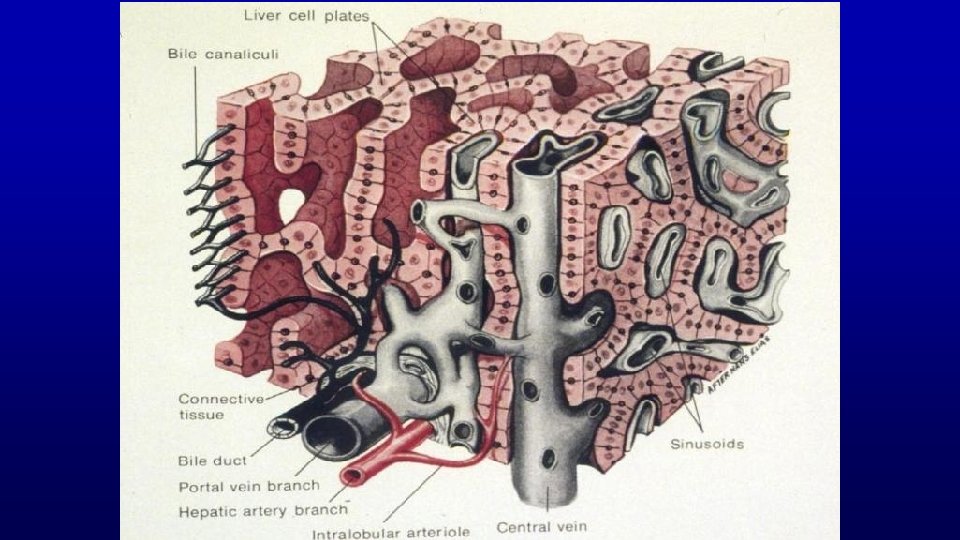
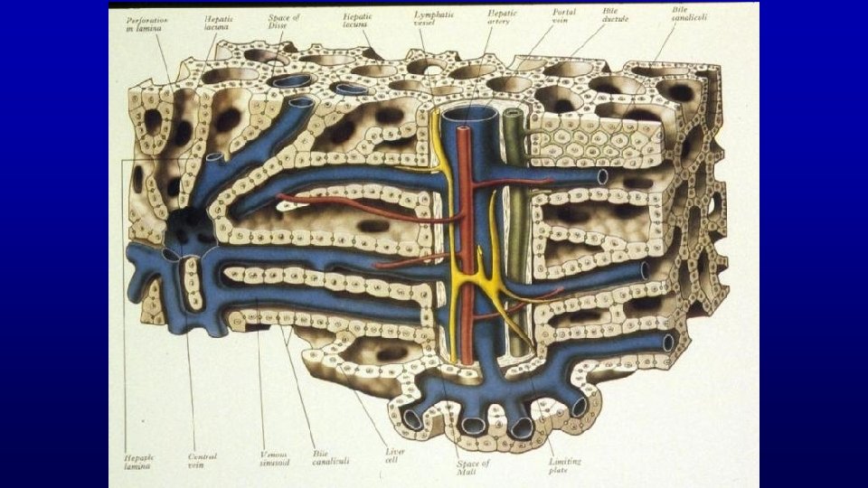
Liver The hepatocyte functions as an endocrine-like cell (e. g. , secretion of glucose and plasma proteins directly into the blood vascular system) and as an exocrine cell (e. g. , secretion of bile into the bile canaliculi). This dual export of secretory products by a single cell requires a unique cellular arrangement in the liver in order to separate and compartmentalize the exocrine and endocrine-like products. Hepatocytes are arranged in fenestrated, anastomosing plates of one cell thick. Also each hepatocyte may have as many as four areas of access to the lumen.

Landscape of the Hepatocyte – Four Luminal Regions

HEPATOCYTE

LIVER FUNCTION - LARGEST GLAND EXOCRINE - BILE ACIDS, BILIRUBIN ENDOCRINE ALBUMIN, FIBRINOGEN, ETC.

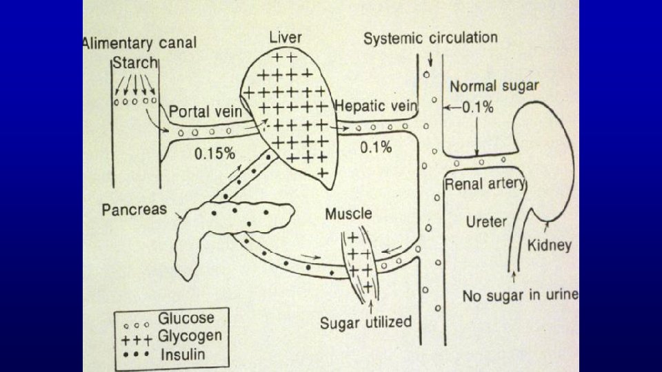
LIVER FUNCTIONS Blood filtration - 1. 2 x 10⁷ Kupffer cells/g Blood storage - liver size and sinusoids expand Maintain normal blood glucose concentrations Metabolism and transport of lipids Secrete plasma proteins - blood clotting Nutritional metabolism and bile secretion Drug metabolism - drug tolerance Excretion of bilirubin - jaundice Secrete bile - emulsifying fats
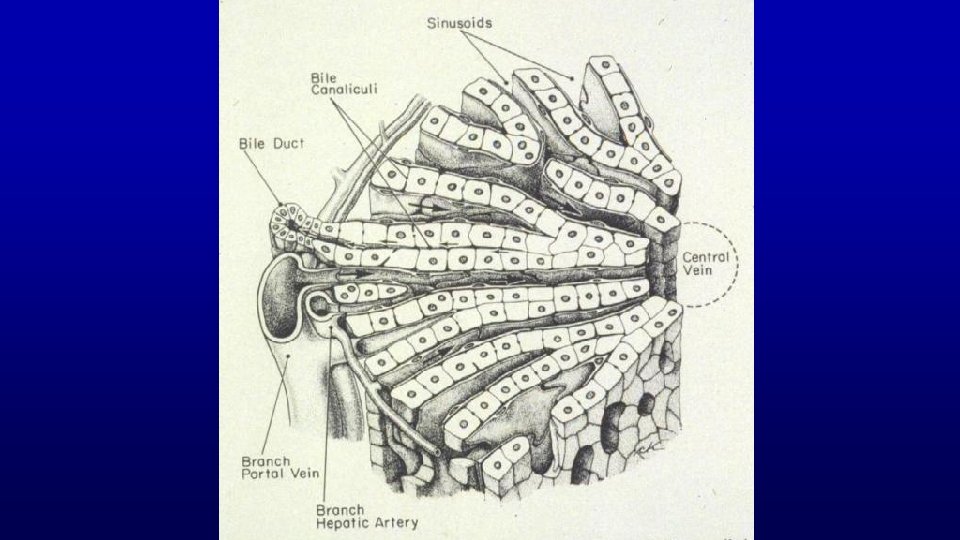


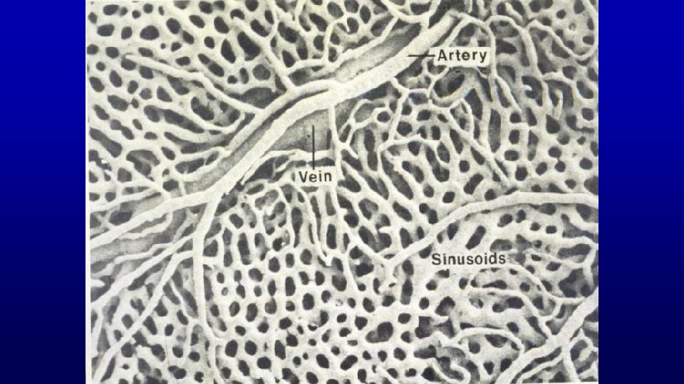
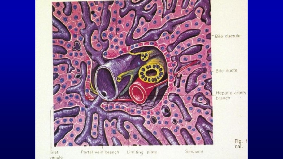
Portal radicles containing: A bile duct Branch of portal vein, Branch of hepatic artery Lymphatic vessel (usually) or portal canals 155 Liver 155 Cords of hepatocytes

454 Liver Central vein Cords of hepatocytes Portal radicles containing: A bile duct Branch of hepatic artery Branch of portal vein Lymphatic vessel (usually)

Bile Canaliculi Bile luminal surfaces 155 Bile duct Blood luminal surface

Liver

Cells of the Liver Lobule A. Hepatocyte B. Kupffer and fat-storing cells C. Endothelial cell Hepatocyte Kupffer cells Endothelial cell

Cells of the Liver Lobule A. Hepatocyte B. Kupffer cells C. Endothelial cell

Triad with bile duct and central vein Liver with colloidal carbon, rat 118

Liver

Liver Lobule Portal triad Blood supply Central vein Hepatic sinusoids Zonation of the liver
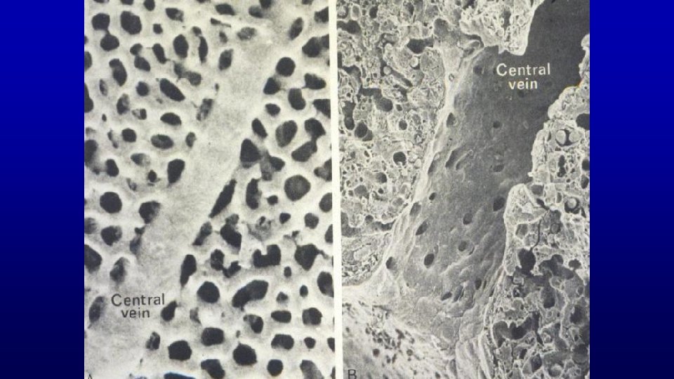

Acinus with portal vein and artery in center Zonation of The Liver 1. Classical lobule 2. Portal lobule with triad in center 3. Acinus layers between two central veins

Zonation of the liver Classical lobule

Portal Lobule with Triad in Center

Acinus with portal vein and artery in center

Acinus with portal vein and artery in center Acinus If liver damage is due to a toxicant, it kills hepatocytes in Zone I first. If liver damage is due to a oxygen deprivation, it will kill the hepatocytes in Zone III first.

HEPATOCYTE

Hepatocyte

Histological Reaction for Peroxidase Hepatocyte

Hepatocyte Space of Disse Bile canaliculi

Space of Disse

Hepatic sinusoid Hepatic parenchymal cells with microvilli Endothelial cell projecting into sinusoid Bile canaliculi with lysosomes close by the canaliculi Liver cells EM 18 Platelet Space of Disse containing reticular fibers

Sugar and protein

Glycogen in Hepatocytes
")
Dietary Differences In Amount Of Glycogen In Hepatocytes 2 -hour Fast (8. 2% Glycogen) 24 -hour Fast (0. 9% Glycogen)

Disease opportunity at each step in a pathway SER

Surface Specializations of Epithelia Transcytosis to get antibodies into secretions

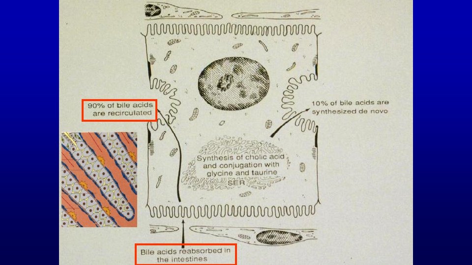
Bile canaliculus Four + compounds that are deposited/secreted into this space. a. Cholesterol b. EGF c. insulin d. Ig. A also bile salts and BILIRUBIN
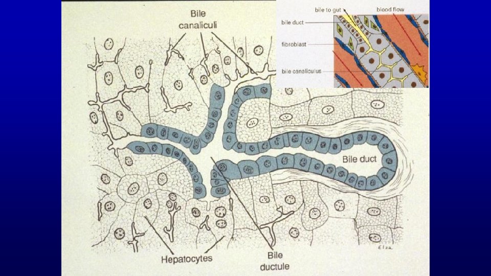
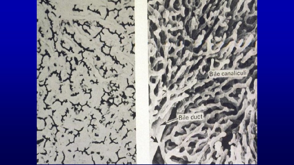

Bile Canaliculi

Bile Duct

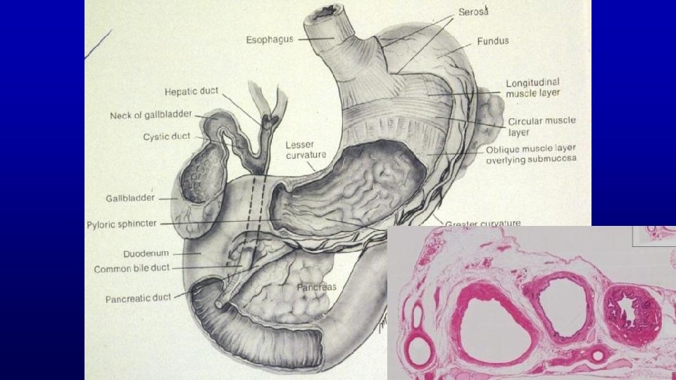
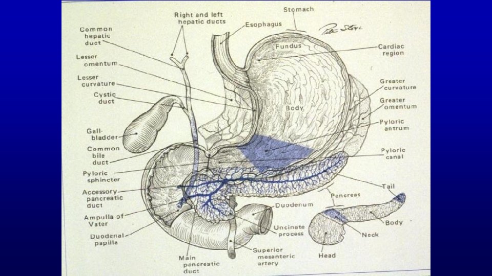
Gallbladder & Bile Ducts Function Biliary tract Organization of gallbladder Epithelium Connective tissue Histophysiology
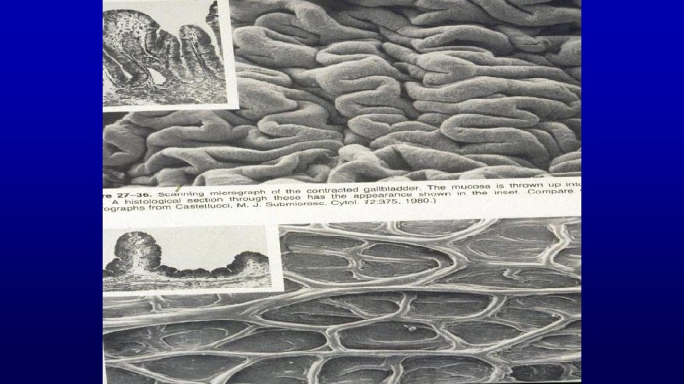
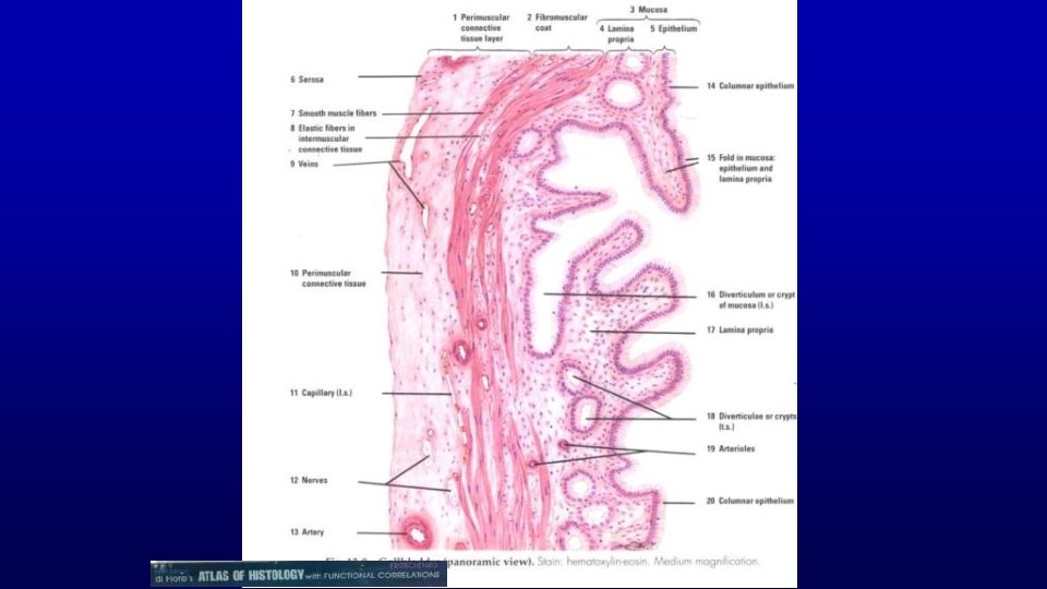

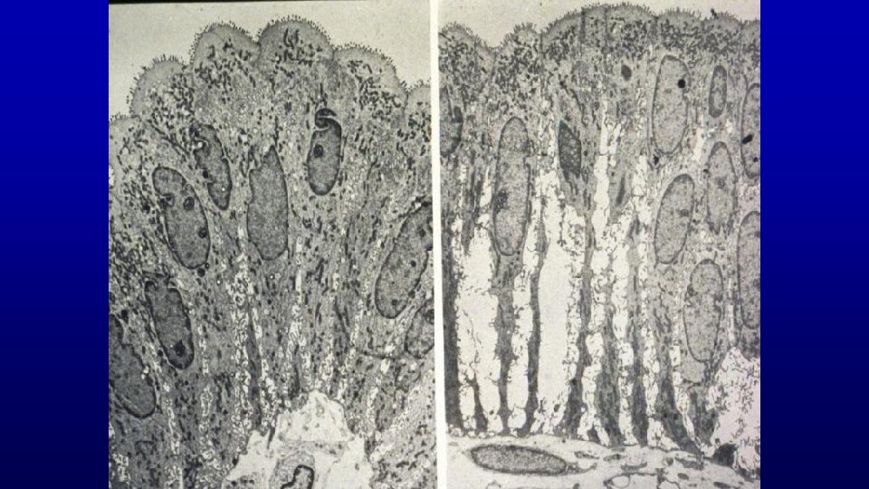
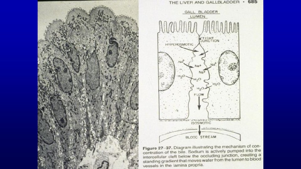
Gallbladder Peritoneal The mucosa is thrown into folds which project into the lumen of the gallbladder. serosal layer 155 Smooth muscle layer or branching layers Lamina propria. A thick perimuscular layer of connective tissue. Simple columnar epithelium

The gallbladder stores and concentrates the 155 Mucosa bile elaborated by the liver Plasma cells In the lamina propria Simple columnar epithelium

126 Bile duct with portal vein, monkey Portal vein Cystic duct Common hepatic duct The wall of the cystic duct is convoluted and contains abundant smooth muscle fibers which represent the spiral valve preventing distention or collapse of the cystic duct when the latter is subject to sudden changes of pressure.

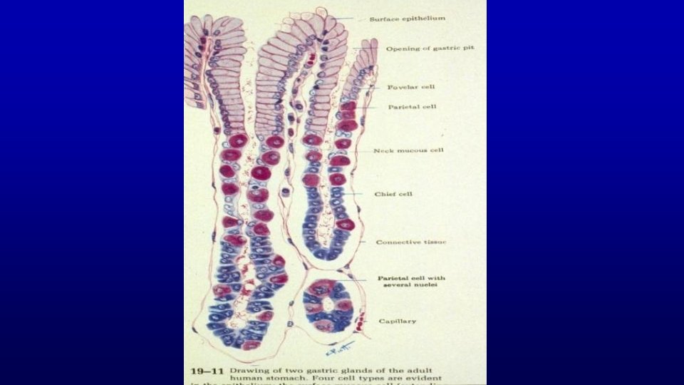
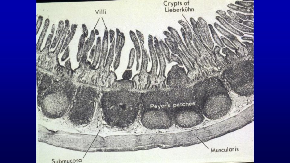
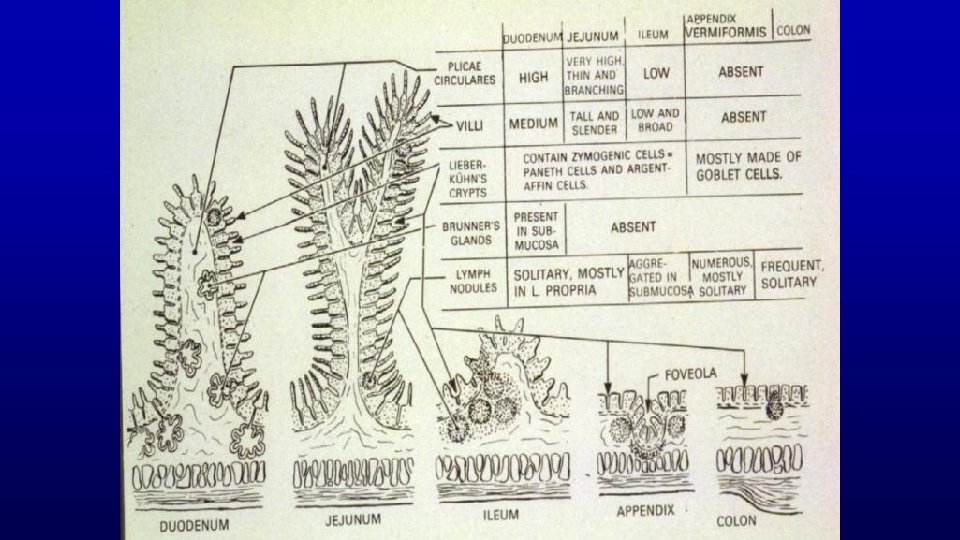
Distinguishing characteristics between the mucosa of the various parts of the stomach, intestines, and gallbladder. Mucosa = surface epithelium, lamina propera, and muscularis mucosa Cardiac stomach Fundic stomach 437 145 Pyloric stomach 141 Intestines 148 Gallbladder 155

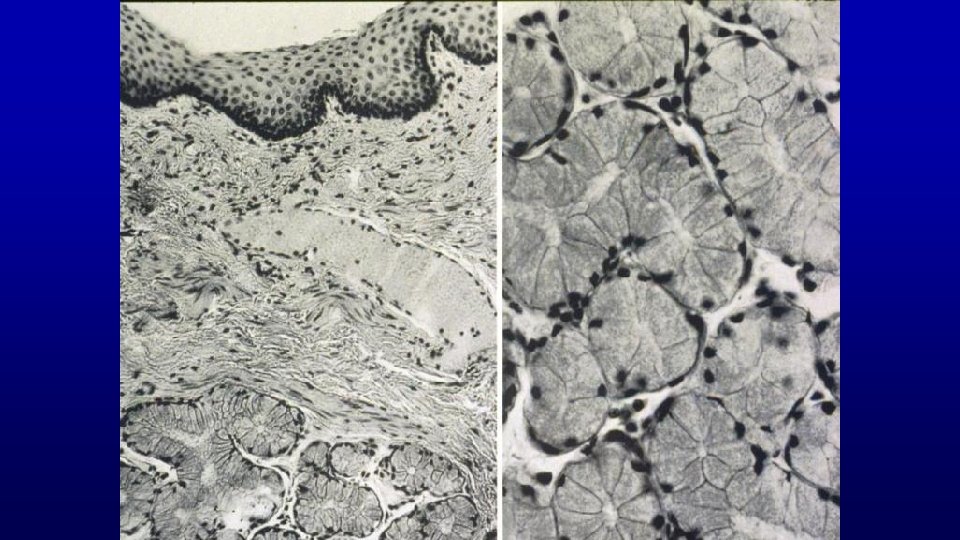
Function Salivary Glands Histological organization Acinus = functional unit Serous Mucous Mixed

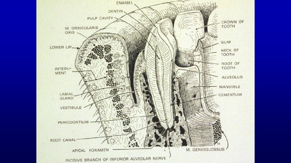
Origin of Salivary Glands? • Ectoderm - oral ectoderm epithelial sheet • Endoderm - alimentary tract

Saliva Helps Prevents Infections Contains secreted Ig. A Contains Lactoferin - bind up iron needed for bacteria division Contains lysosome that kills bacteria Constantly washes mouth to dislodge and sweep bacteria down GI tract

Salivary Glands

Salivary Glands

Ducts of Salivary Glands Intercalated Striated

19758
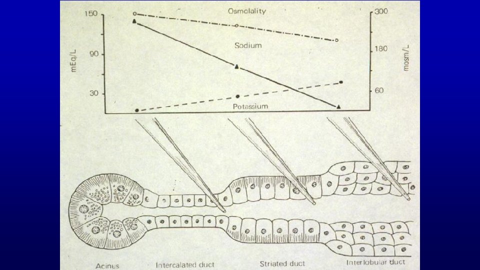
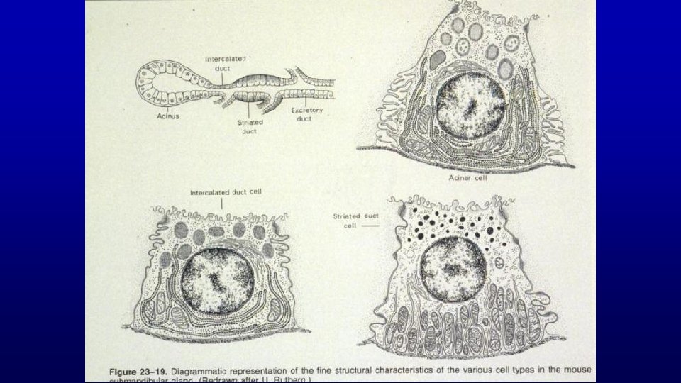

Submandibular gland - intercalated duct runs into Striated duct of salivary gland The salivary gland is a compound, tubuloacinar gland. 130 Striated Ducts These striations reflect vertically arranged mitochondria associated with deep enfolding of the basal plasma membrane Secretory acini Intercalated ducts

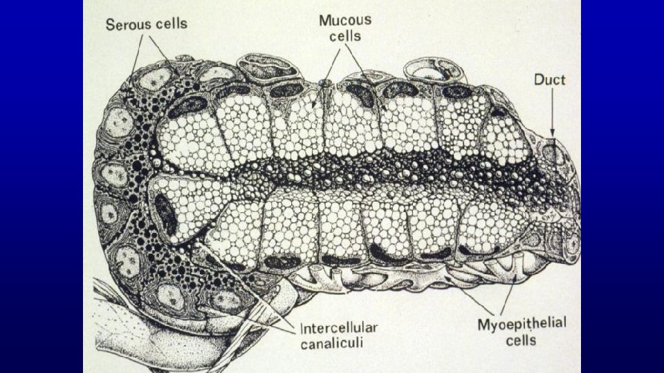
130 Salivary gland Secretory acini are drained by intercalated ducts and join striated ducts Serous and mucous acini Myoepithelial cells

Salivary gland Striated ducts drain into a series of interlobular ducts Lobules Adipose cells Serous and mucous acini Demilune 130 Artery Vein Individual secretory acini are drained by intercalated ducts and join striated ducts

Interlobular ducts Myoepithelial cells Salivary glands 440 Lobules 440 Nerve cell bodies Histo 52 Striated ducts Serous and mucous acini Nerve 19758

Pancreas Function 1. Exocrine 2. Endocrine Histological organization, Exocrine portion 1. Acini 2. Ducts Endocrine portion • Islets of Langerhans Histophysiology

PANCREAS FUNCTION 1. EXOCRINE 2. ENDOCRINE HISTOLOGICAL ORGANIZATION, EXOCRINE PORTION 1. ACINI 2. DUCTS ENDOCRINE PORTION • ISLETS OF LANGERHANS HISTOPHYSIOLOGY
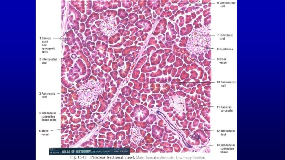

36723 Beginning of intercalated ducts Intercalated duct Islets of Langerhans Interlobular duct
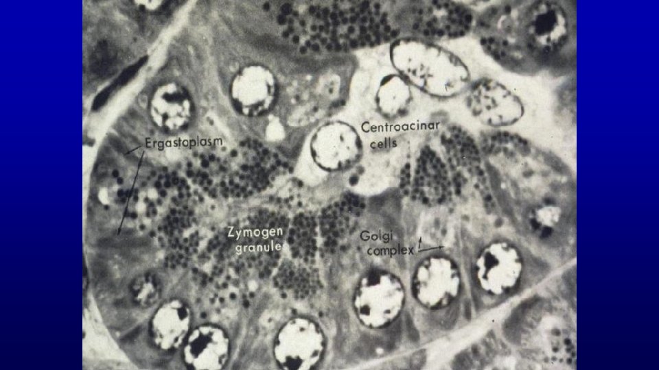
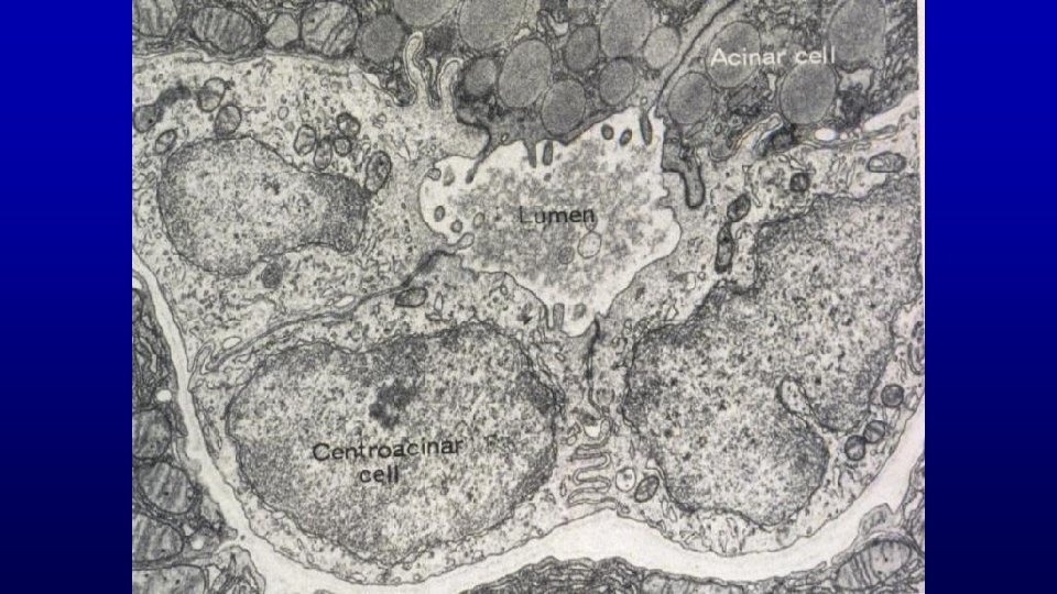
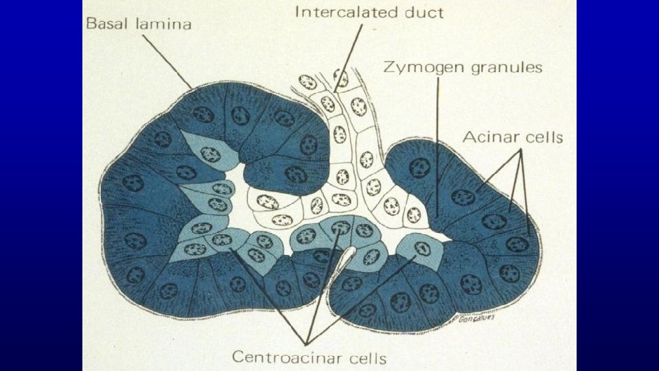

156 and 157 Pancreas 157 156 36723 Intercalated duct 36723 Secretory granules All acini are of the serous type and many contain centroacinar cells initiate the duct inside the acinus. 157

158 Pancreas - Islets of Langerhans Insulin is secreted by the B cells which are most numerous and centrally located in the islets. The pancreas is a compound tubuloalveolar (tubuloacinar) gland which functions in the digestion of food. Lobes composed Intercalated duct Islets of Langerhans of lobules Connective tissue septa. Blood vessels Nerve Interlobular duct
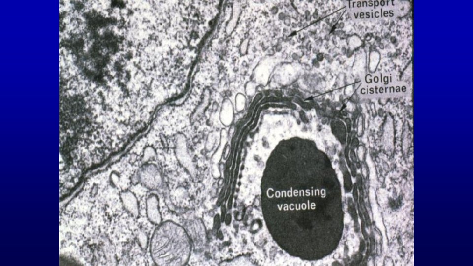
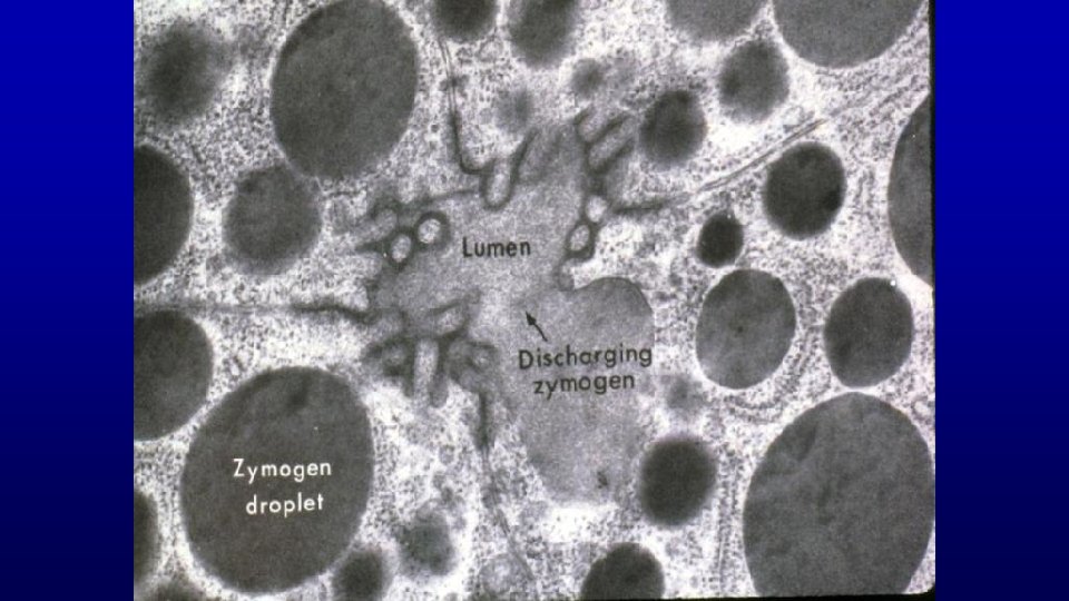
 1. Lumen 2. Zymogen granule 3. Vesicles")
EM 1 Pancreatic acinar cell (EM 1) 1. Lumen 2. Zymogen granule 3. Vesicles 4. Central acinar cell

In summary

• • • • Many illustrations in these VIBS Histology You. Tube videos were modified from the following books and sources: Many thanks to original sources! Bruce Alberts, et al. 1983. Molecular Biology of the Cell. Garland Publishing, Inc. , New York, NY. Bruce Alberts, et al. 1994. Molecular Biology of the Cell. Garland Publishing, Inc. , New York, NY. William J. Banks, 1981. Applied Veterinary Histology. Williams and Wilkins, Los Angeles, CA. Hans Elias, et al. 1978. Histology and Human Microanatomy. John Wiley and Sons, New York, NY. Don W. Fawcett. 1986. Bloom and Fawcett. A textbook of histology. W. B. Saunders Company, Philadelphia, PA. Don W. Fawcett. 1994. Bloom and Fawcett. A textbook of histology. Chapman and Hall, New York, NY. Arthur W. Ham and David H. Cormack. 1979. Histology. J. S. Lippincott Company, Philadelphia, PA. Luis C. Junqueira, et al. 1983. Basic Histology. Lange Medical Publications, Los Altos, CA. L. Carlos Junqueira, et al. 1995. Basic Histology. Appleton and Lange, Norwalk, CT. L. L. Langley, et al. 1974. Dynamic Anatomy and Physiology. Mc. Graw-Hill Book Company, New York, NY. W. W. Tuttle and Byron A. Schottelius. 1969. Textbook of Physiology. The C. V. Mosby Company, St. Louis, MO. Leon Weiss. 1977. Histology Cell and Tissue Biology. Elsevier Biomedical, New York, NY. Leon Weiss and Roy O. Greep. 1977. Histology. Mc. Graw-Hill Book Company, New York, NY. Nature (http: //www. nature. com), Vol. 414: 88, 2001. A. L. Mescher 2013 Junqueira’s Basis Histology text and atlas, 13 th ed. Mc. Graw Internet images and videos on biological presentations

Questions on the Liver, pancreas, and salivary glands The humoral activity of the immune system is illustrated by the transfer of Ig. A immunoglobin by epithelial cells into which of the following body fluids? a. saliva b. milk c. bile d. a and b e. a, b, and c Which function(s) do the gallbladder and urinary bladder have in common? a. temporary storage of waste products b. concentration of their respective luminal contents c. similar type of luminal epithelium d. a and b e. a, b, and c Characteristics of the pancreas include: a. a portal blood vascular system b. endocrine cells of the islets of Langerhans c. acinar cells and striated ducts d. a and b e. a, b, and c

- Slides: 106