Insulin Stimulation test Insulin hypogycemia is a potent


 Na+ (135 -145 mmol/L) K+")

 • Tissue source: acinar cells of pancreas & salivary glands. Lesser concentration")


 Assay by titrimetric method: • Tissue source: primarily in pancreas, little in")
 LPS Hydrolysed fat in solution (Fat particles")


- Slides: 24
Insulin Stimulation test • Insulin hypogycemia is a potent stimulus of acid secretion. • When blood sugar is < 50. 0 mg/dl (2. 8 mmol/L) vagus is stimulated by hypoglycemia. • This test is best limited to those patients suspected to have recurrent ulceration after vagotomy which was probably incomplete.
Plasma Gastrin • • • Valuable in diagnosis of Zolliger- Ellison Syndrome. Normal plasma concentration: 50 – 150 pg/ml. Zolliger- Ellison Syndrome: 1000 – 400, 000 pg/ml. Not increased in simple peptic ulcer. Increased in pernicious anemia.
Test for Occult blood in the feces • Definition: Tests to detect blood in feces in amounts or forms not observable on inspection are referred as occult blood test. • Normal blood loss in the feces 2. 5 ml/day by radiochrome studies. Blood may be introduced from mouth, around teeth, minor abrasion in the GI tract by roughage of food, hemoglobin, myoglobin, their breakdown products, peroxidases of plant & bacterial origin.
• Benzedine test was commonly used, now prevented because of its carcinogenecity. O-toluidine is used with three different concentrations: 4%, 1. 2% & 0. 4% in glacial acetic acid. • Principle: H 2 O 2 hemoglobin & its derivatives O-Toluidine H 2 O + O 2 Coloured product (Measured colorimetrically)
Test procedure • A small portion of feces mixed in 10 ml DW & boil for a minute to destroy peroxidases. Mix fecal suspension + reagent (O-toluidine & H 2 O 2) • Blue colour --- Positive test. • If a single concentration was used 1. 2% recommended. • If all three used 1 st 4% used, positive samples tried with 1. 2%, still positive samples tried with 0. 4%.
Reporting Negative -ve with 4% Weakly positive +ve only with 4% Strongly positive +ve with 4%, 1. 2%, 0. 4%
Interpretation • Test is mainly used in the diagnosis & treament of ulcers, cancer of stomach, gastritis, perpura, lesion in duodenum, small & large intestine. • In case of humorrhoids blood can be seen as streeks of fresh blood on the surface of feces confirmed by misroscopic examinations. • It is also useful practice to do the test on three successive days when the patient is on meat free diet.
Exocrine secretions of Pancrease Inorganic Na. HCO 3(127 mmol/L) Na+ (135 -145 mmol/L) K+ (3. 4 -5. 0 mmol/L) Mg+, Ca+2, Zn+(less) Cl- (155 mmol/L) Organic α - amylase Lipase Trypsin Chymotripsin Carboxipeptidase A & B Ribonucleases Deoxyribonucleases Cholesterol esterases Phospholipases
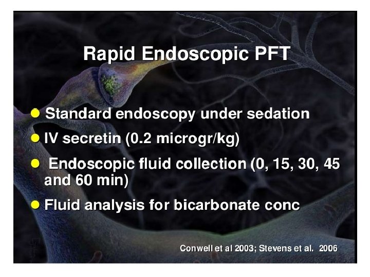
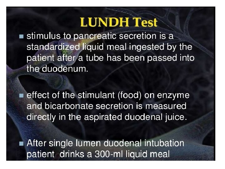
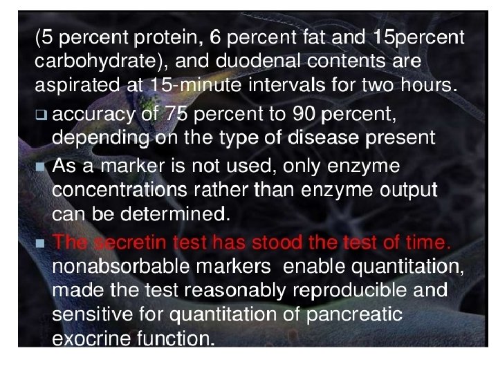
Tests in Pancreatic Diseases • Introduction • Measurement of total volume. • Concentration of HCO 3 • Chemical & cytological examinations performed support suspicion of malignant neoplasm, but exact localization may be unknown. • Secretin/ CCK-PZ test
Amylase (AMS) • Tissue source: acinar cells of pancreas & salivary glands. Lesser concentration in skeletal muscle, small intestine, fallopian tube. • This is the smallest enzyme readily filtered through the renal glomerulus & appears in the urine.
Isoenzymes of AMS • P-type: pancreatic • S- type: salivary, fallopian tube, lung • Isoenzymes of salivary origin migrate most quickly (S 1, S 2, S 3), where as pancreatic origin move slower (P 1, P 2, P 3). • AMS migrate in the regions corresponding to β to α-globulin regions of the protein. • P-type activity, specifically P 3 in acute pancreatitis
Renal clearance of AMS • Useful in detecting minor or intermittent in serum concentration. % AMS clearance UA 100× = Creatinine clearance SA SC × UC • Normal Values: < 3. 1% • Acute pancreatitis: 8% - 9% • Also in burns, sepsis, diabetic ketoacedosis.
Lipase (LPS) Assay by titrimetric method: • Tissue source: primarily in pancreas, little in stomach & small intestine. triglyceride + 2 H 2 O LPS 2 -monoglyceride+2 -fatty acid p. H 8. 6 -9 • Classical Cherry-Crandall method used an olive oil substrate & measured the liberated FA by titration after 24 h incubation. Trioline is one of the substance now used as a more pure form of TAG.
Turbidimetric method Fats in solution (cloudy emulsion) LPS Hydrolysed fat in solution (Fat particles disperse) Rate of clearing of the fat in the solution is measured.
Interpretation • Reference range: 0 – 1. 0 U/ml • This is exclusive for the diagnosis of acute pancreatitis. • Both AMS & LPS levels rise quickly, but LPS elevation persist for 5 days, whereas AMS only for 2 – 3 days. • Elevated also in penetrating duodenal ulcer, intestinal obstruction & acute cholecystitis.
• In contrast to AMS levels, LPS levels are normal in conditions of salivary gland involvement. • Of the three LPS isoenzymes, L 2 is thought to be most clinically specific & sensitive.