Acute and Chronic Pancreatitis Dr Farhanul Huda Additional


 •")
 • Autoimmune (e.")


 • Hemoconcentration •")
 in 10% • is transient •")
, which may herald the onset")

![CT Findings and Grading of Acute Pancreatitis [CT Severity Index (Ctsi)] Grade Findings Score](https://slidetodoc.com/presentation_image_h/7965c955716b5597ba8f8a765b8c929a/image-25.jpg "CT Findings and Grading of Acute Pancreatitis [CT Severity Index (Ctsi)] Grade Findings Score")
 Score 0 0 <33% 2 33")


 perforated viscus, especially peptic ulcer (2) acute cholecystitis")


 • Pancreatic ascites •")


, 20% • Other, 10% • •")


- Slides: 65
Acute and Chronic Pancreatitis Dr Farhanul Huda Additional Professor Department of Surgery
Acute pancreatitis
Acute pancreatitis Interstitial pancreatitis Necrotising pancreatitis
Interstitial pancreatitis
Necrotizing pancreatitis
Etiology Common Causes • Gallstones • Alcohol • Hypertriglyceridemia • ERCP • Trauma • Postoperative • Drugs (azathioprine, 6 -mercaptopurine, sulfonamides, estrogens, tetracycline, valproic acid, anti-HIV medications) • Sphincter of Oddi dysfunction
Etiology Uncommon Causes • Vascular causes and vasculitis (ischemic-hypoperfusion states after cardiac surgery) • Connective tissue disorders • TTP • Cancer of the pancreas • Hypercalcemia • Periampullary diverticulum • Pancreas divisum • Hereditary pancreatitis • Cystic fibrosis • Renal failure
Etiology • Rare Causes • Infections (mumps, coxsackievirus, cytomegalovirus, echovirus, parasites) • Autoimmune (e. g. , Sjögren's syndrome) • Causes to Consider in Patients with Recurrent Bouts of Acute Pancreatitis without an Obvious Etiology • • Occult disease of the biliary tree or pancreatic ducts, especially microlithiasis, sludge Drugs Hypertriglyceridemia Pancreas divisum Pancreatic cancer Sphincter of Oddi dysfunction Cystic fibrosis Idiopathic
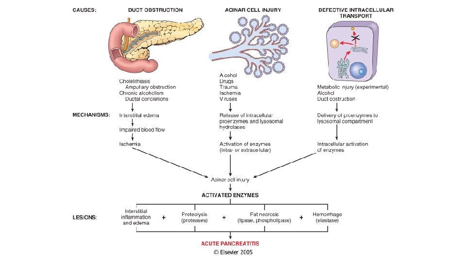
Pathophysiology
Proteolytic enzymes activated Autodigestion • In pancreatic parenchyma • endotoxins, exotoxins, viral infections, ischemia, anoxia, lysosomal calcium, and direct trauma • Activated proteolytic enzymes digest pancreatic and peri pancreatic tissues • also activate elastase and phospholipase A 2
Acute Pancreatitis TNF IL-1 IL-6 IL-8 N. O. PAF Complications: • Vascular leakage • Hypovolemia • Shock • ARDS • Acute renal tubular necrosis Other leucocyte products Oxygen radicals Elastase IFN-α, γ IL-10 IL-2
Clinical features
• Abdominal pain • Nausea, vomiting, and abdominal distention • Low-grade fever • Tachycardia • Hypotension • Jaundice • Erythematous skin nodules • Atelectasis, pleural effusion
• Abdominal tenderness and muscle rigidity • Diminished bowel sounds • A faint blue discoloration around the umbilicus (Cullen's sign) may occur as the result of hemoperitoneum • Blue-red-purple or green-brown discoloration of the flanks (Turner's sign) reflects tissue catabolism of hemoglobin
• Grey Turner sign • Cullen’s sign
Workup
Laboratory Data • serum amylase and lipase • >threefold clinch the diagnosis • No correlation between the severity and the degree of elevation • After three to seven days, values tend to return toward normal • Pancreatic isoamylase and lipase levels may remain elevated for 7 to 14 days
Markers of Severity within 24 Hours • SIRS [temperature >38° or < 36°C, Pulse > 90, Tachypnea > 24, WBC > 12, 000] • Hemoconcentration (Hct >44%) • BISAP (bedside index of severity in acute pancreatitis) • • • (B) Blood urea nitrogen (BUN) >22 mg% (I) Impaired mental status (S) SIRS: 2/4 present (A) Age >60 years (P) Pleural effusion • Organ Failure • • Cardiovascular: systolic BP <90 mm. Hg, heartrate >130 Pulmonary: Pao 2 <60 mm. Hg Renal: serum creatinine >2. 0 mg% Gastrointestinal: bleeding >500 m. L/24 hours
Severity • Risk Factors • Age > 60 years • Obesity, BMI > 30 • Comorbid disease • Markers during Hospitalization • Persistent organ failure • Pancreatic necrosis • Hospital-acquired infection
Laboratory Data • Leukocytosis (15, 000– 20, 000 leukocytes per L) • Hemoconcentration • with hematocrit values >44% • and/or blood urea nitrogen (BUN) level >22 mg/Dl • Hyperglycemia • decreased insulin release, • increased glucagon release, • increased output of adrenal glucocorticoids and catecholamines. • Hypocalcemia
Laboratory Data • Hyperbilirubinemia (>4. 0 mg/d. L) in 10% • is transient • return to normal in four to seven days • Serum alkaline phosphatase and aspartate aminotransferase levels are also transiently elevated • Elevated serum lactic dehydrogenase levels (>500 U/d. L) • Hypertriglyceridemia occurs in 5– 10%
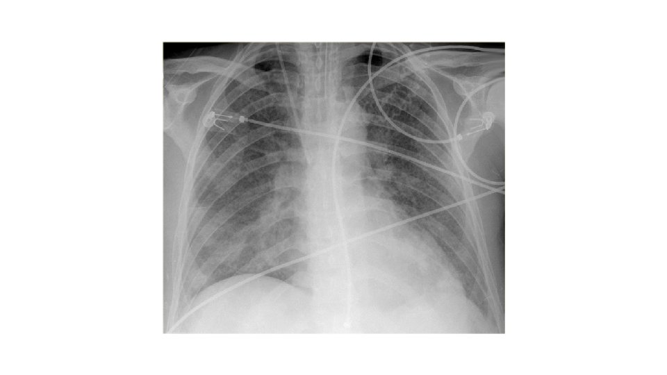
• Hypoxemia (arterial Po 2 60 mm. Hg), which may herald the onset of ARDS • Electrocardiogram ST-segment and T-wave abnormalities simulating myocardial ischemia.
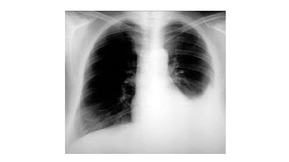
RADIOLOGY • CXR • Sympathetic pleural effusion • Atelectasis • A. R. D. S. • AXR • • • Sentinal loop Colon cutoff sign Duodenal ileus Calcifications Obscured psoas lines • 79% will have radiological signs !!!
CT Findings and Grading of Acute Pancreatitis [CT Severity Index (Ctsi)] Grade Findings Score A 0 Normal pancreas: normal size, sharply defined, smooth contour, homogeneous enhancement, retroperitoneal peripancreatic fat without enhancement B Focal or diffuse enlargement of the pancreas, contour may show irregularity, enhancement may be inhomogeneous but there is no peripancreatic 1 inflammation C Peripancreatic inflammation with intrinsic pancreatic abnormalities 2 D Intrapancreatic or extrapancreatic fluid collections 3 E Two or more large collections or gas in the pancreas or retroperitoneum 4
Necrosis score based on contrast-enhanced CT Necrosis (%) Score 0 0 <33% 2 33 -50% 4 >50% 6 CT severity index equals unenhanced CT score plus necrosis score; 6 = severe disease.
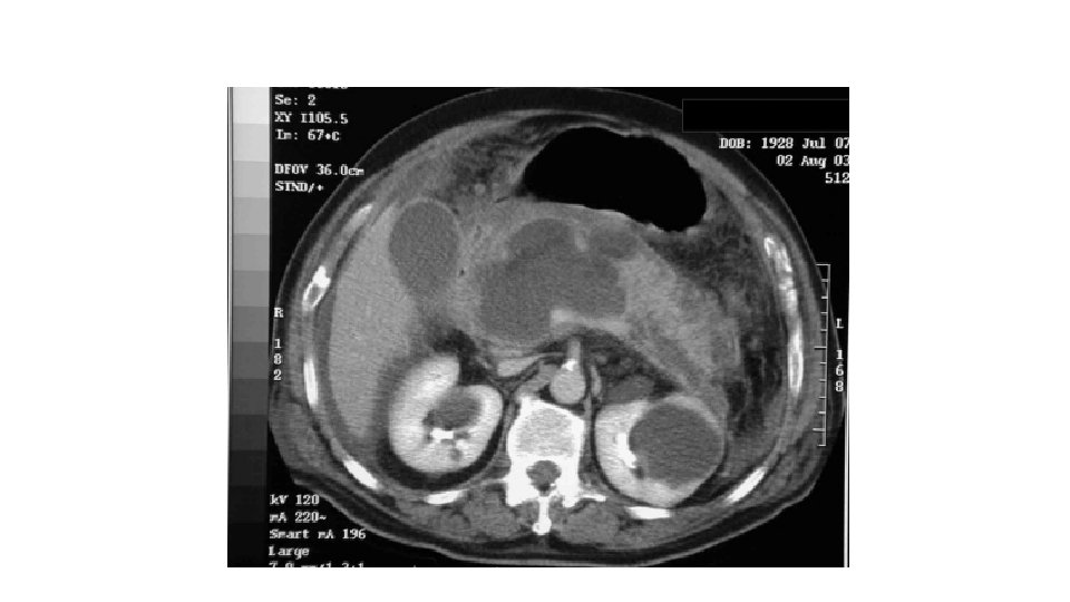
CT Scan of acute pancreatitis • CT shows significant swelling and inflammation of the pancreas
Diagnosis • Requires two of the following: • typical abdominal pain, • threefold or greater elevation in serum amylase and/or lipase level, • and/or confirmatory findings on cross-sectional abdominal imaging.
Differential diagnosis • • • (1) perforated viscus, especially peptic ulcer (2) acute cholecystitis and biliary colic (3) acute intestinal obstruction (4) mesenteric vascular occlusion (5) renal colic (6) myocardial infarction (7) dissecting aortic aneurysm (8) connective tissue disorders with vasculitis (9) pneumonia (10) diabetic ketoacidosis
Complications
Local complications • Necrosis • Sterile • Infected • Walled-off necrosis • Pancreatic fluid collections • Pancreatic abscess • Pancreatic pseudocyst • • Pain Rupture Hemorrhage Infection
Local complications • Obstruction of gastrointestinal tract (stomach, duodenum, colon) • Pancreatic ascites • Disruption of main pancreatic duct • Leaking pseudocyst • Involvement of contiguous organs by necrotizing pancreatitis • Massive intraperitoneal hemorrhage • Thrombosis of blood vessels (splenic vein, portal vein) • Bowel infarction • Obstructive jaundice
Systemic complications • Pulmonary • • • Pleural effusion Atelectasis Mediastinal abscess Pneumonitis Acute respiratory distress syndrome • Cardiovascular • • Hypotension Hypovolemia Sudden death Nonspecific ST-T changes in electrocardiogram simulating myocardial infarction
Systemic complications • Hematologic • Disseminated intravascular coagulation • Gastrointestinal hemorrhage • • Peptic ulcer disease Erosive gastritis Hemorrhagic pancreatic necrosis with erosion into major blood vessels Portal vein thrombosis, variceal hemorrhage • Renal • • Oliguria Azotemia Renal artery and/or renal vein thrombosis Acute tubular necrosis
Systemic complications • Metabolic • Hyperglycemia • Hypertriglyceridemia • Hypocalcemia • Encephalopathy • Sudden blindness (Purtscher's retinopathy) • Central nervous system • Psychosis • Fat emboli • • Fat necrosis Subcutaneous tissues (erythematous nodules) Bone Miscellaneous (mediastinum, pleura, nervous system)
HOW DO WE DETERMINE PROGNOSIS? • 80% Will recover without any complications • 20% Will develop severe cardio-pulmonary complications or septic complications • Prognostic assessment : • • Ranson Imrie APACHE 2 CRP • Classify into mild or severe acute pancreatitis ( Atlanta classification 1992)
Treatment • To limit severity of pancreatic inflammation • To interrupt the pathogenesis of complications • To support the patient and treat complications
TREATMENT • NON-OPERATIVE • To limit severity of pancreatic inflammation • Inhibition of pancreatic secretion • Nasogastric suction • Pharmacologic • Hypothermia • Pancreatic irradiation • Inhibition of pancreatic enzymes • Corticosteroids • Prostaglandins
TREATMENT • To interrupt the pathogenesis of complications • • • Antibiotics Antacids Heparin Low molecular weight dextran Vasopressin Peritoeal lavage
TREATMENT • To support the patient and treat complications • Restoration and maintenance of intravascular volume • Electrolyte replacement • Respiratory support • Nutritional support • Analgesia • Heparin
When to operate? • Diagnostic laparotomy • To limit the severity of the pancreatic inflammation • Biliary procedures • To interrupt the pathogenesis of complications • Pancreatic drainage • Pancreatic resection • Pancreatic debridement • Peritoneal lavage • To support the patient and treat complications • Drainage of pancreatic infection • Feeding jejunostomy • To prevent recurrent pancreatitis
Summary of treatment • All patients • • Nasogastric suction NPO Monitor and maintenance of intravascular volume Respiratory monitoring and support Antibiotics(selective) Early laparotomy only fordiagnosis Estimate prognosis by early signs • Patients with severe pancreatitis • • Peritoneal lavage Nutritional support Suspect and treat pancreatic sepsis Heparin if hypercoagulable
Chronic Pancreatitis 46
Definition and Prevalence • Defined as chronic inflammatory condition that causes irreversible damage to pancreatic structure and function. • Incurable • 5 To 27 Persons Per 100, 000 47
Etiology • Alcohol, 70% • Idiopathic (including tropical), 20% • Other, 10% • • Hereditary Hyperparathyroidism Hypertriglyceridemia Autoimmune pancreatitis Obstruction Trauma Pancreas divisum 48
Classification: 1. Calcific pancreatitis 2. Obstruction pancreatitis 3. Inflammatory pancreatitis 4. Auto immune pancreatitis 5. Asymptomatic fibrosis 6. Tropical pancreatitis 7. Hereditary pancreatitis 8. Idiopathic pancreatitis 49
Signs and Symptoms • Steady and Boring Pain • Not Colicky • Nausea /Vomiting • Anorexia- Most Common • Malabsorption And Weight Loss • Diabetes Mellitus 50
• Classic triad “ pancreatic calcification , steatorrhea , and diabetes mellitus” • Found in <1/3 • secretin stimulation test (abnormal when 60% or more of pancreatic exocrine function has been lost) • decreased serum trypsinogen (<20 ng/ml) or a fecal elastase level of <100 ug/mg of stool strongly suggests severe pancreatic insufficiency
Laboratory Studies Tests for Chronic Pancreatitis I. Measurement of pancreatic products in blood A. Enzymes B. Pancreatic polypeptide II. Measurement of pancreatic exocrine secretion A. Direct measurements 1. Enzymes 2. Bicarbonate B. Indirect measurement 1. Bentiromide test 2. Schilling test 3. Fecal fat, chymotrypsin, or elastase concentration 4. [14 C]-olein absorption 52
Imaging techniques • A. Plain film radiography of abdomen • B. Ultrasonography • C. Computed tomography • D. ERCP • E. MRCP • F. EUS
• Plain films : • Pancreatic calcifications : 30% • most common with alcoholic pancreatitis
Pancreatic calcifications. CT scan showing multiple, calcified, intraductal stones in a patient with hereditary chronic pancreatitis Endoscopic retrograde cholangiopancreatography in chronic pancreatitis. The pancreatic duct and its side branches are irregularly dilated 55
CT features • Pancreatic atrophy, calcifications, and main pancreatic duct dilation 56
ERCP • ERCP is a highly sensitive radiographic test for CP 57
MRCP • MRCP allows a noninvasive alternative to ERCP for imaging the pancreatic duct. 58
EUS is a minimally invasive test that allows simultaneous assessment of ductal and parenchymal structure 59
Treatment • Analgesia • Enzyme Therapy • Antisecretory Therapy • Neurolytic Therapy • Endoscopic Management • Surgical Therapy 60
Complications • Pseudocyst • Pancreatic Ascites • Pancreatic-Enteric Fistula • Head-of-Pancreas Mass • Splenic and Portal Vein Thrombosis 61
62
Management 63
A 45 -year-old man presents to the emergency room complaining of severe epigastric pain that radiates to his back and left scapula. He has had severe nausea and vomiting associated with the pain for 3 days. Nothing seems to relieve his pain. He drinks about a fifth of bourbon a day. He has not had a bowel movement in several days. He also complains of a very dry mouth. He is afebrile and normotensive with a heart rate of 110 beats/min. He is mildly tender on abdominal examination. His serum amylase level is 650 IU/L. Which of the following is the most appropriate initial step in the management of this patient? (A) Exploratory laparotomy and pancreatic débridement (B) Discharge and close follow up as outpatient (C) Obtain an abdominopelvic computed tomography (CT) scan (D) Perform esophagogastroduodenoscopy (EGD) (E) Admit for intravenous (IV) hydration and withhold oral feeding
A 48 -year-old, alcoholic male has been hospitalized over the last 20 years more than 30 times for episodes of acute pancreatitis. He now presents to the emergency department complaining of mid-back pain and horribly malodorous and frequent soft stools. He describes that the stools float in the toilet. He also explains that he has lost 30 lb over the last 6 months. Which of the following therapeutic measures is the best option to improve this patient's health maintenance? (A) Total pancreatectomy (B) Oral pancreatic enzyme replacement (C) Total parenteral nutrition (D) Octreotide therapy (E) High-fat diet