Head and Neck Parotid Gland Prof Dr Mohamed


 Cervical branch of")

 Parotid Posterior Facial Vein (Reteromandibular)")


 nodes Deep Parotid Lymph Nodes inside")


")
- Slides: 26
Head and Neck Parotid Gland Prof Dr. Mohamed El Fiky Associate professor of anatomy
Site, Shape, and Extension of Parotid Gland It is the largest of the salivary glands. Position: This is a wedge-shaped gland which lies below the auricle, between the ramus of the mandible and sternomastoid. The upper end of the gland is in contact with the external acoustic meatus. Surfaces of the gland: 1 - Lateral (superficial ) surface. 2 - Anteromedial surface. 3 - Posteromedial surface.
Divisions of Parotid Gland : Parotid gland is divided into 2 parts by the facial nerve : 1 - superficial part. 2 - deep part. Medial Pterygoid Facial Nerve Deep Lobe Ramus of Mandible Masseter obe L l a i c i f Super Accessory Parotid
Parotid Fascial Compartment Investing layer of Deep Cervical Fascia Parotid Gland
Stylomandibular Ligament Parotid Fascia
Parotid Gland d Glan r a l u ndib a m Sub Stylomandibular Ligament
1 - Surfaces : Posteromedial surface Anteromedial surface a. superficial b. anteromedial C- posteromedial Superficial surface 1 Borders : Base Medial border a. anterior b- posterior c-Medial 3 - Base and Apex Posterior border Apex Anterior border
Relations of Parotid Gland 1 Relations of the Superficial Surface 1 skin 2 superficial fascia 3 branchjes of great auricular nerve 4 - parotid lymph node 1 Skin 2 Superficial Fascia and Platysma 3 - Great Auricular Nerve
2 - Relations of Anteromedial surface Medial pterygoid Ramus of mandible Masseter Mastoid process 1 masseter muscle. 2 ramus of the mandible 3 - medial ptregoid Facial nerve trunk
3 - Relations of Posteromedial surface A- Mastoid Process B- Styloid Process Sternomastoid muscle Stylopharyngeus muscle Styloglossus muscle Posterior belly of digastric muscle Stylohyoid muscle C- Contents of carotid sheath Internal jugular vein Internal carotid artery Glossopharyngeal nerve Accessory nerve Vagus nerve Hypoglossal nerve
1 - relations of anterior Border Zygomatic branch of facial nerve Transverse facial artery Parotid duct and accessory parotid Upper buccal branch of facial nerve Lower buccal branch of facial nerve Mandibular branch of facial nerve
2 -Relatins of Posterior Border Mastoid Process Sternomastoid Muscle
3 -Relations of medial Border The Medial Border Separates anteromedial and posteromedial surfaces and is related to medial wall of pharynx
1 - Relations of the base Temporal branch of facial nerve Superficial temporal vein artery Auricultemporal nerve
2 - Relations of the apex Posterior facial vein (Retromandibular vein) Cervical branch of facial nerve) Anterior and Posterior divisions of posterior facial vein Posterior belly of digastric and stylohyoid muscles External carotid artery)
Structures within the Substance of parotid gland 1 - Terminal part of external carotid artery (deep). 2 - Retromandibular vein (intermediate). 3 - Facial nerve (superficial). a- Facial nerve Facial Nerve Temporal Branch. Zygomatic Branch Upper Buccal Branch Lower Buccal Branch Cervical Branch Mandibular Branch
b- Posterior Facial Vein (Reteromandibular) Parotid Posterior Facial Vein (Reteromandibular)
c- External carotid and superficial temporal arteries Superficial Temporal artery External carotid artery
Parotid Duct Parotid Papilla
Nerve Supply of Parotid Gland Tympanic Plexus Tympanic Branch Lesser Superficial Petrosal Nerve (Nerve of Jacobson) Inferior Otic Ganglion Salivary Nucleus Auriculotemporal Nerve V 3 Parotid Gland External Carotid Artery Glossopharyngeal Nerve Superior Cervical Ganglion
Lymph Drainage of Parotid Gland The pre-auricular (superficial) nodes Deep Parotid Lymph Nodes inside The Gland
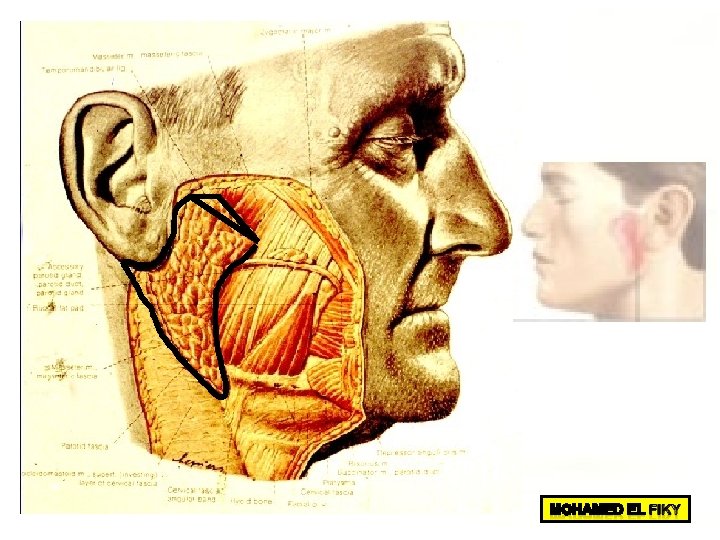
Surface anatomy of Parotid Gland
1 - Affection of Facial nerve within the Gland
1 - Affection of Facial nerve within the Gland
2 - Parotitis (Mumps)