Echocardiography in Right Ventricular Failure and Pulmonary Hypertension


 outflow trabecular apical")

")


 PAH Mean PAP ≥ 25")

 of the pulmonary microvasculature affecting 15 to 50")


 PAH Triad of right heart findings in PVD 98% of")


 Versus Non–WHO Group I: Anatomical Considerations VC US/DS/MAR 11/001 RA")


- Slides: 55
Echocardiography in Right Ventricular Failure and Pulmonary Hypertension Rasoul Azarfarin MD FACC Professor of Anesthesiology 2018
Overview of Right Ventricular Failure and Pulmonary Hypertension Normal Right Ventricle Definition of RV Failure Pathophysiology of RV Failure Etiology Diagnosis Treatment
Normal RV characteristics RV is not simply weak Left Ventricle Normal RV wall thickness: 2 -3 mm Normal LV wall thickness: 8 -11 mm RV contraction: peristaltic-like beginning at apex and moves in a wave toward the outflow tract Coronary perfusion: – Vascular supply : 2/3 RCA, 1/3 left branches – LV confined to diastole – RV with continuous perfusion (low pressure) Less O 2 requirements than the LV : less myocardic mass, less pre load and afterload
RV Anatomy
RV shape is complex and composed of: smooth muscular inflow (body) outflow trabecular apical region cannot image all 3 regions in 1 2 D imaging plane
Comprehensive Imaging of the RV: 14 views
Comprehensive Imaging of the RV: 14 views (2)
Right Ventricular Physiology
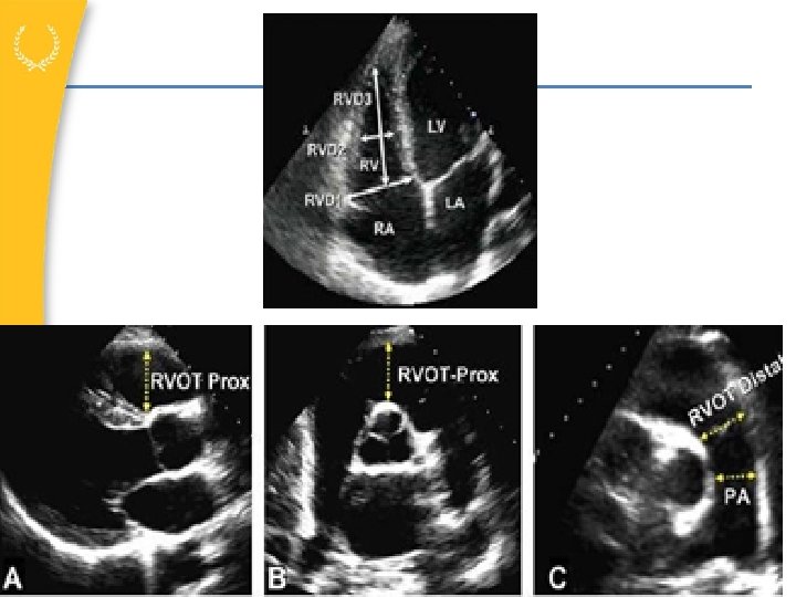
Measure the RV at end diastole from apical 4 chamber image demonstrating the maximum diameter of the RV without foreshortening
Normal RV and LV Hemodynamics
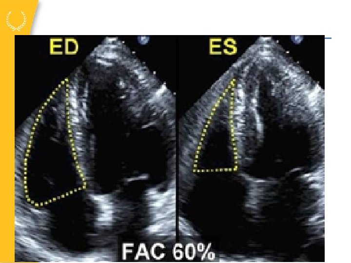
Fractional area change
Measure IVC 0. 5 – 3 cm from RA
Hemodynamic Values 1 -3 at Rest: Normal Ranges 120/80 mm Hg 20 -30/8 -12 mm Hg TPG = m. PAP – mean PCWP Normal TPG = <12 mm Hg TPG PVR = CO x 80 = dyne • sec • cm-5 Normal PVR = <3 Wood units (<240 dyne • sec • cm-5) CO = 4 -7 L/min m. PAP, <12 mm Hg 14 -20 mm Hg 3 -5 mm Hg 120/<8 mm Hg 20 -30/3 -5 mm Hg PCWP = LAP CO, cardiac output; LAP, left atrial pressure; m. PAP, mean pulmonary arterial pressure; PCWP, pulmonary capillary wedge pressure; PVR, pulmonary vascular resistance; TPG, transpulmonary gradient. US/DS/MAR 11/001 1. Kaluski et al. Heart Drug. 2003; 2: 225 -235. 2. Rubin. Chest. 1993; 104: 236 -250. 3. Mc. Laughlin and Mc. Goon. Circulation. 2006; 114: 1417 -1431.
Characterization of the Pulmonary Circulation 1 -3 Low pressure system – One fifth the pressures of systemic circulation, despite same CO as systemic circulation Low resistance – ~One seventh the resistance of systemic circulation High capacitance – Accommodates 5 - to 6 -fold ↑ in blood flow with only 2 -fold ↑ in PAP Dynamic vascular bed – V: Q matching; vasodilatation and recruitment Pulmonary vascular system CO, cardiac output; PAP, pulmonary arterial pressure; V: Q, ventilation-perfusion. US/DS/MAR 11/001 1. Kaluski et al. Heart Drug. 2003; 2: 225 -235. 2. Rubin. Chest. 1993; 104: 236 -250. 3. Mc. Laughlin and Mc. Goon. Circulation. 2006; 114: 1417 -1431.
Right Ventricular Failure Multiple Definitions – Results from structural or functional process decreasing the ability of the RV to pump blood into the pulmonary circulation – The clinical syndrome resulting from the inability of the right ventricle to provide adequate blood flow to the pulmonary circulation at a normal central venous filling pressure
Pathophysiology
Vicious cycle of auto-aggravation Piazza, G. et al. Chest 2005; 128: 1836 -1852
Pathophysiology of Failing RV Piazza, G. et al. Chest 2005; 128: 1836 -1852
Ventricular Interdependence • During systole, LV protrudes in RV • Surrounding pericardium with limited distensibility • Compliance of one ventricle can modify the other = Diastolic ventricular interaction
Etiologies of RV Failure J Am Coll Cardiol 2010; 56: 1435– 46
Factors affecting RV Function
Effects of Mechanical Ventilation Increased RV afterload due to positive pressure ventilation Hemodynamic failure frequently refractory in PAH patient put on MV In ARDS increase in m. PAP while increasing tidal volume and PEEP Permissive hypercapnia is deleterious (increase in m. PAP) Decreased venous return
Effects of PEEP on RV performance
Effect of high PEEP on RV
Pulmonary Hypertension: PAH Differentiation From PH (Dana Point Definitions) PAH Mean PAP ≥ 25 mm Hg + PCWP/LVEDP ≤ 15 mm Hg PH Mean PAP ≥ 25 mm Hg LVEDP, left ventricular end-diastolic pressure; PAP, pulmonary arterial pressure; PCWP, pulmonary capillary wedge pressure; PH, pulmonary hypertension. Badesch et al. J Am Coll Cardiol. 2009; 54: S 55 -S 66.
n RV PAH Definition Mean PAP >25 mm Hg PCWP ≤ 15 mm Hg Increased pressure load o m. PAP, ≥ 25 ≤ 15 mm Hg Increased RVSP m. PAP, mean PAP; PAP, pulmonary arterial pressure; PCWP, pulmonary capillary wedge pressure; RV, right ventricle; RVSP, right ventricular systolic pressure. Mc. Laughlin et al. Circulation. 2009; 119: 2250 -2294.
PAH Background Rare disease (orphan designation) of the pulmonary microvasculature affecting 15 to 50 people per million inhabitants in the Western world 1 Affects all races Affects all ages; however, most prevalent in 4 th and 5 th decades of life Higher prevalence in females Global burden of PAH may be underestimated because of: 1, 2 Underdiagnosis (eg, nondescript symptoms) Misdiagnosis (eg, asthma, left-heart disease) Increasing risk factors (eg, HIV infection, schistosomiasis) HIV, human immunodeficiency virus. 1. Humbert. Eur Respir J. 2007; 30: 1 -2. 2. Humbert et al. Chest. 2007; 132: 365 -367.
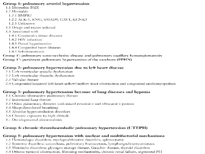
WHO Clinical Classification of PH: Dana Point 2008 Group 2―PH owing to left heart disease Systolic dysfunction Diastolic dysfunction Valvular disease Group 1―PAH Idiopathic PAH Heritable BMPR 2 ALK-1, endoglin (with or without HHT) Unknown Drug- and toxin-induced PAH associated with: Connective tissue diseases HIV infection Portal hypertension Congenital systemic to pulmonary shunts Schistosomiasis Group 3―PH owing to lung diseases or hypoxia Chronic obstructive pulmonary disease Interstitial lung disease Other pulmonary diseases with mixed restrictive and obstructive pattern Sleep-disordered breathing Alveolar hypoventilation disorder Chronic exposure to high altitude Developmental abnormalities Chronic hemolytic anemia Persistent pulmonary hypertension of newborn Pulmonary veno-occlusive disease or pulmonary capillary hemangiomatosis Group 4―Chronic thromboembolic PH Group 5―PH with unclear multifactorial mechanisms Hematologic disordersa Systemic disordersb Metabolic disordersc Othersd ALK-1, activin receptor-like kinase 1; BMPR 2, bone morphogenetic receptor type 2; HHT, hereditary hemorrhagic telangiectasia; HIV, human immunodeficiency virus; PH, pulmonary hypertension; WHO, World Health Organization. a Myeloproliferative disorders, splenectomy. b Sarcoidosis, pulmonary Langerhans cell histiocytosis. c Glycogen storage disease, Gaucher disease, thyroid disorders. d Tumoral obstruction, fibrosing mediastinitis, chronic renal failure. Simonneau et al. J Am Coll Cardiol. 2009; 54(1 suppl S): S 43 -S 54.
Diagnostic Studies Echo MRI Right Heart Cath
Key Role of Echocardiography Screening tool for PAH Diagnostic Approach to PAH – In conjunction with symptoms, chest x-ray, ECG, heart sounds, etc – Exclude secondary causes of PH – Detect preclinical disease Predicts prognosis Monitors efficacy of therapy ABG, arterial blood gas; ANA, antinuclear antibody; cath, catheterization; CHD, congenital heart disease; CPET, cardiopulmonary exercise testing; CT, computed tomographic; CTD, connective tissue disease; CXR, chest x-ray; ECG, electrocardiogram; Echo, echocardiogram; RAE, right atrial enlargement; RV, right ventricle; RVE, right ventricle enlargement; RVSP, right ventricular systolic pressure; TEE, transesophageal echocardiography; VHD, valvular heart disease. US/DS/MAR 11/001 1. Mc. Laughlin et al. Circulation. 2009; 119: 2250 -2294. 2. Bossone et al. Chest. 2005; 127: 1836 -1843. 3. Argiento et al. Eur Respir J. 2010; 35: 1273 -1278.
Echocardiography Assessments Septal position LV size and function Estimated RVSP RV size and function PA size RA and LA size TR Valvular and pericardial disease LA, left atrium; LV, left ventricle; PA, pulmonary artery; RA, right atrium; RV, right ventricle; RVSP, right ventricular systolic pressure; TR, tricuspid regurgitation. US/DS/MAR 11/001 1. Kaluski et al. Heart Drug. 2003; 2: 225 -235. 2. Rubin. Chest. 1993; 104: 236 -250. 3. Mc. Laughlin and Mc. Goon. Circulation. 2006; 114: 1417 -1431.
RV Undervalued in Echocardiography Relative to left heart disease, most echocardiography texts dedicate little space to evaluation of Pulmonary hypertension Right-sided cardiac function Right-sided valvular function Typical echo report provides no objective measurements of RV or RA size or RV function Physician may want to alert the lab if pulmonary hypertension is suspected to ensure appropriate focus on the RV and other important structures and parameters US/DS/MAR 11/001 echo, echocardiography; RA, right atrium; RV, right ventricle.
Representative Echocardiographs: Normal Versus PAH in Apical View Normal PAH Images and video courtesy of Paul Forfia, MD, Hospital of the University of Pennsylvania Heart and Vascular Center. US/DS/MAR 11/001
Echocardiographic Characterization of PAH Disease Progression Early stage PAH Moderate severity PAH Severe PAH Images and video courtesy of Paul Forfia, MD, Hospital of the University of Pennsylvania Heart and Vascular Center. US/DS/MAR 11/001
Echocardiographic Features of (Idiopathic) PAH Triad of right heart findings in PVD 98% of patients demonstrated RV enlargement 92% had RA enlargement 90% exhibit systolic (interventricular) septal flattening 76% had (qualitative) RV systolic dysfunction 70% of patients had “grade I” diastolic dysfunction (E<A) All patients demonstrated normal LV function <2% of patients had >mild mitral regurgitation LV, left ventricular; PVD, pulmonary vascular disease; RA, right atrial; RV, right ventricular. US/DS/MAR 11/001 Bossone et al. J Am Soc Echocardiogr. 1999; 12: 655 -662.
Echocardiography: TR Jet Velocity TR jet velocity = RV pressure – R AP RVSP 4(TR jet velocity)2 + RAP estimated from Doppler TR Jet Velocity – RA size – IVC inspiratory collapse Multicolored high flow jet with severe regurgitation IVC, inferior vena cava; RA, right atrium; RAP, right atrial pressure; RV, right ventricle; RVSP, right ventricular systolic pressure; TR, tricuspid regurgitation. US/DS/MAR 11/001 Image courtesy of Mardi Gomberg-Maitland, MD, MSc, University of Chicago Medical Center.
Advantages of Echocardiography in PAH Readily available Noninvasive Uses ultrasound only No ionizing radiation or contrast dye No contraindications due to pacemakers Early detection of abnormalities Easy to apply serially to monitor disease status Cheitlin et al. Circulation. 1997; 95: 1686 -1744. US/DS/MAR 11/001
Limitations of Echocardiography in PAH Experienced technicians and interpreting physicians are essential Consistency of skilled technicians/readers Applies to all imaging modalities Images can be limited in some patient populations The RV, the chamber of highest concern in PAH, is the least emphasized on “standard” echocardiography exam TR jet may be absent in some patients, thus precluding PASP assessment May overestimate or underestimate actual pulmonary arterial pressure Can estimate LVEDP (PCWP) or CO/CI, but may prove impractical CI, cardiac index; CO, cardiac output; LVEDP, left ventricular end diastolic pressure; PASP, pulmonary arterial systolic pressure; PCWP, pulmonary capillary wedge pressure; RV, right ventricle; TR, tricuspid regurgitation. R 11/0 1. Cheitlin et al. Circulation. 1997; 95: 1686 -1744. 2. Mc. Goon et al. Chest. 2004; 126: 14 S-34 S. US/DS/MA 01
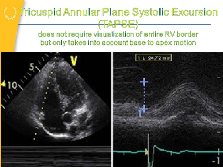
Right ventricle - size & function
Estimation of Pulmonary Pressure PA systolic pressure Tricuspid regurgitation jet velocity
Estimation of Pulmonary Pressure RA pressure IVC size
Cardiac MRI
Key Roles of Right Heart Catheterization Confirm diagnosis – Gold standard Evaluate severity of PAH Assess congenital heart defects Exclude left-sided heart disease Assess response to vasodilator challenge Assess key hemodynamic parameters US/DS/MAR 11/001 Swan-Ganz Catheter 1. Mc. Laughlin et al. Circulation. 2009; 119: 2250 -2294. 2. Tolle et al. Circulation. 2008; 118: 2183 -2189.
Importance of Right Heart Catheterization Vast majority of PH cases are not in WHO group I PAH characterized by – ↑PVR – ↑TPG – Normal left-sided filling pressures PAH PVH characterized by – ↑PCWP, usually normal TPG and PVR US/DS/MAR 11/001 LAP, left atrial pressure; LVEDP, left ventricular end diastolic pressure; PCWP, pulmonary capillary wedge pressure; PVH, pulmonary venous hypertension; PVR, pulmonary vascular resistance; TPG, transpulmonary pressure gradient; WHO. World Health Organization.
WHO Group I (PAH) Versus Non–WHO Group I: Anatomical Considerations VC US/DS/MAR 11/001 RA RV PA PC PV LA LV Ao Ao, aorta; HIV, human immunodeficiency virus; LA, left atrium; LV, left ventricle; PA, pulmonary artery; PC, pulmonary capillary bed; PV, pulmonary vein; RA, right atrium; RV, right ventricle; TE, thromboembolic; VC, vena cava; WHO, World Health Organization. a Glycogen storage disease, Gaucher disease, hereditary hemorrhagic telangiectasia, hemoglobinopathies, myeloproliferative disorders, splenectomy. Simonneau et al. J Am Coll Cardiol. 2004; 43(12 suppl): 5 S-12 S. Graphic adapted from http: //cme. medscape. com/viewarticle/530730.
Key PAH Assessments During RHCa PAP RVSP PCWP Calculations: TPG, PVR, CO, CI CO m. PAP PCWP RAP RVSP CI, cardiac index; CO, cardiac output; m. PAP, mean PAP; PAP, pulmonary arterial pressure; PCWP, pulmonary capillary wedge pressure; PVR, pulmonary vascular resistance; RAP, right atrial pressure; RHC, right heart catheterization; RVSP, right ventricular systolic pressure; TPG, transpulmonary gradient. a RHC to measure oxygen saturations, generate Fick CO, and confirm wedge position. US/DS/MAR 11/001 Mc. Goon et al. Chest. 2004; 126: 14 S-34 S.
RHC: Important Findings Elevation in PCWP PVH: limits treatment for PAH Elevation in RAP Decrease in CO/CI Aggressive early management suggested Elevation of PCWP with vasodilator Pulmonary veno-occlusive disease (PVOD) LV diastolic dysfunction CI, cardiac index; CO, cardiac output; LV, left ventricular; PCWP, pulmonary capillary wedge pressure; PVH, pulmonary venous hypertension; RAP, right atrial pressure. US/DS/MAR 11/001
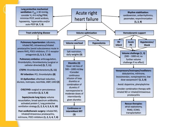
Treatment of RV Failure General Supportive ICU Care – Infection control – DVT Prophylaxis – Daily interruptions of sedation – Na and fluid restriction Optimize volume status – Hemodynamic guidance helpful – Avoid boluses if able Attenuate Hypoxic Pulmonary vasoconstriction Mechanical Ventilation Strategies J Am Coll Cardiol 2010; 56: 1435– 46
Strategies to Improve RV Function
Summary Right Ventricular Failure significant in ICU Setting Diagnosis of Pulmonary Hypertension is very common in obese patient – OSA is predominant underlying condition – Compliance – Echo is good screening tool – RHC is gold standard PAH diagnosis merits referral to specialized treatment center