Good morning THORACIC CAGE THORACIC WALL INTERCOSTAL SPACE


- Slides: 46
Good morning
THORACIC CAGE, THORACIC WALL & INTERCOSTAL SPACE
n n n Thoracic inlet Suprapleural membrane Intercostal space Typical Intercostal nerve Internal thoracic artery
THORAX n The part of the human body between the neck and the abdomen, partially encased by the ribs and containing the heart and lungs.
THORACIC CAGE/BONY CAGE BOUNDARIES n n n Anteriorly: Sternum On either sides: Ribs and costal cartilages. Posteriorly: Thoracic vertebrae & intervertebral discs. Thoracic inlet Thoracic outlet
Shape 10 cm 5 cm
Superior aperture/Inlet of thorax n n n Definition Boundaries Partition at the inlet Structures passing Applied aspect The narrow upper end of the thorax, which is continuous with the neck.
Partition of the inlet
Structures passing
Thoracic inlet syndrome n Compression of brachial plexus, subclavian vessels.
Inferior aperture/outlet of thorax n n Boundaries Thoracoabdominal diaphragm
Functions n n n Protects essential organs of respiration and circulation. Liver, stomach and spleen (subdiaphragmatic organs) are sheltered. Active participation in respiration.
Coverings of thorax n n n Skin Superficial fascia Deep fascia Extrinsic muscles Intrinsic muscles/Intercostal muscles.
Extrinsic muscles
INTERCOSTAL SPACES n n n n The space between 2 adjacent ribs and their costal cartilages. 11 intercostal spaces bounded by 12 ribs on each side. Last 2 spaces are open in front. Each spaces contains: INTERCOSTAL MUSCLES INTERCOSTAL VESSELS INTERCOSTAL NERVE.
RIB - Osteology
Introduction n Intercostal Muscles. n Intercostal Nerves. n Intercostal Vessels. n Intercostal Lymphatics.
Intercostal Muscles.
Intercostal Muscles. n 3 Muscles. n n n External Intercostal muscle. Internal Intercostal muscle. Transversus Thoracis muscle. n n n Subcostalis Intercostalis intimi Sternocostalis
External Intercostal Muscle. n Origin: n n Insertion: n n Lower border of the upper rib. Outer lip of the upper border of the lower rib. Extent: Tubercle of the rib (post) – Costochondral Junction (ant). n Direction of fibers. n Downwards forwards and medially.
Internal Intercostal Muscle. n Origin: n n Insertion: n n Floor of the costal groove of the upper rib. Inner lip of the upper border of the lower rib. Extent: Lateral border of the sternum (ant) – Angle of the rib (post). n Direction of the fibres: Downwards backwards and laterally
Transversus Thoracis muscle n n Subcostalis Origin: n n Inner surface of the rib near the angle. Insertion: n n Inner surface of two or three ribs below. Extent: n Confined to the posterior part of the lower intercostal spaces only.
Transversus Thoracis muscle n Intercostalis intimi. n Origin: n n Insertion: n n Middle two-fourths of the ridge above the costal groove. Inner lip of the upper border of the rib below. Extent: n n Confined to the middle two fourths of the intercostal spaces. Absent in upper two spaces
Transversus Thoracis muscle n Sternocostalis n Origin: n n Lower one third of the posterior surface of the body of the sternum. Posterior surface of the xiphoid Posterior surface of the costal cartilages of the lower 3 or 4 true ribs Insertion: n Costal cartilages of the 2 nd and 6 th ribs.
Intercostal Muscles. n Nerve Supply: Intercostal Nerves. n Actions: n n n Prevent retraction and bulging. Ext. Intercostals, Intercostal help to elevate the rib during inspiration. Intercostals and transversus thoracis depress the ribs during expiration.
Intercostal Nerves
Intercostal Nerves n n n n The anterior primary rami of T 1 – T 11. 1, 2 – Upper limb T 1 –divides two-upper branch-brachial plexus &lower part-intercostal space. T 2 – intercostobrachial nerve 3 -6 – Typical Intercostal nerves. 7 -11 – Abdominal wall Anterior rami of T 12 – Subcostal Nerve.
Typical Intercostal nerve n Below neck of the rib enters the costal groove. n In the groove (VAN) n n Sternum: Crosses the int thoracic artery and the sternocostalis muscle. Pierces the intercostal muscle, ext. Intercostal membrane and the pectoralis major muscle. Terminates as the Anterior Cutaneous nerve of the thorax
course
Typical Intercostal nerve n Muscular branches: n n n Numerous branches, Supplies the intercostal muscles, transverse thoracis and serratus posterior superior Collateral Branch near the angle of the rib. Supplies the parietal pluera, parietal peritoneum and the periosteum of the rib.
Typical Intercostal nerve n Cutaneous branches: n Lateral cutaneous branch n n Anterior cutaneous branch n n Arises from the angle of the rib- mid axillary line. Ant and post branch Supplies skin Arises at the side of the sternum. Medial and Lateral branches. Communicating branches Sympathetic ganglion -White rami communicans & Grey rami communicans.
Intercostal Arteries n n Anterior Intercostal Arteries. Posterior Intercostal Arteries.
Anterior Intercostal Arteries. n n n 9 spaces. 2 in each space 1 -6 – arise from internal thoracic artery. 7 -9 – arise from musculophrenic artery. Upper arteries end by anatamosing with the posterior intercostal arteries at the costochondral junction. Lower arteries end by anastamosing with the collateral branches of the posterior intercostal arteries.
Posterior Intercostal Arteries. n n n 11 in number on each side. 1 st and 2 nd arise form the superior intercostal artery 3 rd to 11 th arise form the descending aorta.
Posterior Intercostal Arteries. n Course and relations: n Front of the vertebrae: n n Right is longer than left Rt: Passes behind the oesophagus, thoracic duct, azygous vein, and sympathetic chain. Lt: Passes behind the hemiazygous vein and the sympathetic chain. Intercostal Space: n Accompanied by the artery and vein [VAN]
Posterior Intercostal Arteries. n Termination: n n Ends at the costochondral junction by anastomosing with the upper anterior intercostal artery of that space. Branches: n n n Dorsal branch Collateral branch Muscular branches Lateral Cutaneous branch Mammary branches Rt. Bronchial branch
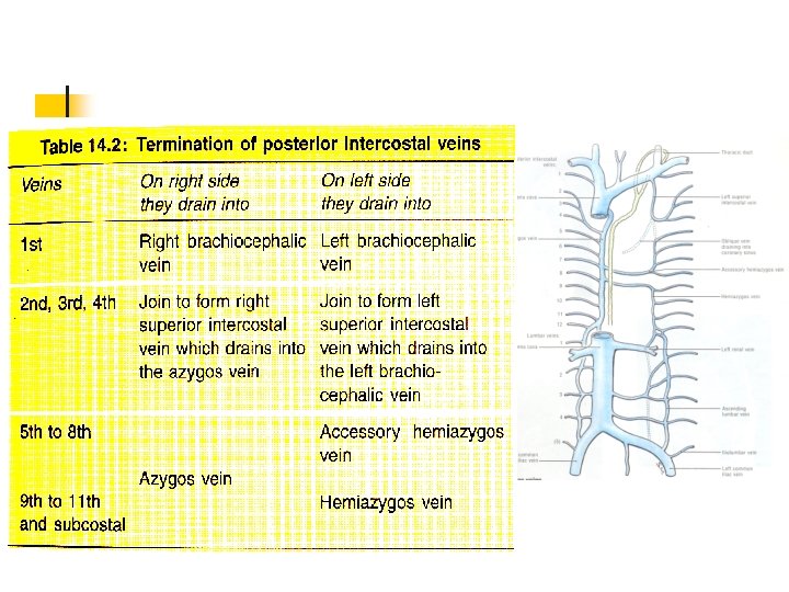
Intercostal Veins n Anterior Intercostal Veins. n n n Upper 9 spaces. 2 in number Upper 6 - Drains into internal thoracic vein Lower 3 –Musculophrenic vein Posterior Intercostal Veins. n One vein and its collateral in each intercostal space.
Intercostal Lymphatics n Lymphatics from the anterior part n n n Anterior intercostal nodes Internal mammary nodes Efferents unite with tracheobronchial and brachiocephalic nodes Rt. Side – Subclavian trunk, Left side – Thoracic duct Lymphatics from the posterior part n n n Posterior intercostal nodes Efferents of upper 4 – Lt - thoracic duct, Rt – Rt. Lymphatic duct. Efferents of the lower 4 – Cisterna Chyli.
Applied anatomy n Root pain / Girdle pain/intercostal neuralgia Irritation of the nerves which refers the pain to the front of the chest & abdomen. Causes n Fracture of the ribs. n Herpes –vesicular eruptions along the entire course. n
APPLIED ANATOMY n n Cardiac pain-referred-medial side of left arm-intercostobrachial nerve. Cold abscess-tuberculosis-neurovascular bundle-three sites of the exit of the branches.
Internal thoracic artery n n n The internal thoracic artery arises from the subclavian artery near its origin. It travels downward on the inside of the ribcage, approximately a centimeter from the sides of the sternum. It is accompanied by the internal thoracic vein.
n n It runs deep to the internal intercostal muscles, but superficial to the transverse thoracic muscles. It continues downward until it divides into the musculophrenic artery and the superior epigastric artery around the sixth intercostal space.
Branches n n n n Mediastinal branches Thymic branches Pericardiacophrenic artery travels with the phrenic nerve Sternal branches Perforating branches Anterior intercostal branches. Musculophrenic artery roughly follows the costal margin Superior epigastric artery
Applied anatomy n graft for heart surgeries