
Heart Failure PARTNERS IN HEALTH C HRONIC C

- Slides: 115
Heart Failure PARTNERS IN HEALTH C HRONIC C ARE TRAINING FOR D ISTRICT H OSPITAL NURSE S 2013
Unit Content �Session 1: Introduction to normal heart function and heart failure �Session 2: Categories of heart failure and treatment �Session 3: Rheumatic Heart Disease �Session 4: Heart failure care at District and Health Center levels
Session 1: Introduction to normal heart function and heart failure By the end of this session, participants will be able to: �Describe the normal function of the heart �Describe heart failure, and its symptoms and signs �Identify decompensated heart failure, and its precipitants �Classify severity of heart failure by functional class
Session 1: Introduction to normal heart function and heart failure By the end of this session, participants will be able to: �Describe the normal function of the heart �Describe heart failure, and its symptoms and signs �Identify decompensated heart failure, and its precipitants �Classify severity of heart failure by functional class
Anatomy of the Heart Chambers Valves Major blood vessels
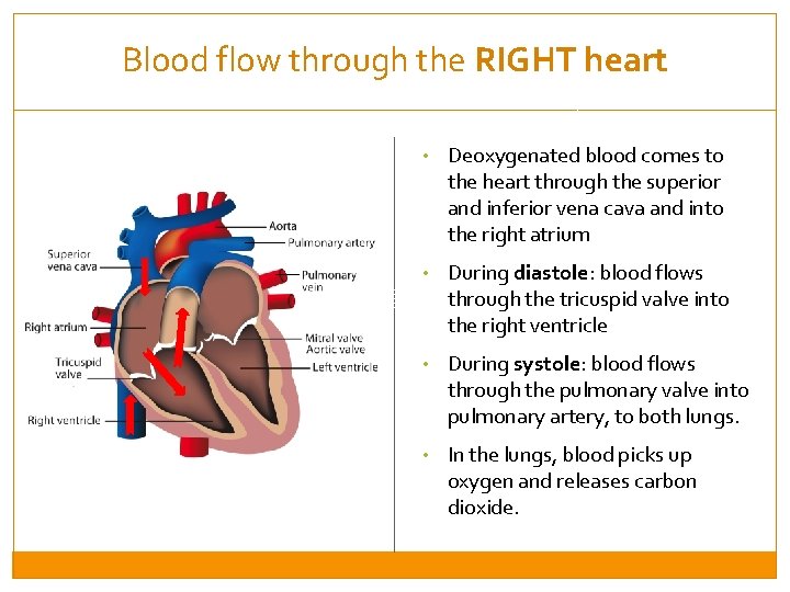
Blood flow through the RIGHT heart • Deoxygenated blood comes to the heart through the superior and inferior vena cava and into the right atrium • During diastole: blood flows through the tricuspid valve into the right ventricle • During systole: blood flows through the pulmonary valve into pulmonary artery, to both lungs. • In the lungs, blood picks up oxygen and releases carbon dioxide.
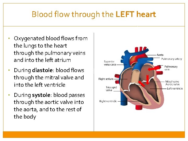
Blood flow through the LEFT heart • Oxygenated blood flows from the lungs to the heart through the pulmonary veins and into the left atrium • During diastole: blood flows through the mitral valve and into the left ventricle • During systole: blood passes through the aortic valve into the aorta, and to the rest of the body
Session 1: Introduction to normal heart function and heart failure By the end of this session, participants will be able to: �Describe the normal function of the heart �Describe heart failure, and its symptoms and signs �Identify decompensated heart failure, and its precipitants �Classify severity of heart failure by functional class
Heart Failure When the heart fails, cardiac output decreases! • Blood pressure drops • Heart rate increases as heart tries to compensate • Fluid leaks from the veins into the tissues (typically in legs, lungs and abdomen) • Volume overload worsened by high salt intake • In very severe cases, the heart can no longer pump enough blood to meet body’s demands, resulting in: o cold extremities, kidney failure, confusion
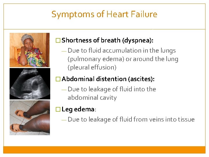
Symptoms of Heart Failure � Shortness of breath (dyspnea): — Due to fluid accumulation in the lungs (pulmonary edema) or around the lung (pleural effusion) � Abdominal distention (ascites): — Due to leakage of fluid into the abdominal cavity � Leg edema: — Due to leakage of fluid from veins into tissue
Symptoms of Heart Failure �Orthopnea/Paroxysmal Nocturnal Dyspnea Difficulty breathing while laying flat Due to more blood going to the kidneys when the patient is laying flat Patients may use several pillows under their head when sleeping Patients may wake up suddenly in the night with SOB �Nocturia Urination during the night Due to more blood going to the kidneys when the patient is laying flat �In children Can present as cyanosis, lethargy, poor feeding, or poor weight gain
Signs of Heart Failure: Vital Signs and General Appearance Rapid breathing rate (tachypnea) Low or high BP Low or high heart rate Low oxygen (hypoxia, cyanosis) General appearance, severity of dyspnea: • Dyspnea at rest • Coughing • Inability to lie flat • Use of accessory muscles • Confusion, hypotonia, lethargy Hypoxic respiratory failure is an emergency!
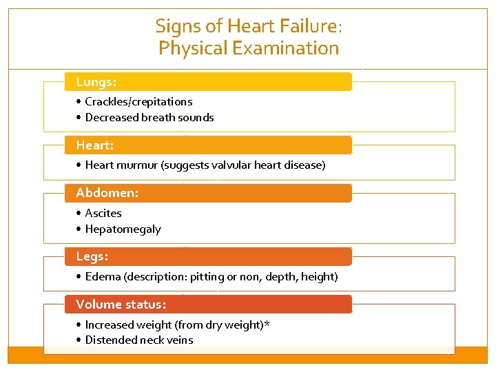
Signs of Heart Failure: Physical Examination Lungs: • Crackles/crepitations • Decreased breath sounds Heart: • Heart murmur (suggests valvular heart disease) Abdomen: • Ascites • Hepatomegaly Legs: • Edema (description: pitting or non, depth, height) Volume status: • Increased weight (from dry weight)* • Distended neck veins
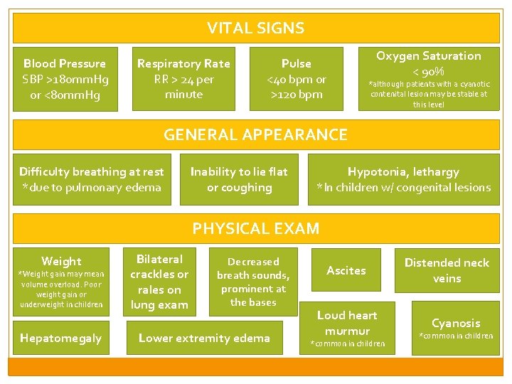
VITAL SIGNS Blood Pressure SBP >180 mm. Hg or <80 mm. Hg Respiratory Rate RR > 24 per minute Pulse <40 bpm or >120 bpm Oxygen Saturation < 90% *although patients with a cyanotic contenital lesion may be stable at this level GENERAL APPEARANCE Difficulty breathing at rest *due to pulmonary edema Inability to lie flat or coughing Hypotonia, lethargy *In children w/ congenital lesions PHYSICAL EXAM Weight *Weight gain may mean volume overload. Poor weight gain or underweight in children Hepatomegaly Bilateral crackles or rales on lung exam Decreased breath sounds, prominent at the bases Lower extremity edema Ascites Loud heart murmur *common in children Distended neck veins Cyanosis *common in children
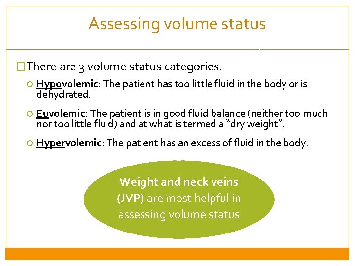
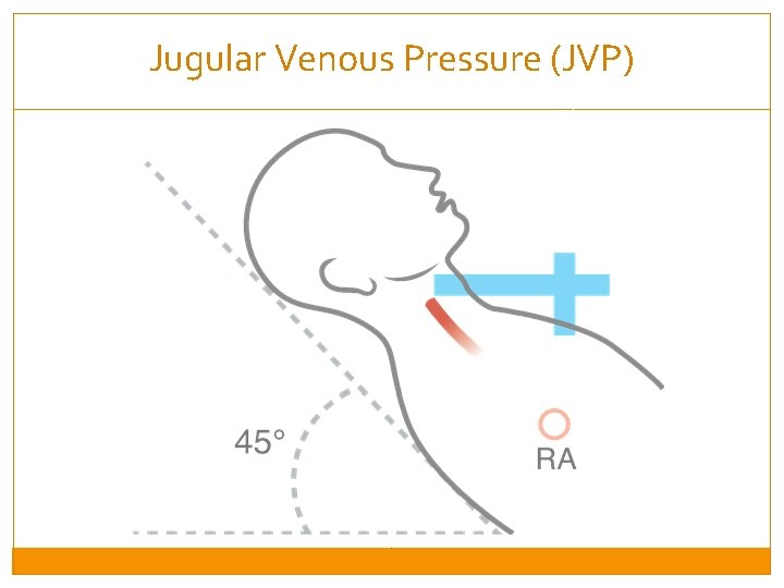
Assessing volume status �There are 3 volume status categories: Hypovolemic: The patient has too little fluid in the body or is dehydrated. Euvolemic: The patient is in good fluid balance (neither too much nor too little fluid) and at what is termed a “dry weight”. Hypervolemic: The patient has an excess of fluid in the body. Weight and neck veins (JVP) are most helpful in assessing volume status
Jugular Venous Pressure (JVP)
Session 1: Introduction to normal heart function and heart failure By the end of this session, participants will be able to: �Describe the normal function of the heart �Describe heart failure, and its symptoms and signs �Identify decompensated heart failure, and its precipitants �Classify severity of heart failure by functional class
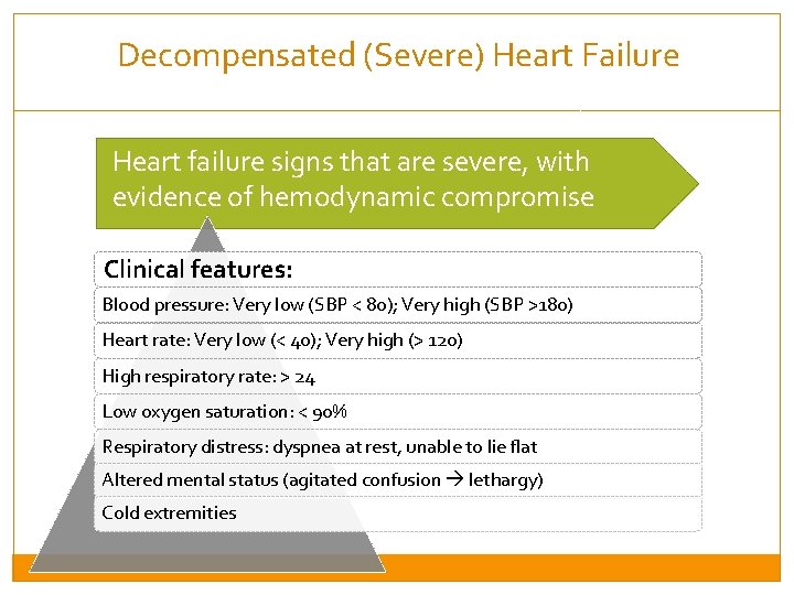
Decompensated (Severe) Heart Failure Heart failure signs that are severe, with evidence of hemodynamic compromise Clinical features: Blood pressure: Very low (SBP < 80); Very high (SBP >180) Heart rate: Very low (< 40); Very high (> 120) High respiratory rate: > 24 Low oxygen saturation: < 90% Respiratory distress: dyspnea at rest, unable to lie flat Altered mental status (agitated confusion lethargy) Cold extremities
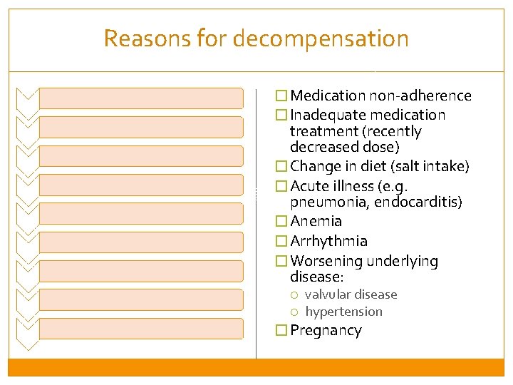
Reasons for decompensation � Medication non-adherence � Inadequate medication treatment (recently decreased dose) � Change in diet (salt intake) � Acute illness (e. g. pneumonia, endocarditis) � Anemia � Arrhythmia � Worsening underlying disease: valvular disease hypertension � Pregnancy
Session 1: Introduction to normal heart function and heart failure By the end of this session, participants will be able to: �Describe the normal function of the heart �Describe heart failure, and its symptoms and signs �Identify decompensated heart failure, and its precipitants �Classify severity of heart failure by functional class
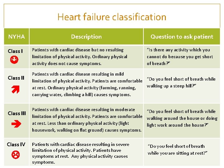
Heart failure classification NYHA Class III Class IV Description Patients with cardiac disease but no resulting limitation of physical activity. Ordinary physical activity does not cause symptoms. Question to ask patient “Is there any activity which you cannot do because you get short of breath? ” Patients with cardiac disease resulting in mild limitation of physical activity. Patients are comfortable “Do you feel short of breath while walking up a steep hill? ” at rest. Ordinary physical activity (farming, running, carrying water, climbing a hill) causes symptoms. Patients with cardiac disease resulting in moderate “Do you feel short of breath while limitation of physical activity. Patients are comfortable walking around the house or doing at rest. Less than ordinary physical activity (light work around the house? ” housework, walking on flat ground) causes symptoms. Patients with cardiac disease resulting in severe limitation of physical activity. Patients have symptoms at rest. Any physical activity causes symptoms. “Do you feel short of breath while you are sitting at rest? ”
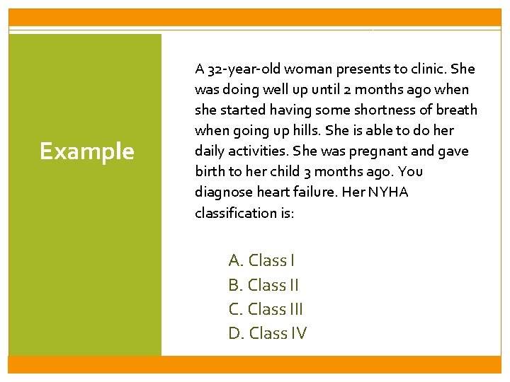
Example A 32 -year-old woman presents to clinic. She was doing well up until 2 months ago when she started having some shortness of breath when going up hills. She is able to do her daily activities. She was pregnant and gave birth to her child 3 months ago. You diagnose heart failure. Her NYHA classification is: A. Class I B. Class II C. Class III D. Class IV
Session 2: Types of Heart Failure and Treatment By the end of this session, participants will be able to: �Understand the major categories of heart failure �Understand the principles of fluid management, and use of diuretics �Understand the mortality-reducing medications, and adjunctive therapy �Understand management of specific types of heart failure
Session 2: Types of Heart Failure and Treatment By the end of this session, participants will be able to: �Understand the major categories of heart failure �Understand the principles of fluid management, and use of diuretics �Understand the mortality-reducing medications, and adjunctive therapy �Understand management of specific types of heart failure
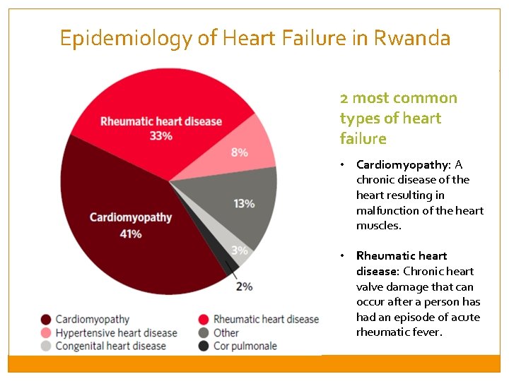
Epidemiology of Heart Failure in Rwanda 2 most common types of heart failure • Cardiomyopathy: A chronic disease of the heart resulting in malfunction of the heart muscles. • Rheumatic heart disease: Chronic heart valve damage that can occur after a person has had an episode of acute rheumatic fever.
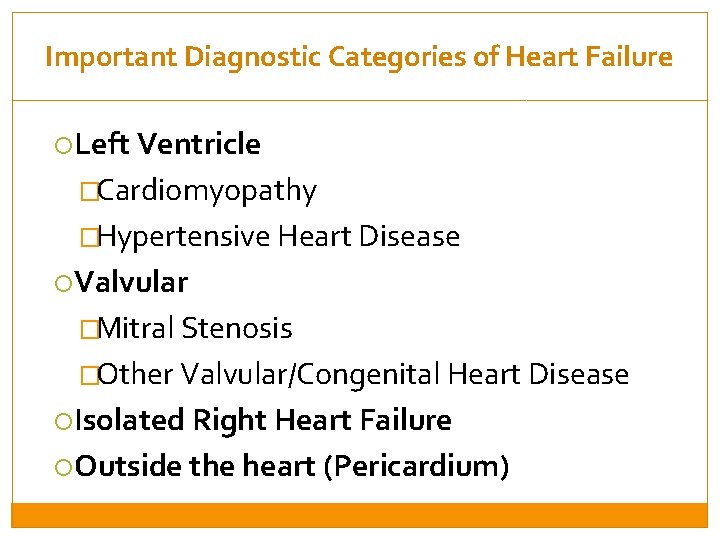
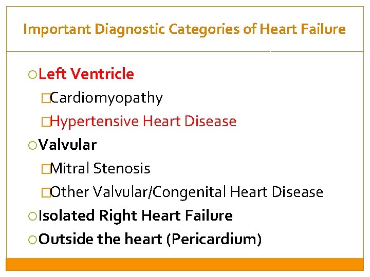
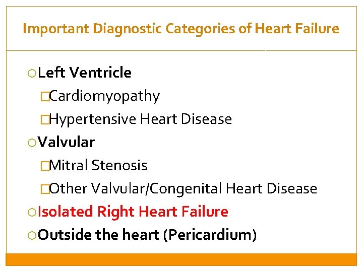
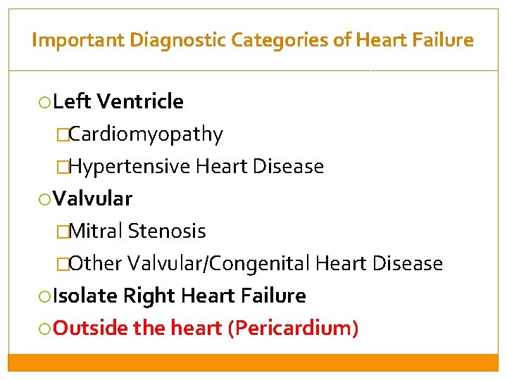
Important Diagnostic Categories of Heart Failure Left Ventricle �Cardiomyopathy �Hypertensive Heart Disease Valvular �Mitral Stenosis �Other Valvular/Congenital Heart Disease Isolated Right Heart Failure Outside the heart (Pericardium)
Important Diagnostic Categories of Heart Failure Left Ventricle �Cardiomyopathy �Hypertensive Heart Disease Valvular �Mitral Stenosis �Other Valvular/Congenital Heart Disease Isolated Right Heart Failure Outside the heart (Pericardium)
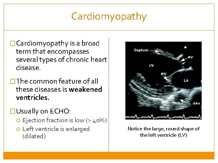
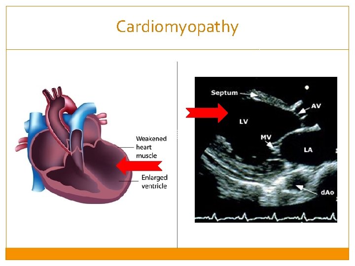
Cardiomyopathy �Cardiomyopathy is a broad term that encompasses several types of chronic heart disease. �The common feature of all these diseases is weakened ventricles. �Usually on ECHO: Ejection fraction is low (> 40%) Left ventricle is enlarged (dilated) Notice the large, round shape of the left ventricle (LV)
Cardiomyopathy
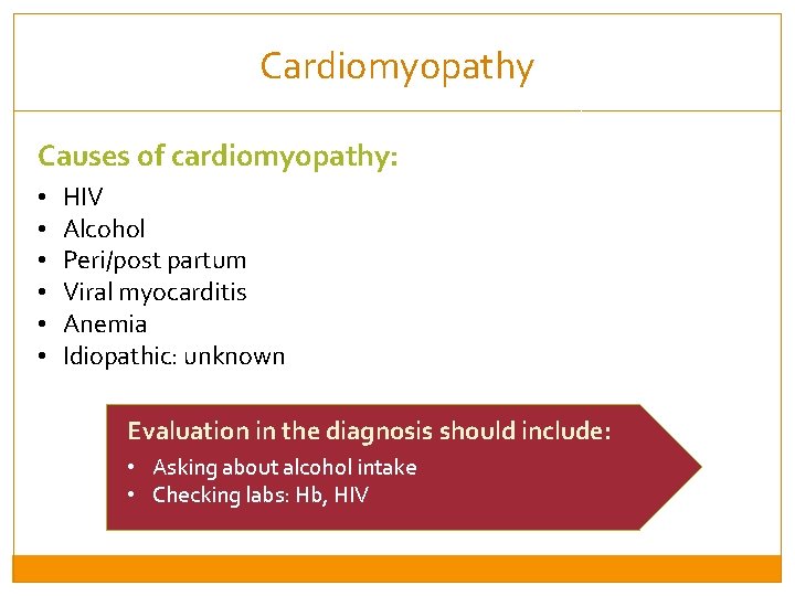
Cardiomyopathy Causes of cardiomyopathy: • • • HIV Alcohol Peri/post partum Viral myocarditis Anemia Idiopathic: unknown Evaluation in the diagnosis should include: • Asking about alcohol intake • Checking labs: Hb, HIV
Important Diagnostic Categories of Heart Failure Left Ventricle �Cardiomyopathy �Hypertensive Heart Disease Valvular �Mitral Stenosis �Other Valvular/Congenital Heart Disease Isolated Right Heart Failure Outside the heart (Pericardium)
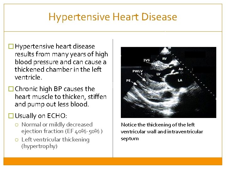
Hypertensive Heart Disease � Hypertensive heart disease results from many years of high blood pressure and can cause a thickened chamber in the left ventricle. � Chronic high BP causes the heart muscle to thicken, stiffen and pump out less blood. � Usually on ECHO: Normal or mildly decreased ejection fraction (EF 40%-50% ) Left ventricular thickening (hypertrophy) Notice thickening of the left ventricular wall and intraventricular septum
Important Diagnostic Categories of Heart Failure Left Ventricle �Cardiomyopathy �Hypertensive Heart Disease Valvular �Mitral Stenosis �Other Valvular/Congenital Heart Disease Isolated Right Heart Failure Outside the heart (Pericardium)
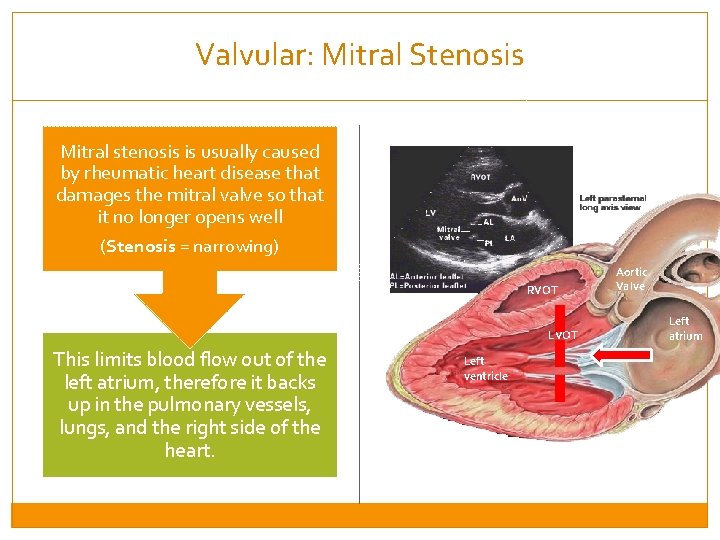
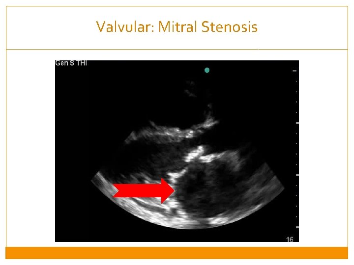
Valvular: Mitral Stenosis Mitral stenosis is usually caused by rheumatic heart disease that damages the mitral valve so that it no longer opens well (Stenosis = narrowing) RVOT LVOT This limits blood flow out of the left atrium, therefore it backs up in the pulmonary vessels, lungs, and the right side of the heart. Left ventricle Aortic Valve Left atrium
Valvular: Mitral Stenosis
Important Diagnostic Categories of Heart Failure Left Ventricle �Cardiomyopathy �Hypertensive Heart Disease Valvular �Mitral Stenosis �Other Valvular/Congenital Heart Disease Isolated Right Heart Failure Outside the heart (Pericardium)
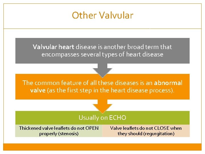
Other Valvular heart disease is another broad term that encompasses several types of heart disease The common feature of all these diseases is an abnormal valve (as the first step in the heart disease process). Usually on ECHO Thickened valve leaflets do not OPEN properly (stenosis) Valve leaflets do not CLOSE when they should (regurgitation)
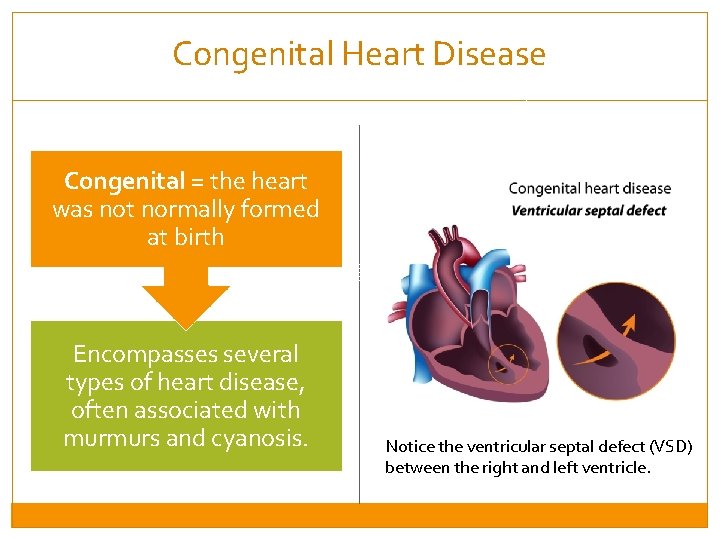
Congenital Heart Disease Congenital = the heart was not normally formed at birth Encompasses several types of heart disease, often associated with murmurs and cyanosis. Notice the ventricular septal defect (VSD) between the right and left ventricle.
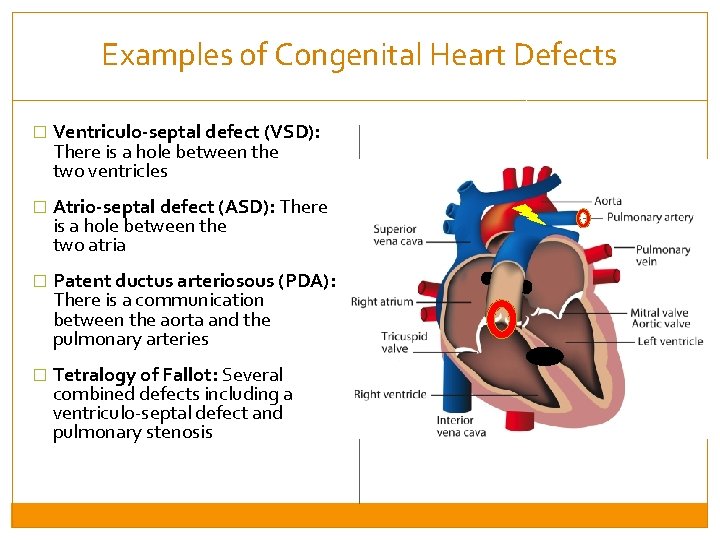
Examples of Congenital Heart Defects � Ventriculo-septal defect (VSD): There is a hole between the two ventricles � Atrio-septal defect (ASD): There is a hole between the two atria � Patent ductus arteriosous (PDA): There is a communication between the aorta and the pulmonary arteries � Tetralogy of Fallot: Several combined defects including a ventriculo-septal defect and pulmonary stenosis
Important Diagnostic Categories of Heart Failure Left Ventricle �Cardiomyopathy �Hypertensive Heart Disease Valvular �Mitral Stenosis �Other Valvular/Congenital Heart Disease Isolated Right Heart Failure Outside the heart (Pericardium)
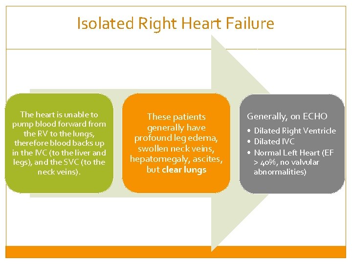
Isolated Right Heart Failure The heart is unable to pump blood forward from the RV to the lungs, therefore blood backs up in the IVC (to the liver and legs), and the SVC (to the neck veins). These patients generally have profound leg edema, swollen neck veins, hepatomegaly, ascites, but clear lungs Generally, on ECHO • Dilated Right Ventricle • Dilated IVC • Normal Left Heart (EF > 40%, no valvular abnormalities)
Important Diagnostic Categories of Heart Failure Left Ventricle �Cardiomyopathy �Hypertensive Heart Disease Valvular �Mitral Stenosis �Other Valvular/Congenital Heart Disease Isolate Right Heart Failure Outside the heart (Pericardium)
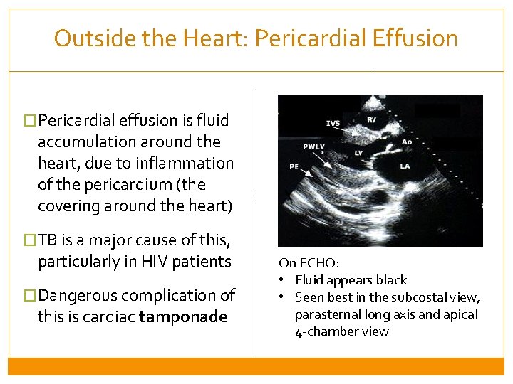
Outside the Heart: Pericardial Effusion �Pericardial effusion is fluid accumulation around the heart, due to inflammation of the pericardium (the covering around the heart) �TB is a major cause of this, particularly in HIV patients �Dangerous complication of this is cardiac tamponade On ECHO: • Fluid appears black • Seen best in the subcostal view, parasternal long axis and apical 4 -chamber view
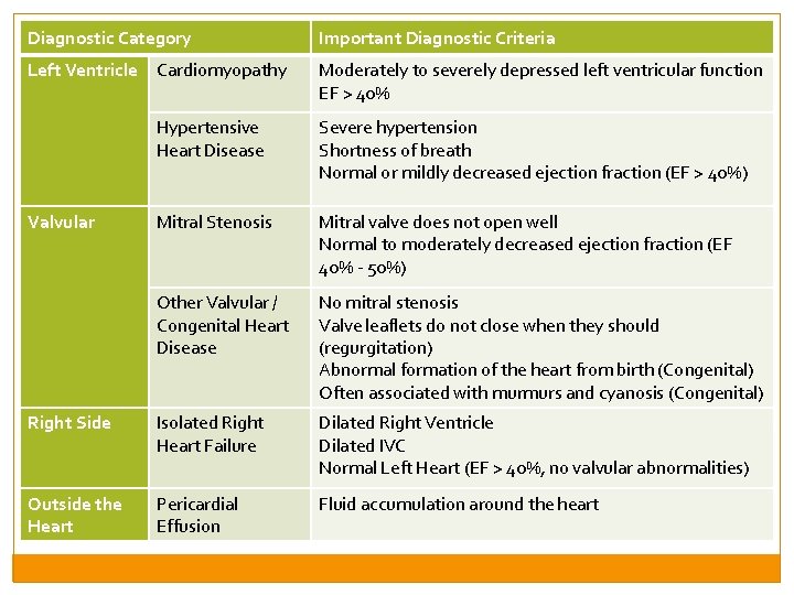
Diagnostic Category Important Diagnostic Criteria Left Ventricle Cardiomyopathy Moderately to severely depressed left ventricular function EF > 40% Hypertensive Heart Disease Severe hypertension Shortness of breath Normal or mildly decreased ejection fraction (EF > 40%) Mitral Stenosis Mitral valve does not open well Normal to moderately decreased ejection fraction (EF 40% - 50%) Other Valvular / Congenital Heart Disease No mitral stenosis Valve leaflets do not close when they should (regurgitation) Abnormal formation of the heart from birth (Congenital) Often associated with murmurs and cyanosis (Congenital) Right Side Isolated Right Heart Failure Dilated Right Ventricle Dilated IVC Normal Left Heart (EF > 40%, no valvular abnormalities) Outside the Heart Pericardial Effusion Fluid accumulation around the heart Valvular
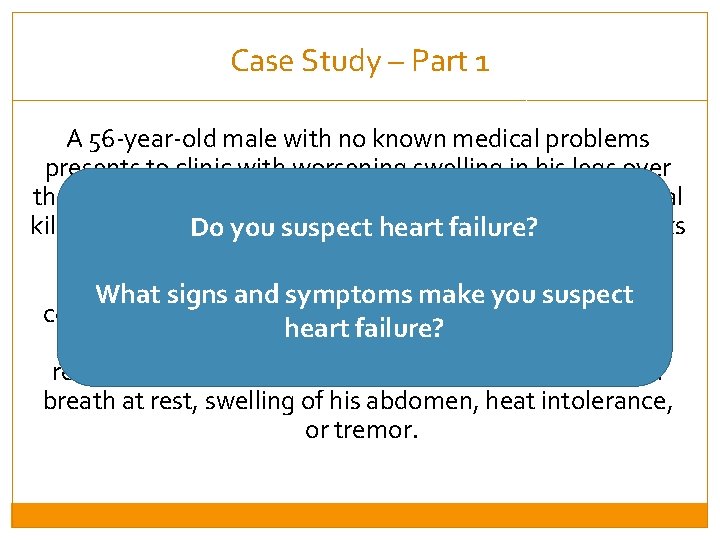
Case Study – Part 1 A 56 -year-old male with no known medical problems presents to clinic with worsening swelling in his legs over the past month. Until recently, he was able to walk several kilometers without stopping, but over the past three weeks Do you suspect heart failure? he has become fatigued after walking much shorter distances or up hills. He has also felt short of breath or What signs and symptoms make you suspect coughs while lying in bed at night, and currently needs to heart failure? use two pillows in order to fall asleep. He denies any recent chest pain or pressure, palpitations, shortness of breath at rest, swelling of his abdomen, heat intolerance, or tremor.
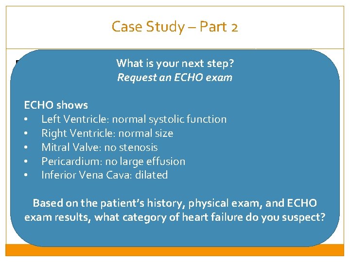
Case Study – Part 2 What is your next step? Physical Exam Request an ECHO exam � Temperature 37. 1 o. C, BP 185/110 mm. Hg, Pulse 80 beats per minute, Respiratory rate 12 breaths per minute , oxygen saturation of 95% ECHO shows on room air, weight 75 kg Left Ventricle: normal systolic function � • Neck veins visibly distended to 3 cm above the clavicle in the • upright position Right Ventricle: normal size • Mitral Valve: no stenosis � Cardiac exam with regular rate and rhythm and no loud murmers • Pericardium: no large effusion � Lung exam with crackles at the bases bilaterally; Good air • Inferior Vena Cava: dilated movement throughout; No wheezes � Extremities are warm and well-perfused; Pitting edema is present Based on the patient’s history, physical exam, and ECHO to the knees bilaterally exam results, what category of heart failure do you suspect?
Session 2: Types of Heart Failure and Treatment By the end of this session, participants will be able to: �Understand the major categories of heart failure �Understand the principles of fluid management, and use of diuretics �Understand the mortality-reducing medications, and adjunctive therapy �Understand management of specific types of heart failure
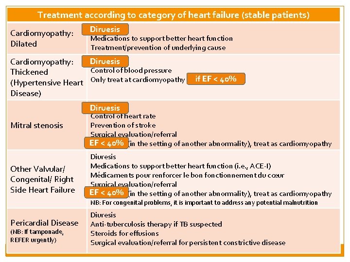
Treatment according to category of heart failure (stable patients) Cardiomyopathy: Dilated Cardiomyopathy: Thickened (Hypertensive Heart Disease) Diruesis Medications to support better heart function Treatment/prevention of underlying cause Diruresis Diruesis Control of blood pressure if EF < 40% Only treat at cardiomyopathy if EF < 40% Diuresis Diruesis Mitral stenosis Other Valvular/ Congenital/ Right Side Heart Failure Control of heart rate Prevention of stroke Surgical evaluation/referral EF < 40% If EF < 40% (in the setting of another abnormality), treat as cardiomyopathy Diuresis Medications to support better heart function (i. e. , ACE-I) Médicaments pour renforcer le bon fonctionnement du cœur Surgical evaluation/referral EF < 40% If EF < 40% (in the setting of another abnormality), treat as cardiomyopathy NB: For congenital problems, it is important to address any potential malnutrition Pericardial Disease (NB: If tamponade, REFER urgently) Diuresis Anti-tuberculosis therapy if TB suspected Steroids for effusions Surgical evaluation/referral for persistent constrictive disease
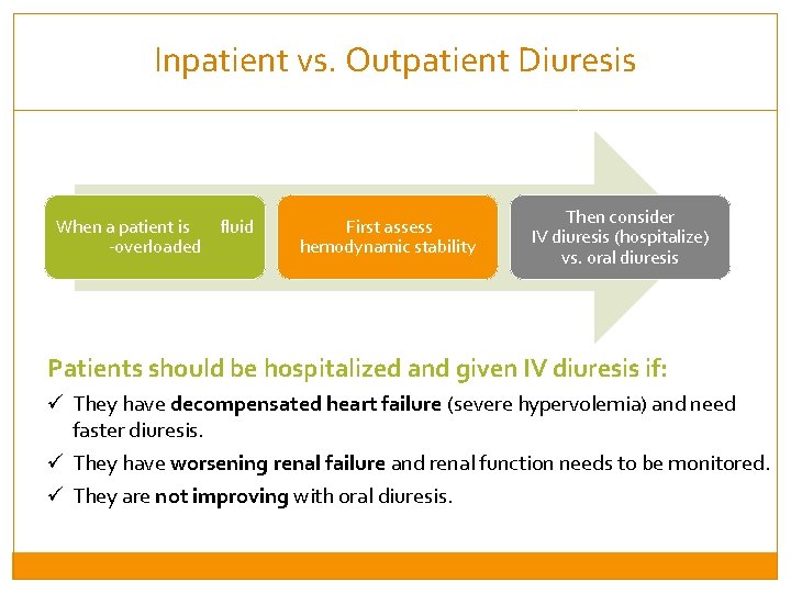
Inpatient vs. Outpatient Diuresis When a patient is fluid -overloaded First assess hemodynamic stability Then consider IV diuresis (hospitalize) vs. oral diuresis Patients should be hospitalized and given IV diuresis if: ü They have decompensated heart failure (severe hypervolemia) and need faster diuresis. ü They have worsening renal failure and renal function needs to be monitored. ü They are not improving with oral diuresis.
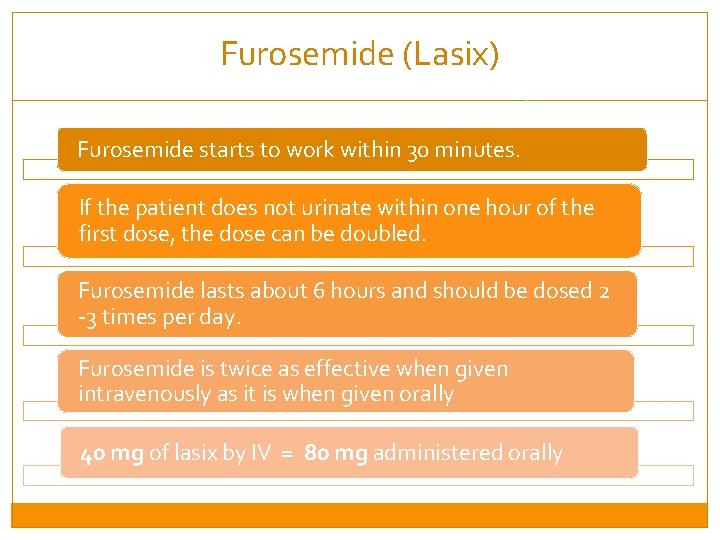
Furosemide (Lasix) Furosemide starts to work within 30 minutes. If the patient does not urinate within one hour of the first dose, the dose can be doubled. Furosemide lasts about 6 hours and should be dosed 2 -3 times per day. Furosemide is twice as effective when given intravenously as it is when given orally 40 mg of lasix by IV = 80 mg administered orally
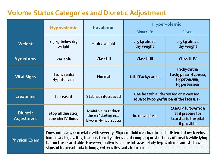
Volume Status Categories and Diuretic Adjustment Hypovolemic Euvolemic Weight > 5 kg below dry weight Symptoms Variable Moderate Severe At dry weight > 5 kg above dry weight Class I-II Class II-III Class III-IV Mild Tachycardia, Tachypnea, Hypoxia, Hypotension, Hypertension Vital Signs Tachycardia Hypotension Normal Creatinine Increased Stable or decreased Diuretic Adjustment Physical Exam Stop all diuretics, consider IV fluids Hypervolemic Maintain or reduce dose (if starting beta blocker, do not reduce) Can be stable, decreased or increased (due to hypo perfusion of the kidneys) Increase dose Start IV furosemide and prepare for transfer to hospital if possible Does not always correlate with severity. Signs of fluid overload include distended neck veins, lung crackles, ascites, lower extremity edema and coughing or shortness of breath while lying flat on the exam table. However, patients can be intravascularly hypovolemic and still have signs of hypervolemia in lungs, extremities and abdomen.
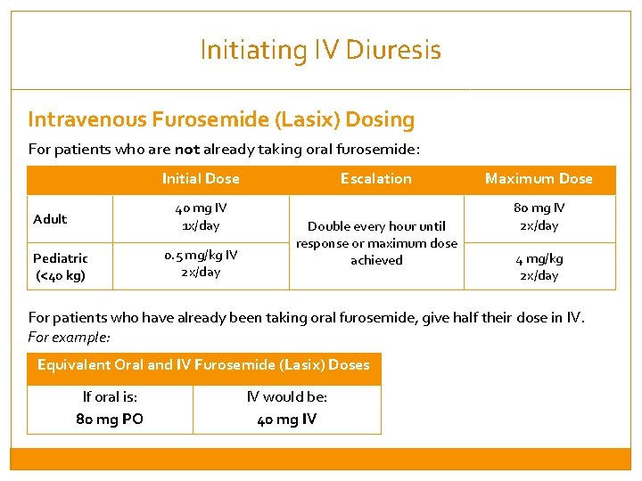
Initiating IV Diuresis Intravenous Furosemide (Lasix) Dosing For patients who are not already taking oral furosemide: Initial Dose 40 mg IV 1 x/day Adult Pediatric (<40 kg) 0. 5 mg/kg IV 2 x/day Escalation Double every hour until response or maximum dose achieved Maximum Dose 80 mg IV 2 x/day 4 mg/kg 2 x/day For patients who have already been taking oral furosemide, give half their dose in IV. For example: Equivalent Oral and IV Furosemide (Lasix) Doses If oral is: 80 mg PO IV would be: 40 mg IV
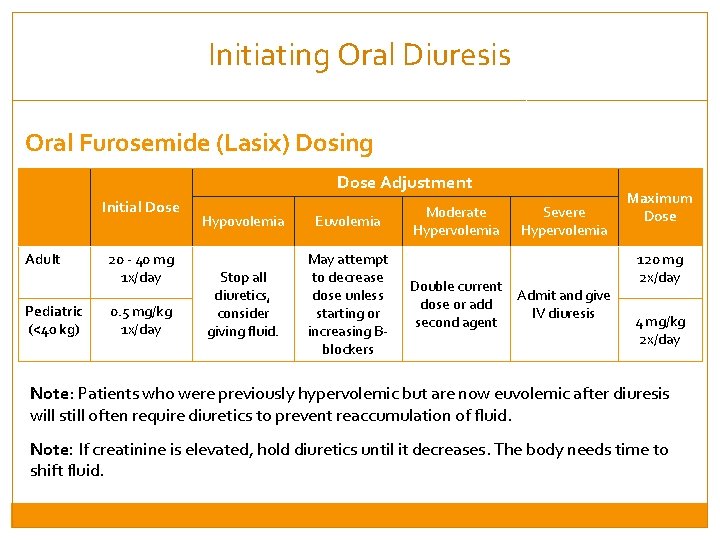
Initiating Oral Diuresis Oral Furosemide (Lasix) Dosing Dose Adjustment Initial Dose Adult Pediatric (<40 kg) 20 - 40 mg 1 x/day 0. 5 mg/kg 1 x/day Hypovolemia Euvolemia Stop all diuretics, consider giving fluid. May attempt to decrease dose unless starting or increasing Bblockers Moderate Hypervolemia Severe Hypervolemia Double current Admit and give dose or add IV diuresis second agent Maximum Dose 120 mg 2 x/day 4 mg/kg 2 x/day Note: Patients who were previously hypervolemic but are now euvolemic after diuresis will still often require diuretics to prevent reaccumulation of fluid. Note: If creatinine is elevated, hold diuretics until it decreases. The body needs time to shift fluid.
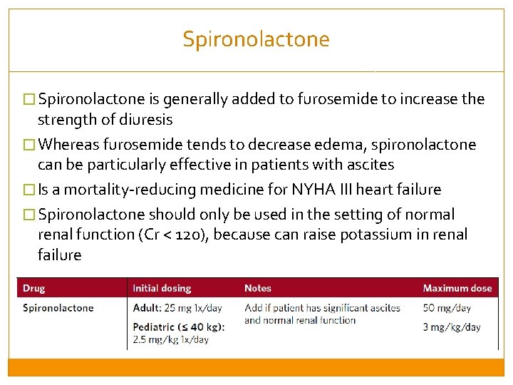
Spironolactone � Spironolactone is generally added to furosemide to increase the strength of diuresis � Whereas furosemide tends to decrease edema, spironolactone can be particularly effective in patients with ascites � Is a mortality-reducing medicine for NYHA III heart failure � Spironolactone should only be used in the setting of normal renal function (Cr < 120), because can raise potassium in renal failure
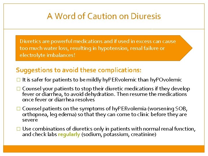
A Word of Caution on Diuresis Diuretics are powerful medications and if used in excess can cause too much water loss, resulting in hypotension, renal failure or electrolyte imbalances! Suggestions to avoid these complications: � It is safer for patients to be mildly hy. PERvolemic than hy. POvolemic � Counsel your patients to stop their diuretic medications if they develop fever or diarrhea, to avoid dehydration. Then resume the medications once fever or diarrhea resolves � Counsel patients on the symptoms of hy. PERvolemia (worsening SOB, orthopnea, leg edema) so that they can come to clinic before they are severe � Use combinations of diuretics only in patients with normal renal function, and check labs regularly (sodium, potassium, creatinine)
Session 2: Types of Heart Failure and Treatment By the end of this session, participants will be able to: �Understand the major categories of heart failure �Understand the principles of fluid management, and use of diuretics �Understand the mortality-reducing medications, and adjunctive therapy �Understand management of specific types of heart failure
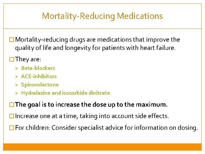
Mortality-Reducing Medications � Mortality-reducing drugs are medications that improve the quality of life and longevity for patients with heart failure. � They are: Ø Beta-blockers Ø ACE-inhibitors Ø Spironolactone Ø Hydralazine and isosorbide dinitrate � The goal is to increase the dose up to the maximum. � Increase one at a time, taking into account side effects. � For children: Consider specialist advice for information on dosing.
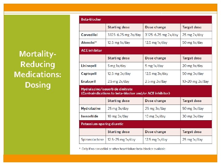
Mortality. Reducing Medications: Dosing
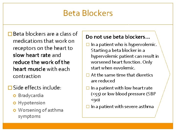
Beta Blockers � Beta blockers are a class of medications that work on receptors on the heart to slow heart rate and reduce the work of the heart muscle with each contraction � Side effects include: Bradycardia Hypotension Worsening of asthma symptoms Do not use beta blockers… � In a patient who is hypervolemic. Starting a beta blocker in a hypervolemic patient can result in worsened heart function. Only start when euvolemic. � At the same time that diuretics are reduced � In a patient with low heart rate (<55) or low blood pressure (SBP <90) � In a patient with severe asthma
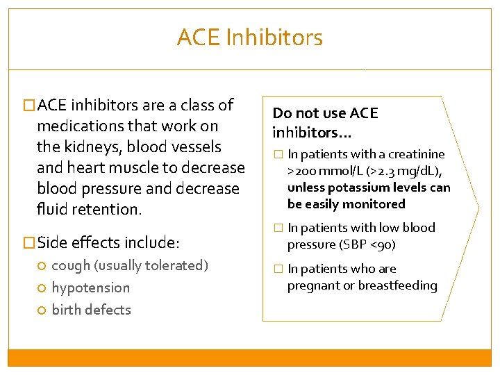
ACE Inhibitors �ACE inhibitors are a class of medications that work on the kidneys, blood vessels and heart muscle to decrease blood pressure and decrease fluid retention. �Side effects include: cough (usually tolerated) hypotension birth defects Do not use ACE inhibitors… � In patients with a creatinine >200 mmol/L (>2. 3 mg/d. L), unless potassium levels can be easily monitored � In patients with low blood pressure (SBP <90) � In patients who are pregnant or breastfeeding
Hydralazine and Isosorbide Dinitrate �Hydralazine is an arteriolar dilator and isosorbide is a venodilator �These medications do not affect heart rate or renal function �Side effects of this medication combination include: headaches These drugs should only be used for patients with contraindications to beta -blockers and/or ACE inhibitors because… � It requires an inconvenient three-times-a-day dosing � This medication combination is expensive
Spironolactone �Spironolactone is an antihormone drug that has a neurohormonal stimulation effects �It decreases edema, swelling, and the strain on the heart �It also acts as a diuretic (antihypertensive) as described in the previous section Spironolactone should NOT be increased to maximum because. . � It can increase potassium and lead to hyperkaliemia
Other (Adjunct) Therapies DIGOXIN ASPIRIN FAMILY PLANNING
Digoxin �Increases pumping function of the heart and can also slow down the heart rate if atrial fibrillation is present �Side effects: at high doses, toxicity can develop which results in visual changes, dizziness or nausea. Risk of toxicity is worse with renal failure (if renal function worsens, stop or reduce dose) �Starting dose is 0. 125 mg/day. Monitor creatinine.
Aspirin �Aspirin works as a blood thinner, decreasing the risk of blood clots �Heart failure can lead to blood clots in the heart that travel to other parts of the body, causing stroke �Patients with peripartum cardiomyopathy and atrial fibrillilation are at particularly high risk for blood clots. These patients should be started on aspirin 100 mg 1 x/day. �Side effects: Aspirin is generally safe, but at high doses can cause GI bleeding and bruising
Family Planning �All female patients of childbearing age should be counseled on the dangers of pregnancy in the setting of heart disease, and be advised to use birth control �The physiologic changes during pregnancy, including increased fluid retention and smaller lung volumes, usually result in worsened cardiac symptoms �In the case of peripartum cardiomyopathy, subsequent pregnancies can in some cases be lethal
Session 2: Types of Heart Failure and Treatment By the end of this session, participants will be able to: �Understand the major categories of heart failure �Understand the principles of fluid management, and use of diuretics �Understand the mortality-reducing medications, and adjunctive therapy �Understand management of specific types of heart failure
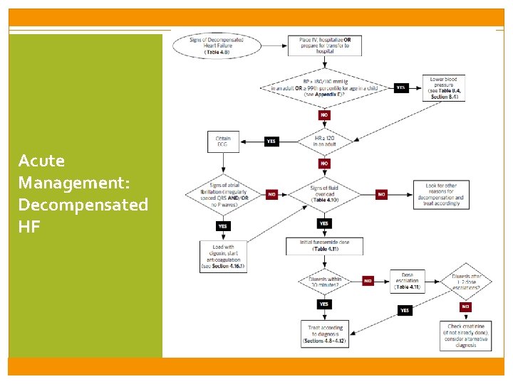
Acute Management: Decompensated HF
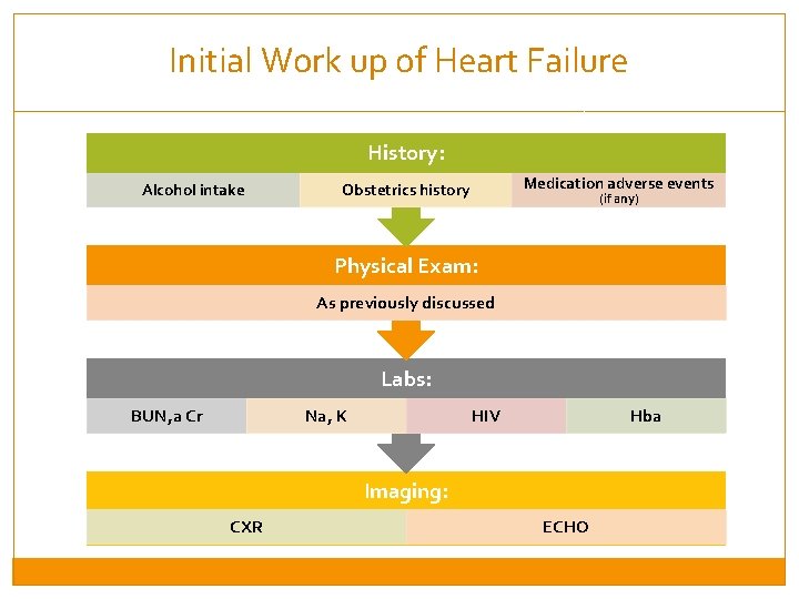
Initial Work up of Heart Failure History: Alcohol intake Medication adverse events Obstetrics history (if any) Physical Exam: As previously discussed Labs: BUN, a Cr Na, K HIV Hba Imaging: CXR ECHO
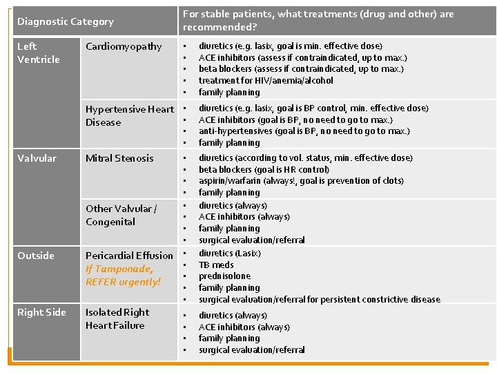
Diagnostic Category Left Ventricle Cardiomyopathy For stable patients, what treatments (drug and other) are recommended? • • • Hypertensive Heart • • Disease Valvular Outside Right Side • • • Mitral Stenosis • • Other Valvular / • Congenital • • Pericardial Effusion • • If Tamponade, • REFER urgently! • • Isolated Right Heart Failure • • diuretics (e. g. lasix, goal is min. effective dose) ACE inhibitors (assess if contraindicated, up to max. ) beta blockers (assess if contraindicated, up to max. ) treatment for HIV/anemia/alcohol family planning diuretics (e. g. lasix, goal is BP control, min. effective dose) ACE inhibitors (goal is BP, no need to go to max. ) anti-hypertensives (goal is BP, no need to go to max. ) family planning diuretics (according to vol. status, min. effective dose) beta blockers (goal is HR control) aspirin/warfarin (always!, goal is prevention of clots) family planning diuretics (always) ACE inhibitors (always) family planning surgical evaluation/referral diuretics (Lasix) TB meds prednisolone family planning surgical evaluation/referral for persistent constrictive disease diuretics (always) ACE inhibitors (always) family planning surgical evaluation/referral
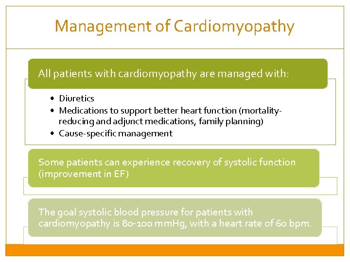
Management of Cardiomyopathy All patients with cardiomyopathy are managed with: • Diuretics • Medications to support better heart function (mortalityreducing and adjunct medications, family planning) • Cause-specific management Some patients can experience recovery of systolic function (improvement in EF) The goal systolic blood pressure for patients with cardiomyopathy is 80 -100 mm. Hg, with a heart rate of 60 bpm.
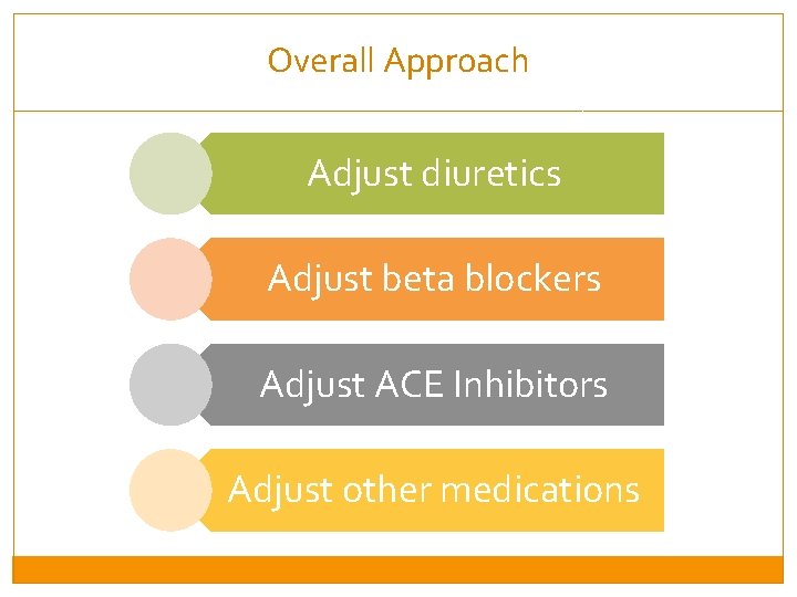
Overall Approach Adjust diuretics Adjust beta blockers Adjust ACE Inhibitors Adjust other medications
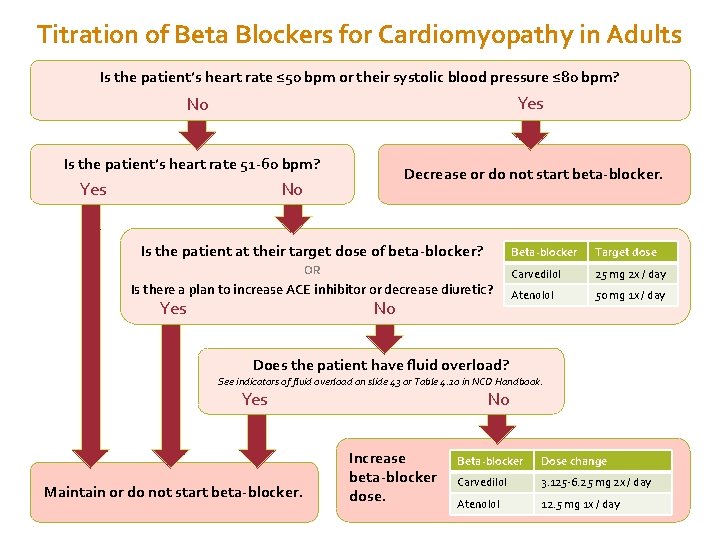
Titration of Beta Blockers for Cardiomyopathy in Adults Is the patient’s heart rate ≤ 50 bpm or their systolic blood pressure ≤ 80 bpm? Yes No Is the patient’s heart rate 51 -60 bpm? Yes Decrease or do not start beta-blocker. No Is the patient at their target dose of beta-blocker? OR Is there a plan to increase ACE inhibitor or decrease diuretic? Yes No Beta-blocker Target dose Carvedilol 25 mg 2 x / day Atenolol 50 mg 1 x / day Does the patient have fluid overload? See indicators of fluid overload on slide 43 or Table 4. 10 in NCD Handbook. Yes Maintain or do not start beta-blocker. No Increase beta-blocker dose. Beta-blocker Dose change Carvedilol 3. 125 -6. 25 mg 2 x / day Atenolol 12. 5 mg 1 x / day
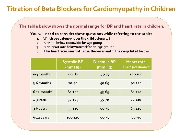
Titration of Beta Blockers for Cardiomyopathy in Children The table below shows the normal range for BP and heart rate in children. You will need to consider these questions while referring to the table: 1. 2. 3. 4. Which age category does the child belong to? Is his BP below normal for his age group? Is his heart rate below normal for his age group? If his heart rate is normal, is it in the lower end of the range listed below? Systolic BP (mm. Hg) Diastolic BP (mm. Hg) Heart rate beats per minute 0 -3 months 60 -80 45 -55 120 -160 3 -6 months 70 -90 50 -65 90 -120 6 -12 months 80 -100 55 -65 80 -120 1 -3 years 90 -105 55 -70 70 -110 3 -6 years 95 -110 60 -75 65 -110 6 -12 years 100 -120 60 -75 60 -95
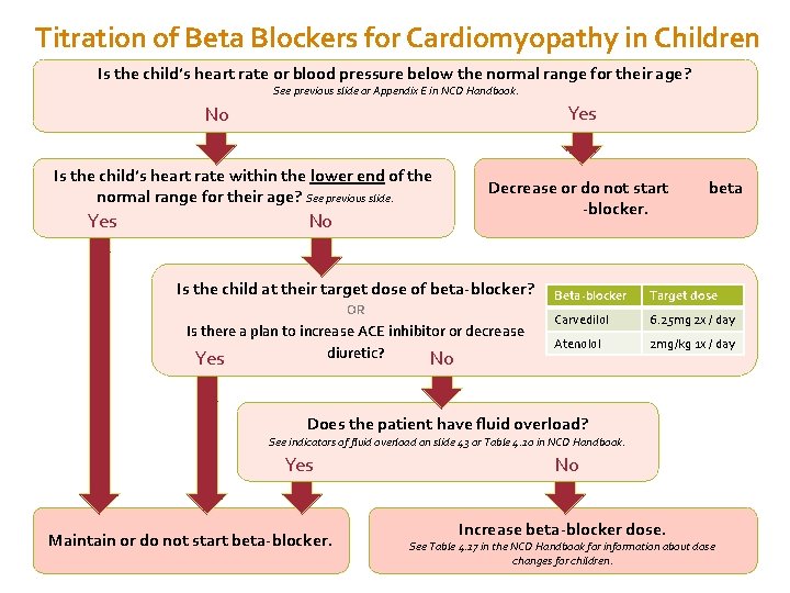
Titration of Beta Blockers for Cardiomyopathy in Children Is the child’s heart rate or blood pressure below the normal range for their age? See previous slide or Appendix E in NCD Handbook. Yes No Is the child’s heart rate within the lower end of the normal range for their age? See previous slide. Yes No Decrease or do not start beta -blocker. Is the child at their target dose of beta-blocker? OR Is there a plan to increase ACE inhibitor or decrease diuretic? Yes No Beta-blocker Target dose Carvedilol 6. 25 mg 2 x / day Atenolol 2 mg/kg 1 x / day Does the patient have fluid overload? See indicators of fluid overload on slide 43 or Table 4. 10 in NCD Handbook. Yes Maintain or do not start beta-blocker. No Increase beta-blocker dose. See Table 4. 17 in the NCD Handbook for information about dose changes for children.
Titration of ACEinhibitors in Cardiomyopathy
Other Medications in Cardiomyopathy
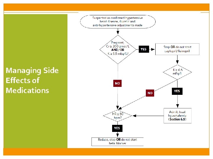
Managing Side Effects of Medications
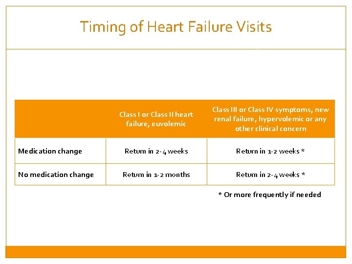
Timing of Heart Failure Visits Class I or Class II heart failure, euvolemic Class III or Class IV symptoms, new renal failure, hypervolemic or any other clinical concern Medication change Return in 2 -4 weeks Return in 1 -2 weeks * No medication change Return in 1 -2 months Return in 2 -4 weeks * * Or more frequently if needed
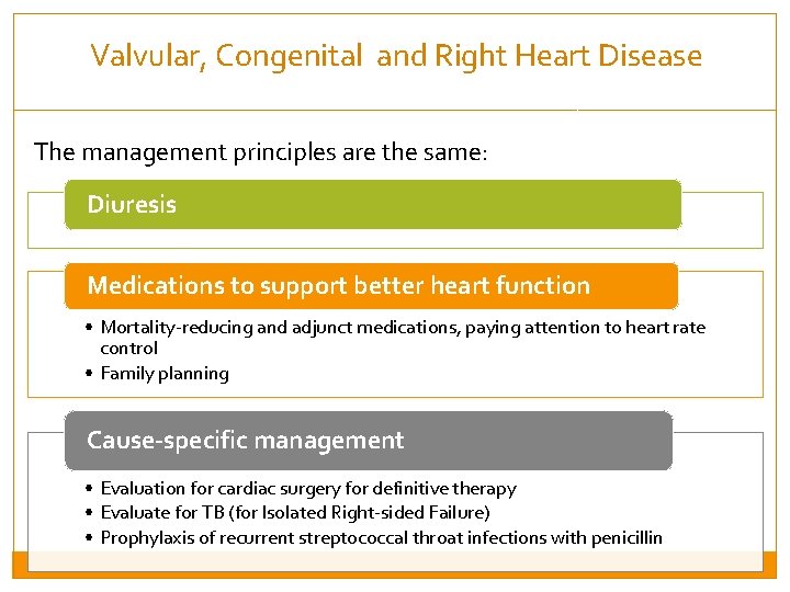
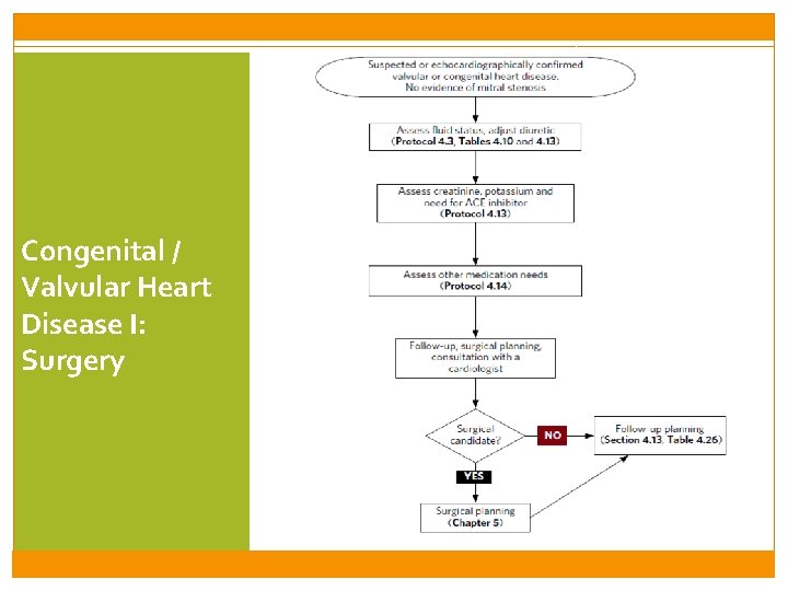
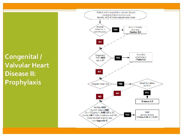
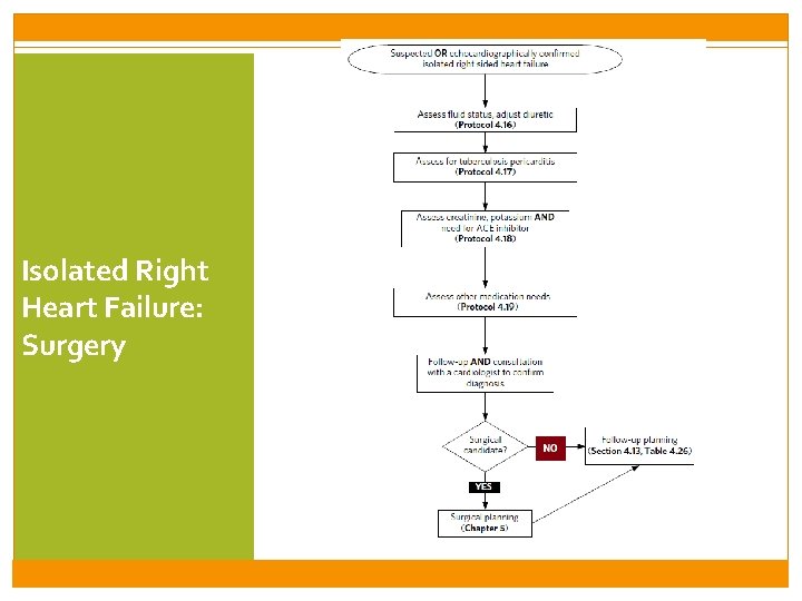
Valvular, Congenital and Right Heart Disease The management principles are the same: Diuresis Medications to support better heart function • Mortality-reducing and adjunct medications, paying attention to heart rate control • Family planning Cause-specific management • Evaluation for cardiac surgery for definitive therapy • Evaluate for TB (for Isolated Right-sided Failure) • Prophylaxis of recurrent streptococcal throat infections with penicillin
Congenital / Valvular Heart Disease I: Surgery
Congenital / Valvular Heart Disease II: Prophylaxis
Isolated Right Heart Failure: Surgery
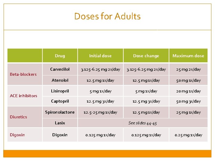
Doses for Adults Beta-blockers ACE inhibitors Diuretics Drug Initial dose Dose change Maximum dose Carvedilol 3. 125 -6. 25 mg 2 x/day Atenolol 12. 5 mg 1 x/day 50 mg 1 x/day Lisinopril 5 mg 1 x/day 20 mg 1 x/day Captopril 12. 5 mg 3 x/day 50 mg 3 x/day Spironolactone 12. 5 -25 mg 1 x/day 12. 5 mg 1 x/day 25 mg 1 x/day Lasix Digoxin See slides 44 -45 0. 125 mg 1 x/day 0. 25 mg 1 x/day
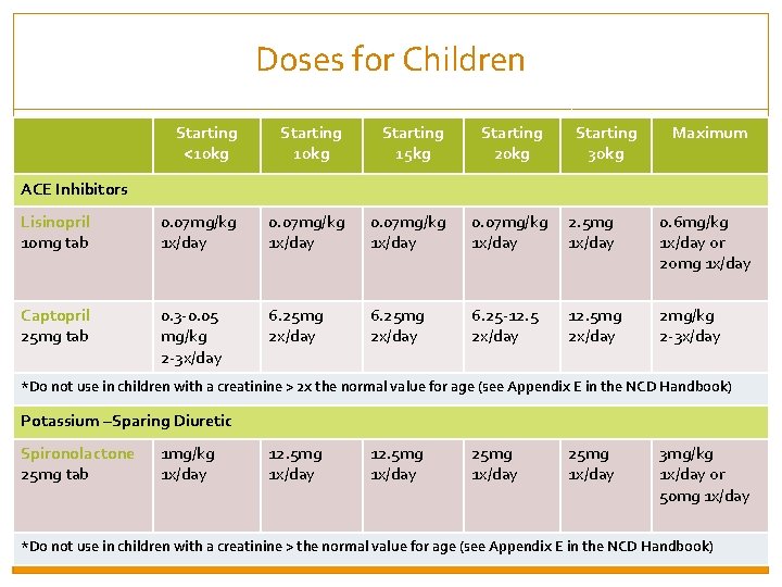
Doses for Children Starting <10 kg Starting 15 kg Starting 20 kg Starting 30 kg Maximum ACE Inhibitors Lisinopril 10 mg tab 0. 07 mg/kg 1 x/day 0. 07 mg/kg 2. 5 mg 1 x/day 0. 6 mg/kg 1 x/day or 2 omg 1 x/day Captopril 25 mg tab 0. 3 -0. 05 mg/kg 2 -3 x/day 6. 25 mg 2 x/day 6. 25 -12. 5 2 x/day 2 mg/kg 2 -3 x/day 12. 5 mg 2 x/day *Do not use in children with a creatinine > 2 x the normal value for age (see Appendix E in the NCD Handbook) Potassium –Sparing Diuretic Spironolactone 25 mg tab 1 mg/kg 1 x/day 12. 5 mg 1 x/day 25 mg 1 x/day 3 mg/kg 1 x/day or 50 mg 1 x/day *Do not use in children with a creatinine > the normal value for age (see Appendix E in the NCD Handbook)
Session 3: Rheumatic Heart Disease By the end of this session, participants will be able to: �Discuss the relationship between streptococcal pharyngitis and rheumatic heart disease, and rationale for antibiotic prophylaxis �Describe the clinical and echocardiographic features of mitral stenosis. �Understand basic approach to anticoagulation, and its specific role in mitral stenosis and post-cardiac surgery �Understand the role of cardiac surgery in the management of mitral stenosis
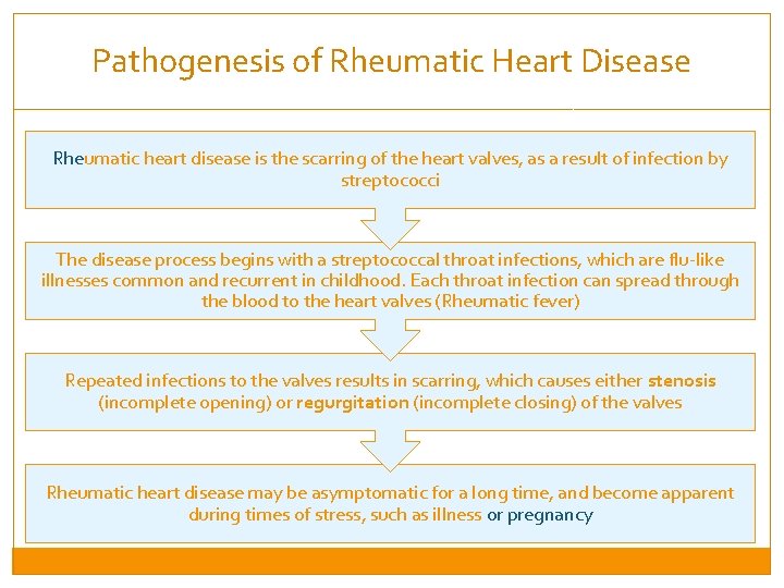
Pathogenesis of Rheumatic Heart Disease Rheumatic heart disease is the scarring of the heart valves, as a result of infection by streptococci The disease process begins with a streptococcal throat infections, which are flu-like illnesses common and recurrent in childhood. Each throat infection can spread through the blood to the heart valves (Rheumatic fever) Repeated infections to the valves results in scarring, which causes either stenosis (incomplete opening) or regurgitation (incomplete closing) of the valves Rheumatic heart disease may be asymptomatic for a long time, and become apparent during times of stress, such as illness or pregnancy
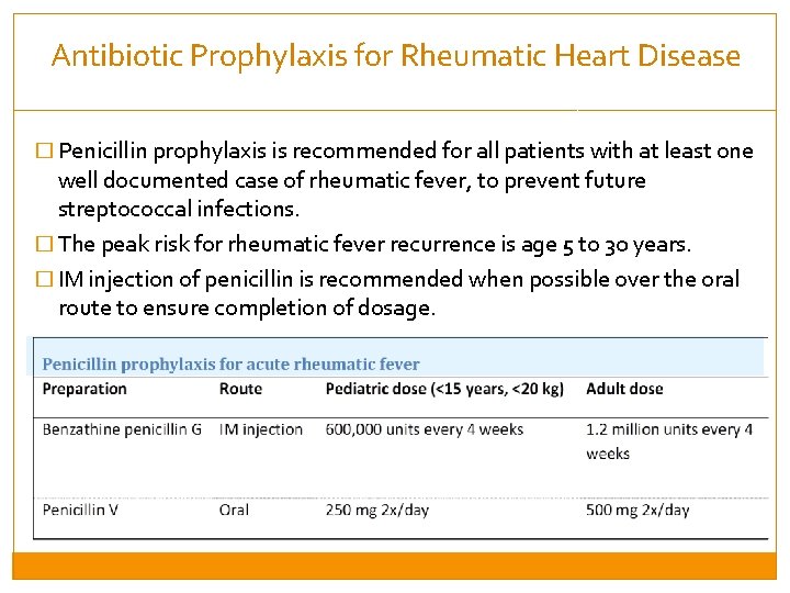
Antibiotic Prophylaxis for Rheumatic Heart Disease � Penicillin prophylaxis is recommended for all patients with at least one well documented case of rheumatic fever, to prevent future streptococcal infections. � The peak risk for rheumatic fever recurrence is age 5 to 30 years. � IM injection of penicillin is recommended when possible over the oral route to ensure completion of dosage.
Session 3: Rheumatic Heart Disease By the end of this session, participants will be able to: �Discuss the relationship between streptococcal pharyngitis and rheumatic heart disease, and rationale for antibiotic prophylaxis �Describe the clinical and echocardiographic features of mitral stenosis. �Understand basic approach to anticoagulation, and its specific role in mitral stenosis and post-cardiac surgery �Understand the role of cardiac surgery in the management of mitral stenosis
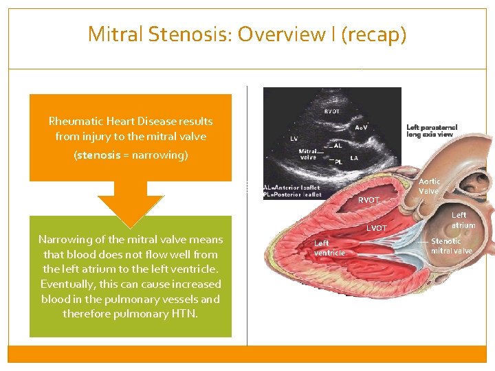
Mitral Stenosis: Overview I (recap) Rheumatic Heart Disease results from injury to the mitral valve (stenosis = narrowing) RVOT Narrowing of the mitral valve means that blood does not flow well from the left atrium to the left ventricle. Eventually, this can cause increased blood in the pulmonary vessels and therefore pulmonary HTN. LVOT Left ventricle Aortic Valve Left atrium Stenotic mitral valve
Mitral Stenosis: Overview II Back up of blood into the left atrium results in: • Stretching of the atrium, which is prone to developing arrhythmias such as atrial fibrillation. • Back up of blood to the lungs (causing lung edema and hypertension) and right heart (causing strain on the right ventricle, leg edema) Mitral stenosis almost always results from untreated rheumatic heart disease, typically many years after the infections (most commonly diagnosed at age 30 and 50 years old) The most common presenting symptom is dyspnea, due to pulmonary edema.
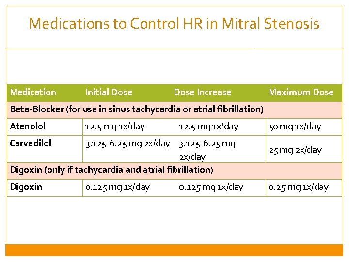
Management of Mitral Stenosis: HR Control Ventricular filling occurs during diastole. Decreasing heart rate increases the amount of time the heart spends filling up the left ventricle, thus increasing how much blood the left heart pumps to the body with each contraction (systole). Ideal heart rate in mitral stenosis is 50 -60 bpm, without reducing the systolic blood pressure below 90 mm. Hg. HR Control medications are: - Beta-blockers (first line) - Digoxin can be used in addition to a beta blocker if the patient has a creatinine < 200 mmol/L AND is in atrial fibrillation
Medications to Control HR in Mitral Stenosis Medication Initial Dose Increase Maximum Dose Beta-Blocker (for use in sinus tachycardia or atrial fibrillation) Atenolol 12. 5 mg 1 x/day Carvedilol 3. 125 -6. 25 mg 2 x/day Digoxin (only if tachycardia and atrial fibrillation) Digoxin 0. 125 mg 1 x/day 50 mg 1 x/day 25 mg 2 x/day 0. 25 mg 1 x/day
Session 3: Rheumatic Heart Disease By the end of this session, participants will be able to: �Discuss the relationship between streptococcal pharyngitis and rheumatic heart disease, and rationale for antibiotic prophylaxis �Describe the clinical and echocardiographic features of mitral stenosis. �Understand basic approach to anticoagulation, and its specific role in mitral stenosis and post-cardiac surgery �Understand the role of cardiac surgery in the management of mitral stenosis
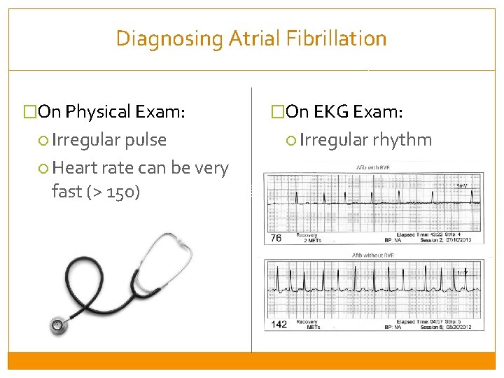
Diagnosing Atrial Fibrillation �On Physical Exam: Irregular pulse Heart rate can be very fast (> 150) �On EKG Exam: Irregular rhythm
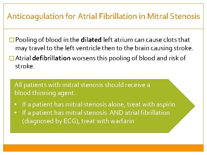
Anticoagulation for Atrial Fibrillation in Mitral Stenosis � Pooling of blood in the dilated left atrium can cause clots that may travel to the left ventricle then to the brain causing stroke. � Atrial defibrillation worsens this pooling of blood and risk of stroke. All patients with mitral stenosis should receive a blood thinning agent. • If a patient has mitral stenosis alone, treat with aspirin • If a patient has mitral stenosis AND atrial fibrillation (diagnosed by ECG), treat with warfarin
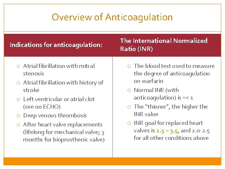
Overview of Anticoagulation Indications for anticoagulation: Atrial fibrillation with mitral stenosis Atrial fibrillation with history of stroke Left ventricular or atrial clot (see on ECHO) Deep venous thrombosis After heart valve replacements (lifelong for mechanical valve; 3 months for bioprosthenic valve) The International Normalized Ratio (INR) The blood test used to measure the degree of anticoagulation on warfarin Normal INR (with anticoagulation) is =< 1 The “thinner”, the higher the INR value INR goal for replaced heart valves is 2. 5 – 3. 5, and 2. 0 -2. 5 for all other conditions above
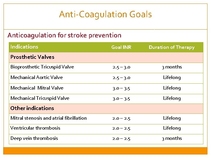
Anti-Coagulation Goals Anticoagulation for stroke prevention Indications Goal INR Duration of Therapy Bioprosthetic Tricuspid Valve 2. 5 – 3. 0 3 months Mechanical Aortic Valve 2. 5 – 3. 0 Lifelong Mechanical Mitral Valve 3. 0 – 3. 5 Lifelong Mechanical Tricuspid Valve 3. 0 – 3. 5 Lifelong Mitral stenosis and atrial fibrillation 2. 0 – 2. 5 Lifelong Ventricular thrombosis 2. 0 – 2. 5 Lifelong Deep vein thrombosis 2. 0 – 2. 5 3 months Prosthetic Valves Other indications
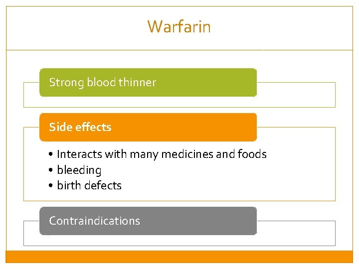
Warfarin Strong blood thinner Side effects • Interacts with many medicines and foods • bleeding • birth defects Contraindications
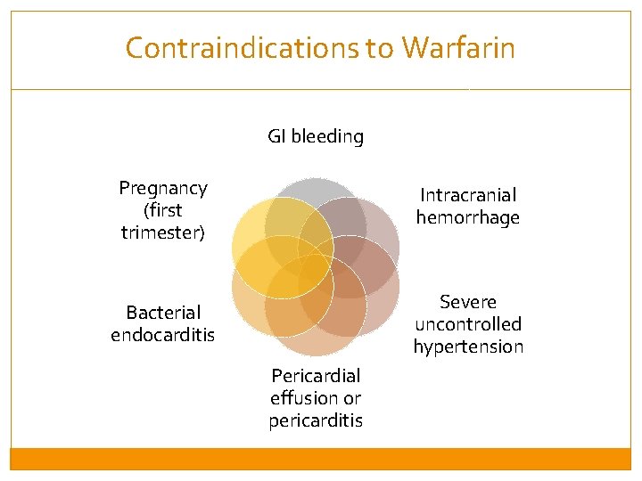
Contraindications to Warfarin GI bleeding Pregnancy (first trimester) Intracranial hemorrhage Bacterial endocarditis Severe uncontrolled hypertension Pericardial effusion or pericarditis
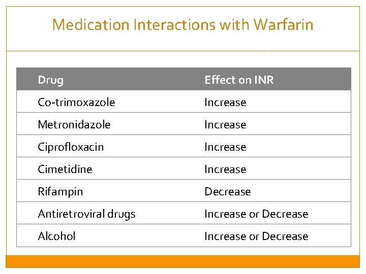
Medication Interactions with Warfarin Drug Effect on INR Co-trimoxazole Increase Metronidazole Increase Ciprofloxacin Increase Cimetidine Increase Rifampin Decrease Antiretroviral drugs Increase or Decrease Alcohol Increase or Decrease
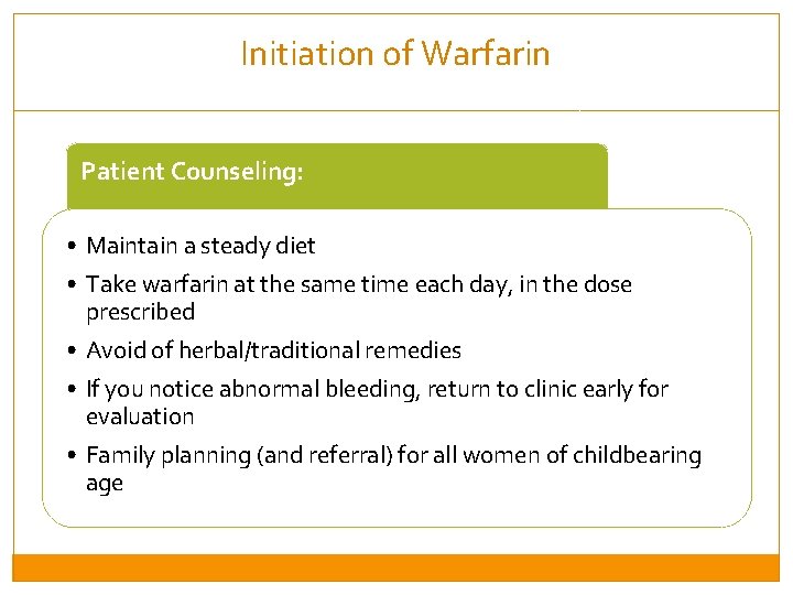
Initiation of Warfarin Patient Counseling: • Maintain a steady diet • Take warfarin at the same time each day, in the dose prescribed • Avoid of herbal/traditional remedies • If you notice abnormal bleeding, return to clinic early for evaluation • Family planning (and referral) for all women of childbearing age
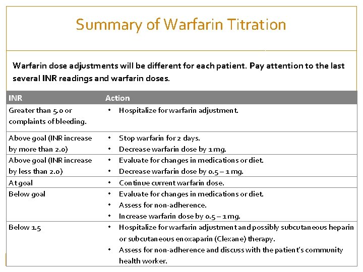
Summary of Warfarin Titration Warfarin dose adjustments will be different for each patient. Pay attention to the last several INR readings and warfarin doses. INR Action Greater than 5. 0 or complaints of bleeding. • Hospitalize for warfarin adjustment. Above goal (INR increase by more than 2. 0) Above goal (INR increase by less than 2. 0) At goal Below goal • • • Stop warfarin for 2 days. Decrease warfarin dose by 1 mg. Evaluate for changes in medications or diet. Decrease warfarin dose by 0. 5 – 1 mg. Continue current warfarin dose. Evaluate for changes in medications or diet. Assess for non-adherence. Increase warfarin dose by 0. 5 – 1 mg. Hospitalize for warfarin adjustment and possibly subcutaneous heparin or subcutaneous enoxaparin (Clexane) therapy. Assess for non-adherence and discuss with the patient’s community health worker. Below 1. 5 •
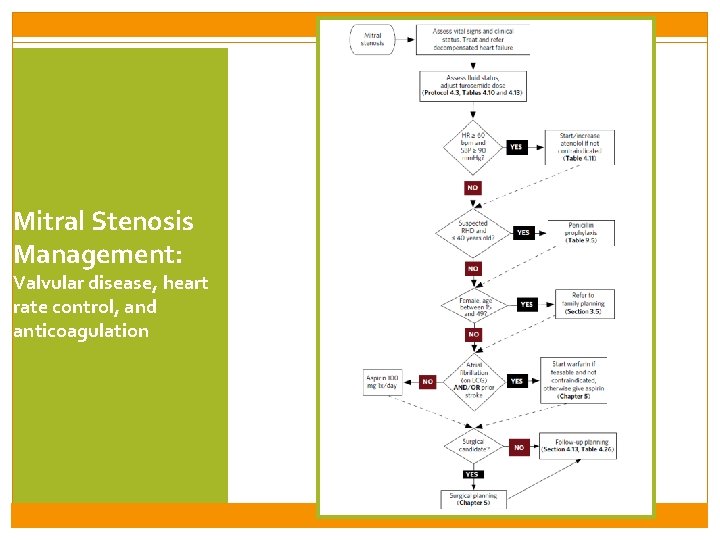
Mitral Stenosis Management: Valvular disease, heart rate control, and anticoagulation
Session 3: Rheumatic Heart Disease By the end of this session, participants will be able to: �Discuss the relationship between streptococcal pharyngitis and rheumatic heart disease, and rationale for antibiotic prophylaxis �Describe the clinical and echocardiographic features of mitral stenosis. �Understand basic approach to anticoagulation, and its specific role in mitral stenosis and post-cardiac surgery �Understand the role of cardiac surgery in the management of mitral stenosis
Cardiac Surgery for Valve Repair �Many patients with mitral stenosis can be managed with medications but some can be definitively treated with surgery. �Surgery options are: Valve replacement (minor surgery) Valve repair (major surgery) �All patients with mitral stenosis should be referred to a cardiologist within 6 months �The decision of referral to cardiac surgery should be made in consultation with a cardiologist; in general all mitral stenosis patients under age 40 should be referred to cardiac surgery.
Rwanda’s National Cardiac Surgery Program �All patients with these conditions should see a cardiologist within six months of diagnosis: Mitral stenosis Other valvular heart disease Pericardial disease Congenital heart disease
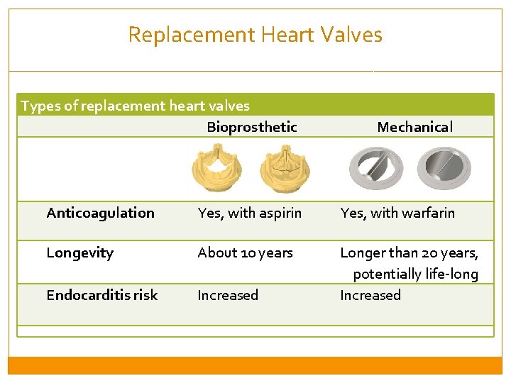
Replacement Heart Valves Types of replacement heart valves Bioprosthetic Mechanical Anticoagulation Yes, with aspirin Yes, with warfarin Longevity About 10 years Endocarditis risk Increased Longer than 20 years, potentially life-long Increased
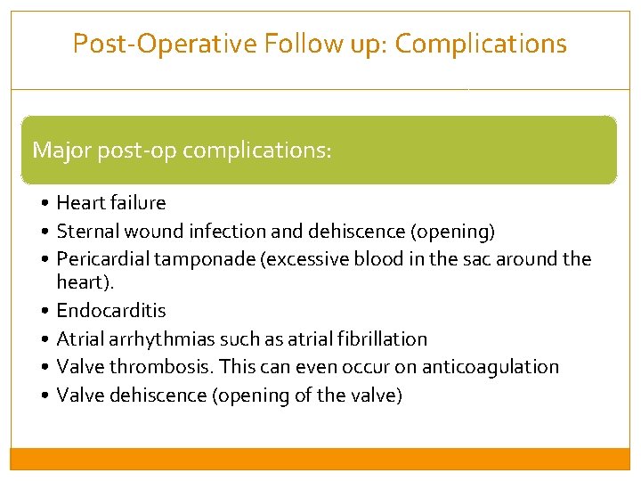
Post-Operative Follow up: Complications Major post-op complications: • Heart failure • Sternal wound infection and dehiscence (opening) • Pericardial tamponade (excessive blood in the sac around the heart). • Endocarditis • Atrial arrhythmias such as atrial fibrillation • Valve thrombosis. This can even occur on anticoagulation • Valve dehiscence (opening of the valve)
Session 4: Heart Failure care at District and Health Center levels By the end of this session, participants will be able to: �Understand the role of health center care in management of Heart failure �Understand the role of CHW in management of Heart Failure
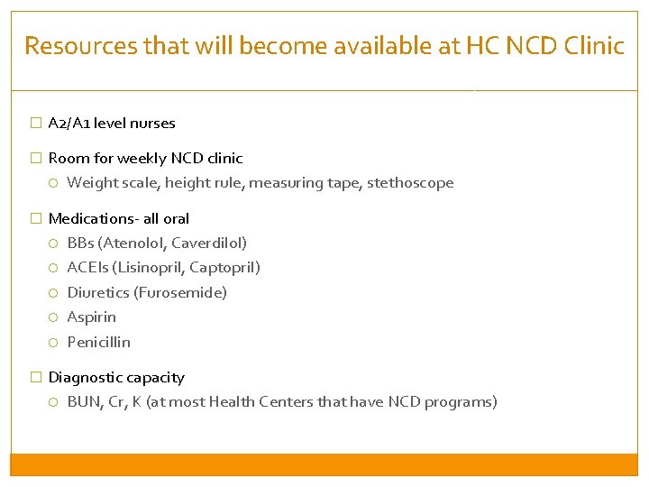
Resources that will become available at HC NCD Clinic � A 2/A 1 level nurses � Room for weekly NCD clinic Weight scale, height rule, measuring tape, stethoscope � Medications- all oral BBs (Atenolol, Caverdilol) ACEIs (Lisinopril, Captopril) Diuretics (Furosemide) Aspirin Penicillin � Diagnostic capacity BUN, Cr, K (at most Health Centers that have NCD programs)
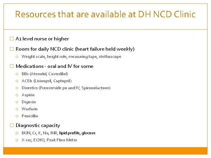
Resources that are available at DH NCD Clinic � A 1 level nurse or higher � Room for daily NCD clinic (heart failure held weekly) Weight scale, height rule, measuring tape, stethoscope � Medications - oral and IV for some BBs (Atenolol, Caverdilol) ACEIs (Lisinopril, Captopril) Diuretics (Furosemide po and IV, Spironolactone) Aspirin Digoxin Warfarin Penicillin � Diagnostic capacity BUN, Cr, K, Na, INR, lipid profile, glucose X-ray, ECHO, Peak Flow Meter
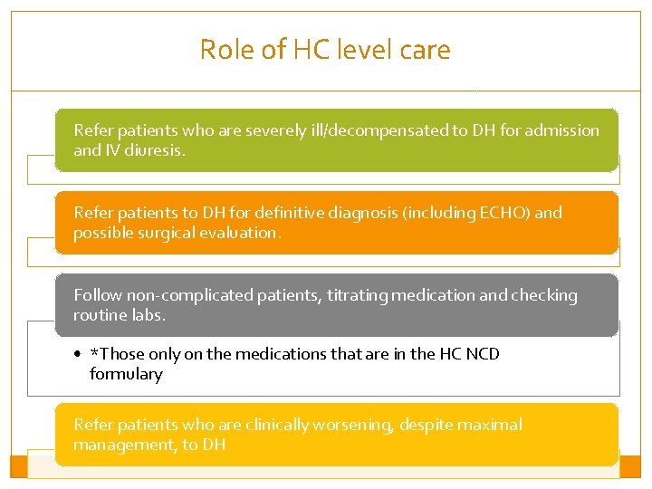
Role of HC level care Refer patients who are severely ill/decompensated to DH for admission and IV diuresis. Refer patients to DH for definitive diagnosis (including ECHO) and possible surgical evaluation. Follow non-complicated patients, titrating medication and checking routine labs. • *Those only on the medications that are in the HC NCD formulary Refer patients who are clinically worsening, despite maximal management, to DH
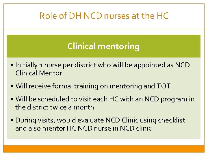
Role of DH NCD nurses at the HC Clinical mentoring • Initially 1 nurse per district who will be appointed as NCD Clinical Mentor • Will receive formal training on mentoring and TOT • Will be scheduled to visit each HC with an NCD program in the district twice a month • During visits, would evaluate NCD Clinic using checklist and also mentor HC NCD nurse in NCD clinic
Thank you for your hard work and dedication to health and human rights! Matilda Nikolasi talks with PIH clinician Joe Lusaka and was the first of many to be served during the new Dambe Health Center’s first day on April 25, 2016 in Malawi. She was screened for Hypertension, HIV and Diabetes during this visit through the SHARF (Screening for health and referrals at the facilities) initiative. (Photo by Nandi Bwanali / Partners In Health)