Structure Function and Disorders of the Integument Anatomy





- Slides: 69
Structure, Function, and Disorders of the Integument
Anatomy and Physiology • The skin (combined with its appendages) is the largest organ of the body – Hair – Nails – Sebaceous glands – Eccrine and apocrine sweat glands
Function of Skin • Cover- protects from germs, dehydration, injury • First line of defense • Regulates body temperature • Site of many nerve endings
Skin Functions • Regulates body temperature – regulates heat loss • Helps regulate fluid balance – absorbs water – prevents excessive water & electrolyte loss. – Slow loss up to 600 ml daily by evaporation • Immune Response Function – inflammatory process
Skin Functions • Vitamin production – exposure to UV light allows for the conversion of substances necessary for synthesizing vitamin D – Necessary to prevent osteoporosis, rickets
• Temporary storage of glucose, fat, water and salt. • Can absorb chemical substances – Nitroglycerin patch – Ointment for rashes
Functional Review • Protector and barrier between internal organs and external environment • Barrier against foreign body intrusions – against invading bacteria and foreign matter • Transmits sensation – nerve receptors – allows for feelings of temperature, pain, light touch and pressure
Anatomy and Physiology • Maintains body surface integrity by ongoing cell replacement and increased regeneration for wound repair • Maintains a buffered protective skin film by eccrine and sebaceous glands to protect against microbial and fungal agents
Anatomy and Physiology • Delays hypersensitivity reactions to foreign substances • Indicates emotion through color change
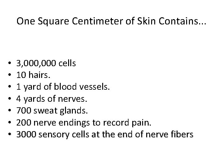
One Square Centimeter of Skin Contains. . . • • 3, 000 cells 10 hairs. 1 yard of blood vessels. 4 yards of nerves. 700 sweat glands. 200 nerve endings to record pain. 3000 sensory cells at the end of nerve fibers
Skin- 3 basic layers • Epidermis- epithelial cells with no blood – Avascular • Dermis- True skin made of connective tissue and is vascular • Hypodermis- aka subcutaneous. • Attaches integument to muscle
Layers of the Skin • Dermis – Collagen, elastin, reticulum, and a gel-like ground substance – Hair follicles, sebaceous glands, sweat glands, blood vessels, lymphatic vessels, nerves – Fibroblasts, mast cells, macrophages • Subcutaneous layer – Adipocytes – Dermal and subcutaneous collagen are continuous
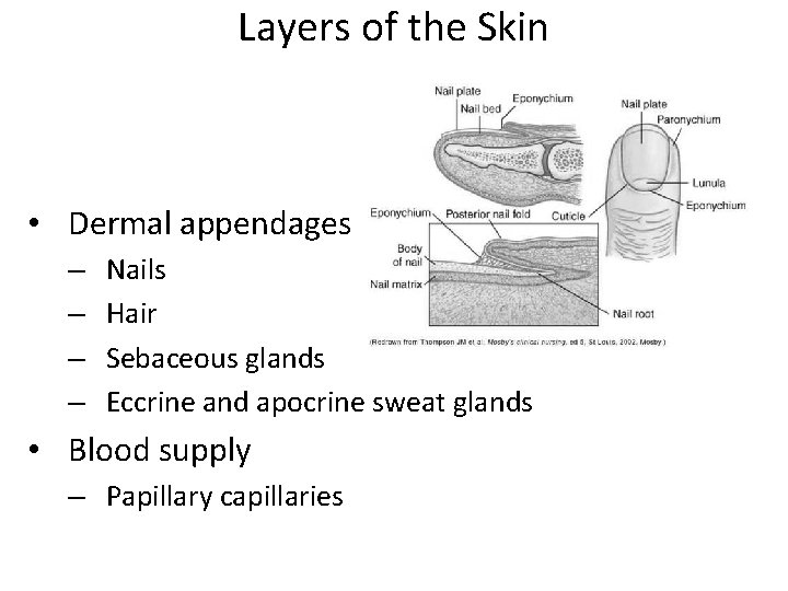
Layers of the Skin • Dermal appendages – – Nails Hair Sebaceous glands Eccrine and apocrine sweat glands • Blood supply – Papillary capillaries
Aging and Skin Integrity • The integumentary system reflects numerous changes from genetic and environmental factors – The skin becomes thinner, drier, wrinkled, and demonstrates a changes in pigmentation – Shortening and decrease in the number of capillary loops – Fewer melanocytes and Langerhans cells
Aging and Skin Integrity – Atrophy of the sebaceous, eccrine, and apocrine glands – Changes in hair color – Fewer hair follicles and growth of thinner hair
Gerontology Considerations Watch for significant changes in aging: • Decrease immunity functions • Susceptibility to infections • Poor nutrition • Decrease collagen production – loss of subcutaneous • Thinning of epidermal skin layers • Increase skin problems
Gerontology Considerations • Taking more medications • Excessive environmental exposure • Dryness, wrinkling • Uneven pigmentation • Various proliferative lesions
Characteristics of skin lesions • ATROPHY – Thinning of the skin surface and loss of skin markings – Skin appears translucent and paperlike
Cherry Angiomas • Elderly • bright red moles on the skin • Papules • Benign
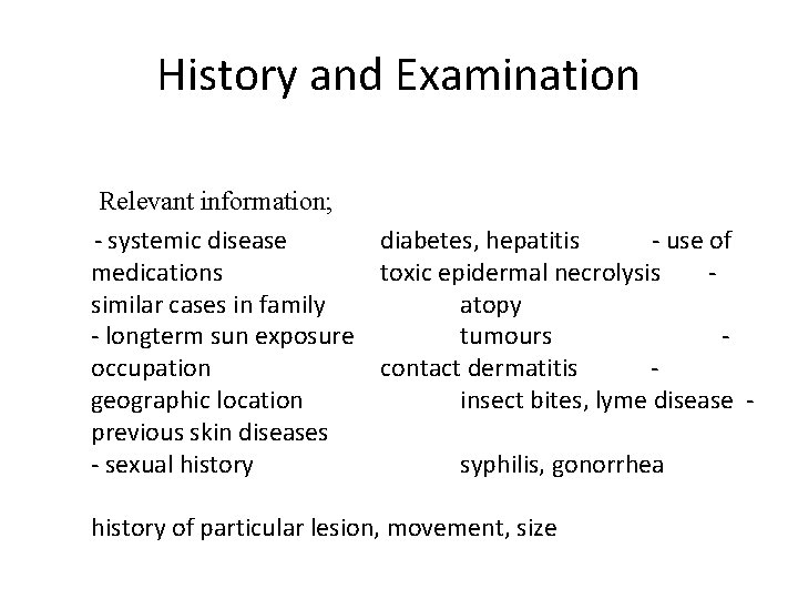
History and Examination Relevant information; - systemic disease diabetes, hepatitis - use of medications toxic epidermal necrolysis similar cases in family atopy - longterm sun exposure tumours occupation contact dermatitis geographic location insect bites, lyme disease previous skin diseases - sexual history syphilis, gonorrhea history of particular lesion, movement, size
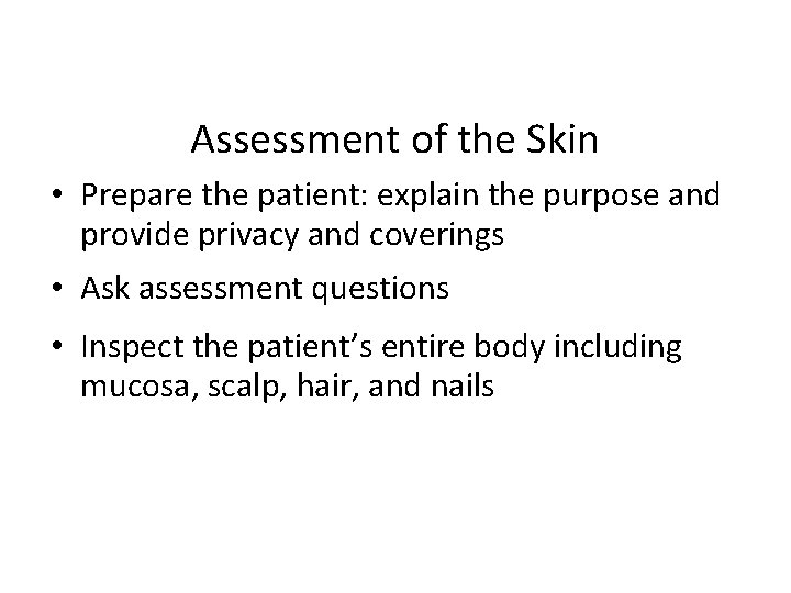
Assessment of the Skin • Prepare the patient: explain the purpose and provide privacy and coverings • Ask assessment questions • Inspect the patient’s entire body including mucosa, scalp, hair, and nails
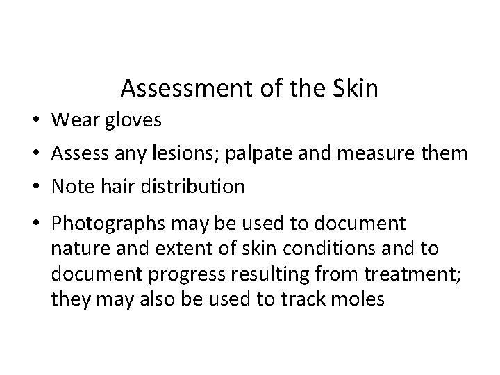
Assessment of the Skin • Wear gloves • Assess any lesions; palpate and measure them • Note hair distribution • Photographs may be used to document nature and extent of skin conditions and to document progress resulting from treatment; they may also be used to track moles
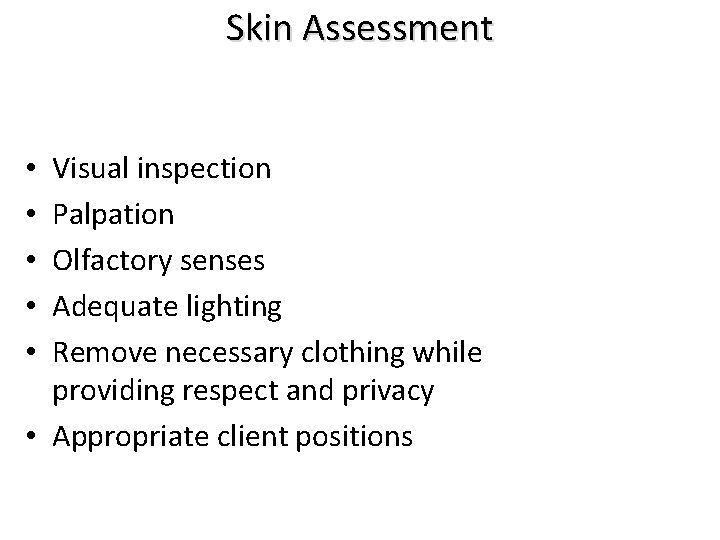
Skin Assessment Visual inspection Palpation Olfactory senses Adequate lighting Remove necessary clothing while providing respect and privacy • Appropriate client positions • • •
Assessing the Skin • Lesions: Lightly palpate any lesions to detect tenderness, firmness, and depth. Measure length, width, and depth also. • Birthmarks/Moles: Note location, color, shape, and size. Assess with the following four warning signs (ABCD) that might indicate the presence of skin cancer: – A little later
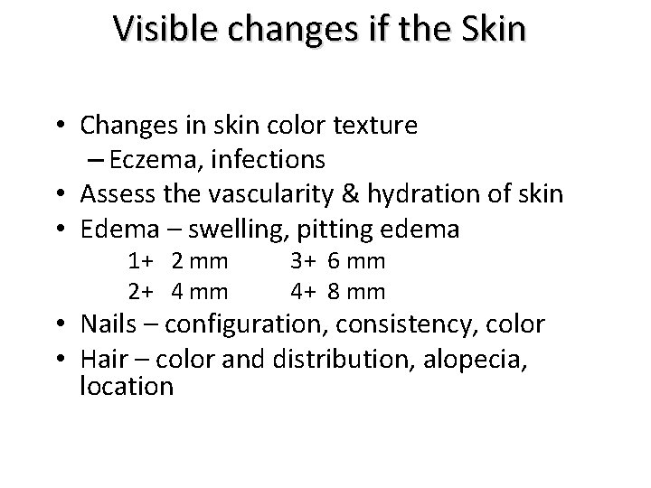
Visible changes if the Skin • Changes in skin color texture – Eczema, infections • Assess the vascularity & hydration of skin • Edema – swelling, pitting edema 1+ 2 mm 2+ 4 mm 3+ 6 mm 4+ 8 mm • Nails – configuration, consistency, color • Hair – color and distribution, alopecia, location
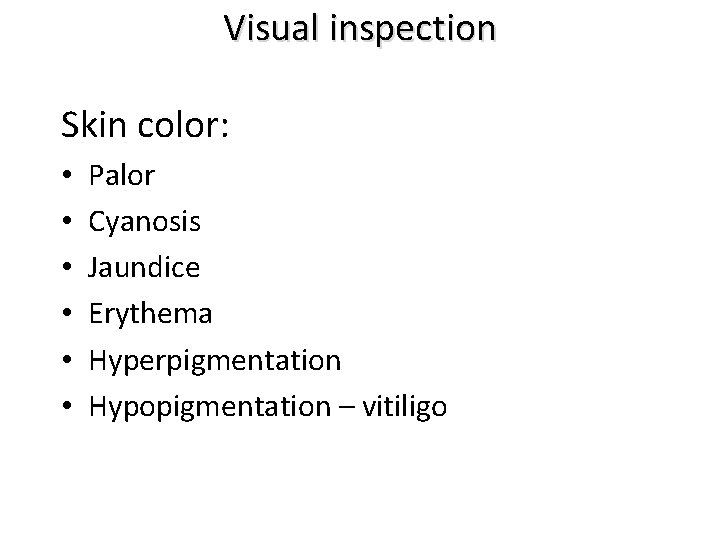
Visual inspection Skin color: • • • Palor Cyanosis Jaundice Erythema Hyperpigmentation Hypopigmentation – vitiligo
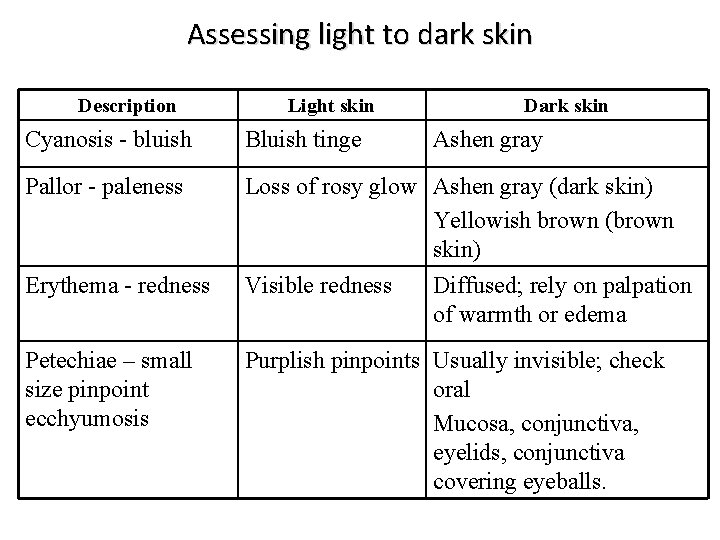
Assessing light to dark skin Description Light skin Dark skin Cyanosis - bluish Bluish tinge Pallor - paleness Loss of rosy glow Ashen gray (dark skin) Yellowish brown (brown skin) Visible redness Diffused; rely on palpation of warmth or edema Erythema - redness Petechiae – small size pinpoint ecchyumosis Ashen gray Purplish pinpoints Usually invisible; check oral Mucosa, conjunctiva, eyelids, conjunctiva covering eyeballs.
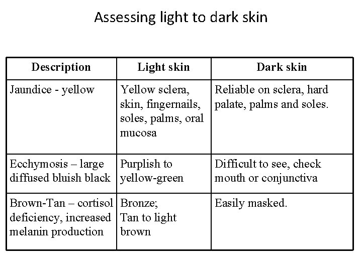
Assessing light to dark skin Description Jaundice - yellow Light skin Dark skin Yellow sclera, Reliable on sclera, hard skin, fingernails, palate, palms and soles, palms, oral mucosa Ecchymosis – large Purplish to diffused bluish black yellow-green Difficult to see, check mouth or conjunctiva Brown-Tan – cortisol Bronze; Easily masked. deficiency, increased Tan to light melanin production brown
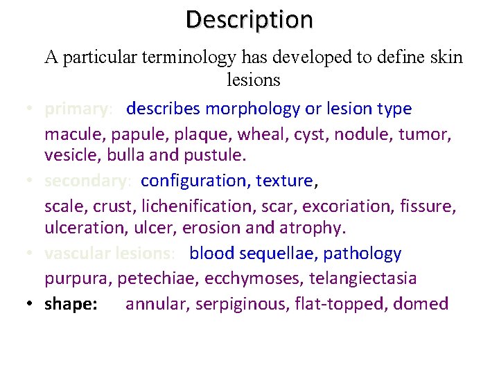
Description • • A particular terminology has developed to define skin lesions primary: describes morphology or lesion type macule, papule, plaque, wheal, cyst, nodule, tumor, vesicle, bulla and pustule. secondary: configuration, texture, scale, crust, lichenification, scar, excoriation, fissure, ulceration, ulcer, erosion and atrophy. vascular lesions: blood sequellae, pathology purpura, petechiae, ecchymoses, telangiectasia shape: annular, serpiginous, flat-topped, domed
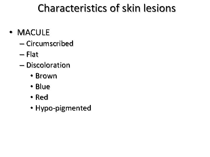
Macule A macule is a change in the color of the skin. It is flat, if you were to close your eyes and run your fingers over the surface of a purely macular lesion, you could not detect it. A macule greater than 1 cm. may be referred to as a patch
Characteristics of skin lesions • MACULE – Circumscribed – Flat – Discoloration • Brown • Blue • Red • Hypo-pigmented
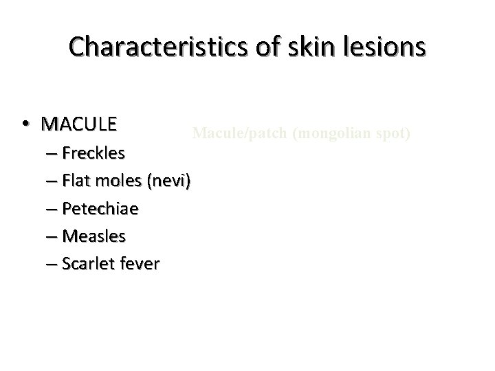
Characteristics of skin lesions • MACULE – Freckles – Flat moles (nevi) – Petechiae – Measles – Scarlet fever Macule/patch (mongolian spot)
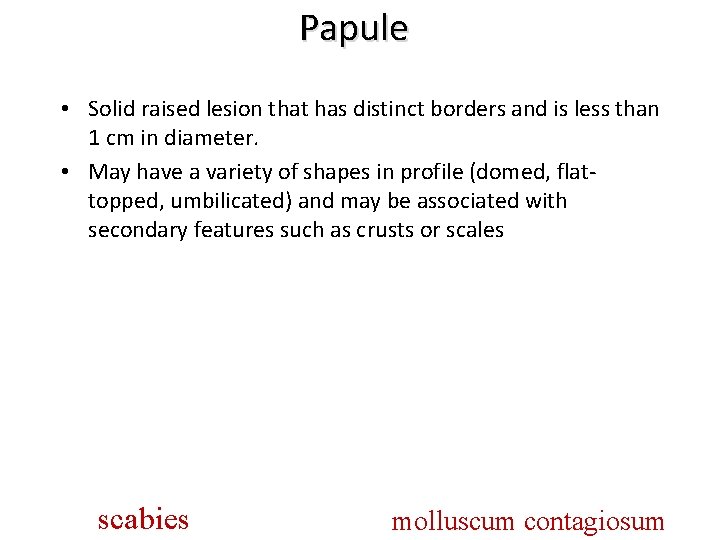
Papule • Solid raised lesion that has distinct borders and is less than 1 cm in diameter. • May have a variety of shapes in profile (domed, flattopped, umbilicated) and may be associated with secondary features such as crusts or scales scabies molluscum contagiosum
Characteristics of skin lesions • PAPULE – Circumscribed – Elevated – Superficial – Solid lesion – > 0. 5 cm in diameter – Often confluent
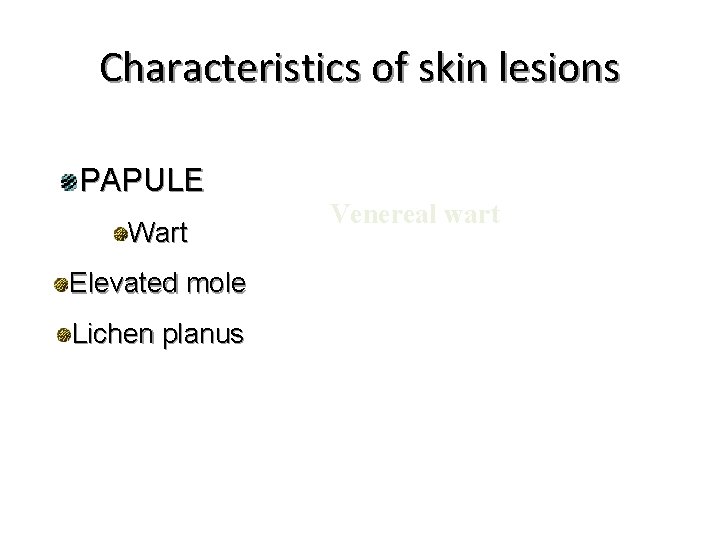
Characteristics of skin lesions PAPULE Wart Elevated mole Lichen planus Venereal wart
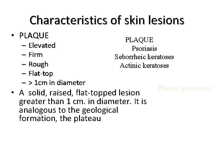
Characteristics of skin lesions • PLAQUE – Elevated – Firm – Rough – Flat-top – > 1 cm in diameter PLAQUE Psoriasis Seborrheic keratoses Actinic keratoses • A solid, raised, flat-topped lesion greater than 1 cm. in diameter. It is analogous to the geological formation, the plateau Plaque (psoriasis)
Wheal (hive) An area of edema in the upper epidermis “Edematous, transient papule or plaque caused by infiltration of dermis by fluid WHEAL Elevated Irregular shaped Cutaneous edema Solid Transient Variable diameter An elevated, white to pink edematous lesion that is unstable and associated with pruritus. Wheals are evanescent – they appear and disappear quickly. Seen in mosquito bites and hives.
Characteristics of skin lesions • WHEAL – Urticaria – Insect bite – Allergic reaction – Desired technique
Nodule is a raised solid lesion more than 1 cm. and may be in the epidermis, or subcutaneous tissue NODULE Elevated Firm Circumscribed Deeper in dermis than a papule 1 -2 cm in diameter
Characteristics of skin lesions • NODULE – Erythema nodosum – Lipoma
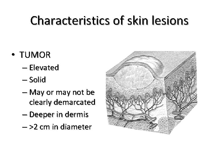
Characteristics of skin lesions • TUMOR – Elevated – Solid – May or may not be clearly demarcated – Deeper in dermis – >2 cm in diameter
Characteristics of skin lesions • TUMOR – Neoplasm – Benign – Lipoma – hemangioma
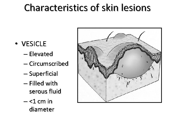
Characteristics of skin lesions • VESICLE – Elevated – Circumscribed – Superficial – Filled with serous fluid – <1 cm in diameter
Vesicle Raised lesions less than 5 mm. in diameter that are filled with clear fluid (blister) http: //z. about. com/d/dermatology/1/0/M/5/three_lesions. jpg
Characteristics of skin lesions • VESICLE – Varicella – (chicken pox) – Herpes Zoster – (shingles)
Characteristics of skin lesions • BULLA – Vesicle greater than 1 cm in diameter
Characteristics of skin lesions • BULLA – Blister – Pemphigus vulgaris
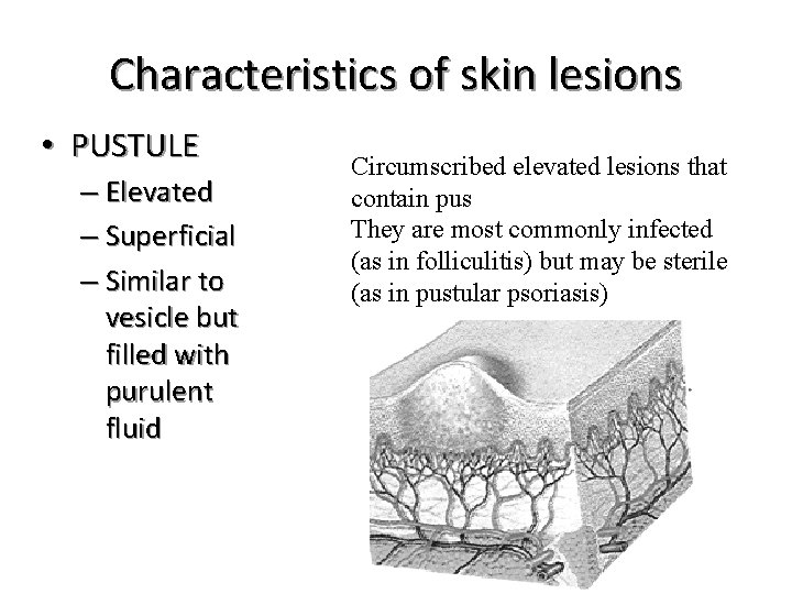
Characteristics of skin lesions • PUSTULE – Elevated – Superficial – Similar to vesicle but filled with purulent fluid Circumscribed elevated lesions that contain pus They are most commonly infected (as in folliculitis) but may be sterile (as in pustular psoriasis)
Characteristics of skin lesions • PUSTULE – Impetigo – acne
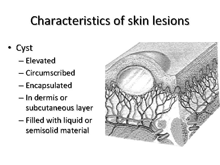
Characteristics of skin lesions • Cyst – Elevated – Circumscribed – Encapsulated – In dermis or subcutaneous layer – Filled with liquid or semisolid material
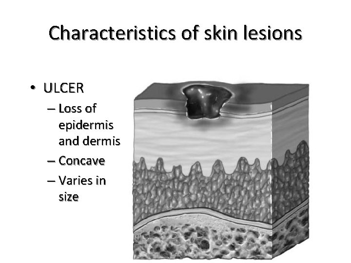
Characteristics of skin lesions • ULCER – Loss of epidermis and dermis – Concave – Varies in size
Characteristics of skin lesions • FISSURE – Linear crack or break from the epidermis to the dermis – May be moist or dry
Characteristics of skin lesions • FISSURE – Athlete’s foot – Crack at the corner of the mouth
Characteristics of skin lesions • SCALE – Heaped-up – Keratinized cells – Flaky skin – Irregular – Thick or thin – Dry or oily – Variation in size
Characteristics of skin lesions • SCALE – Flaking of skin with seborrheic dermatitis following Scarlet fever – Drug reaction – Dry skin
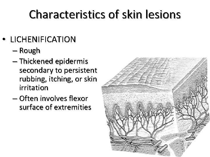
Characteristics of skin lesions • LICHENIFICATION – Rough – Thickened epidermis secondary to persistent rubbing, itching, or skin irritation – Often involves flexor surface of extremities
Characteristics of skin lesions • LICHENIFICATION – Chronic dermatitis – Eczema – Contact dermatitis.
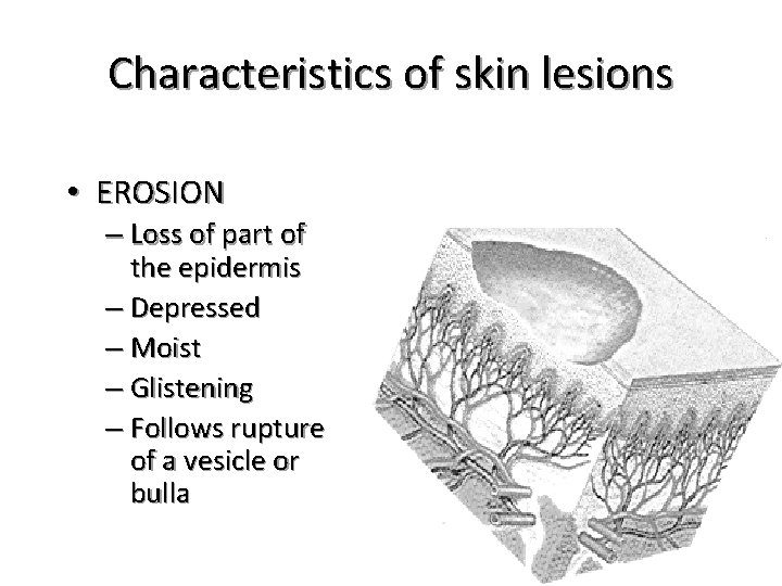
Characteristics of skin lesions • EROSION – Loss of part of the epidermis – Depressed – Moist – Glistening – Follows rupture of a vesicle or bulla
Characteristics of skin lesions • EROSION – Varicella – Variola after rupture
Characteristics of skin lesions • TELANGIECTASIA – Fine – Dilation of capillaries – Irregular red line
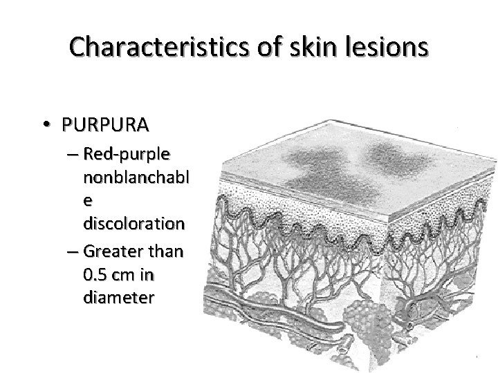
Characteristics of skin lesions • PURPURA – Red-purple nonblanchabl e discoloration – Greater than 0. 5 cm in diameter
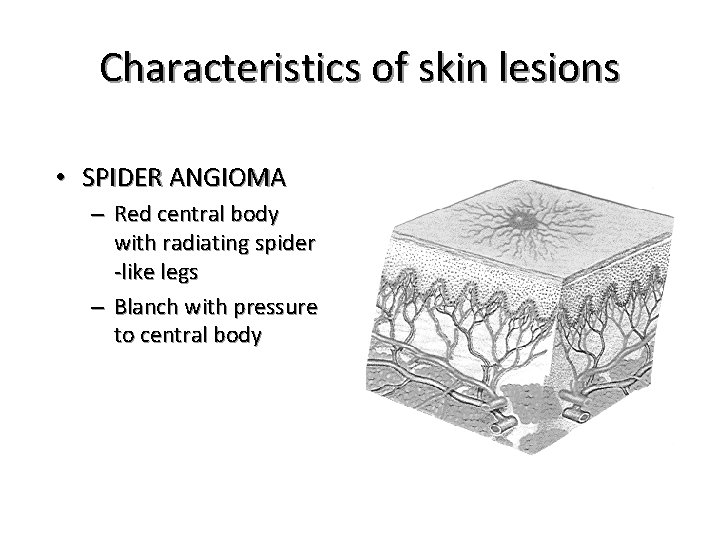
Characteristics of skin lesions • SPIDER ANGIOMA – Red central body with radiating spider -like legs – Blanch with pressure to central body
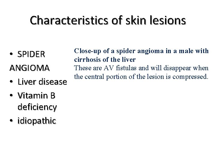
Characteristics of skin lesions • SPIDER ANGIOMA • Liver disease • Vitamin B deficiency • idiopathic Close-up of a spider angioma in a male with cirrhosis of the liver These are AV fistulas and will disappear when the central portion of the lesion is compressed.
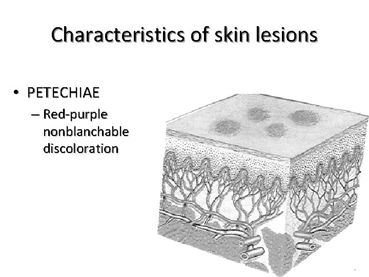
Characteristics of skin lesions • PETECHIAE – Red-purple nonblanchable discoloration
Characteristics of skin lesions • PETECHIAE – Intravascular defects – Infection Found in the mouth Vancomycin-Induced Thrombocytopenia
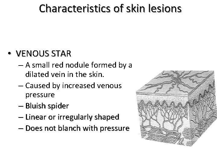
Characteristics of skin lesions • VENOUS STAR – A small red nodule formed by a dilated vein in the skin. – Caused by increased venous pressure – Bluish spider – Linear or irregularly shaped – Does not blanch with pressure
Characteristics of skin lesions • VENOUS STAR – Increased pressure in superficial veins
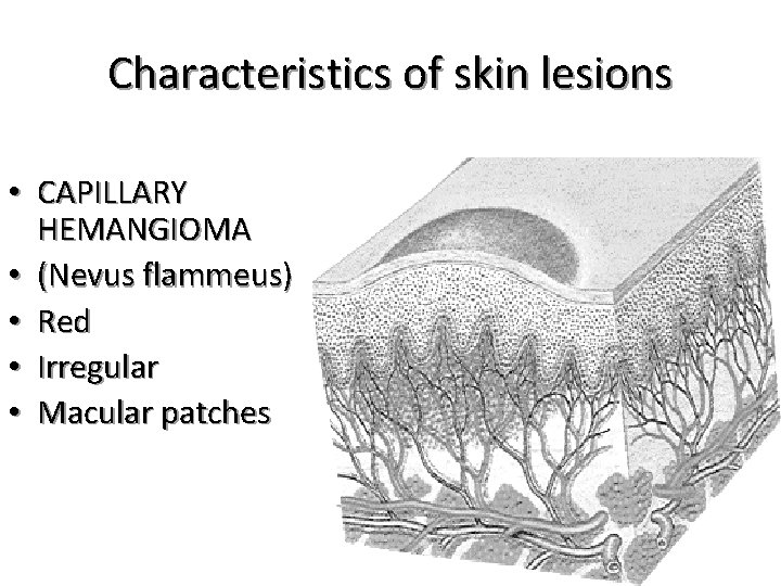
Characteristics of skin lesions • CAPILLARY HEMANGIOMA • (Nevus flammeus) • Red • Irregular • Macular patches
Characteristics of skin lesions • CAPILLARY HEMANGIOMA • (Nevus flammeus) – Dilation of dermal capillaries