
Integument and appendages Dr M Asam Riaz Assistant
- Slides: 31
Integument and appendages Dr. M. Asam Riaz Assistant Professor Entomology, College of Agriculture, University of Sargodha, Pakistan Dr M Asam Riaz, Assistant Professor, Entomology, College of Agri, UOS 1
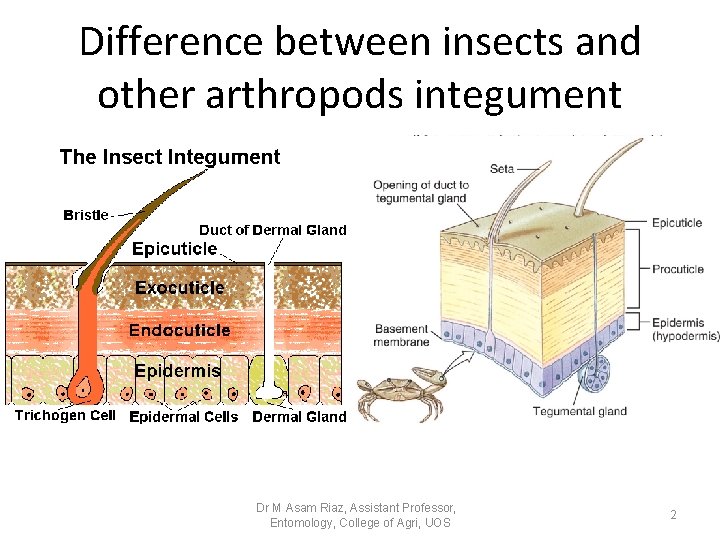
Difference between insects and other arthropods integument Dr M Asam Riaz, Assistant Professor, Entomology, College of Agri, UOS 2
Movie before lecture • https: //www. youtube. com/watch? v=f. Zk. Nlv. Kd K 3 g • https: //www. youtube. com/watch? v=BXpwh. U CDzc 8 Dr M Asam Riaz, Assistant Professor, Entomology, College of Agri, UOS 3
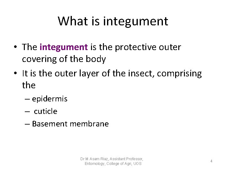
What is integument • The integument is the protective outer covering of the body • It is the outer layer of the insect, comprising the – epidermis – cuticle – Basement membrane Dr M Asam Riaz, Assistant Professor, Entomology, College of Agri, UOS 4
Epidermis • It is the outer cell layer of the insect. It is one cell thick, but the cell densities and cell depth changes during development. • This specialized regions are known as plasma membrane plaques and they are the sites of secretion of the outer epicuticle and of chitin fibers. • the epidermal cells form pore canal of the cuticle Dr M Asam Riaz, Assistant Professor, Entomology, College of Agri, UOS 5
Epidermis • All epidermal cells are glandular in the sense. • They secret cuticle and the enzyme concerned in its production and its digestion at the time of molting. • Some epidermal cells have additional specialized glandular functions. • Trichogen, tormogen cells, dermal gland cells Dr M Asam Riaz, Assistant Professor, Entomology, College of Agri, UOS 6
Basal lamina/Basement membrane • The epidermal cells stand on a basal lamina or basement membrane. – The primary components of it are • • fibrous protein, collagen, glycoprotein and glycosaminoglycans. – The basal lamina acts as a molecular sieve. – It forms a sheet where muscles are attached. – It may be produced by the epidermal cells, but plasmatocytes also contribute to it. Dr M Asam Riaz, Assistant Professor, Entomology, College of Agri, UOS 7
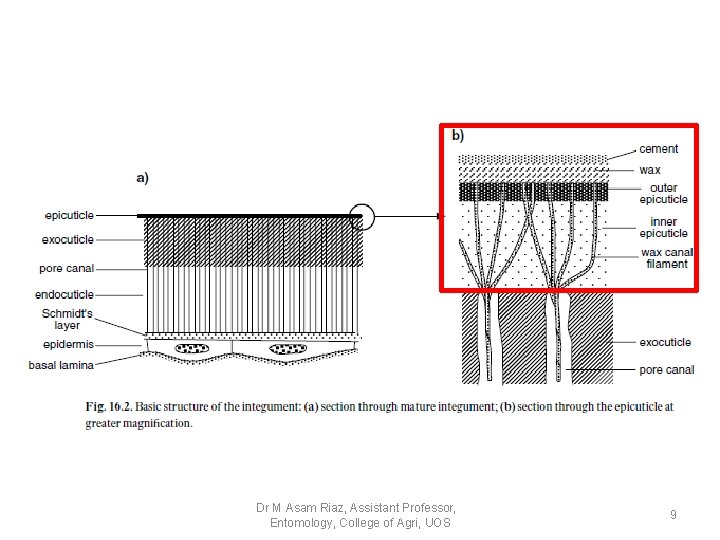
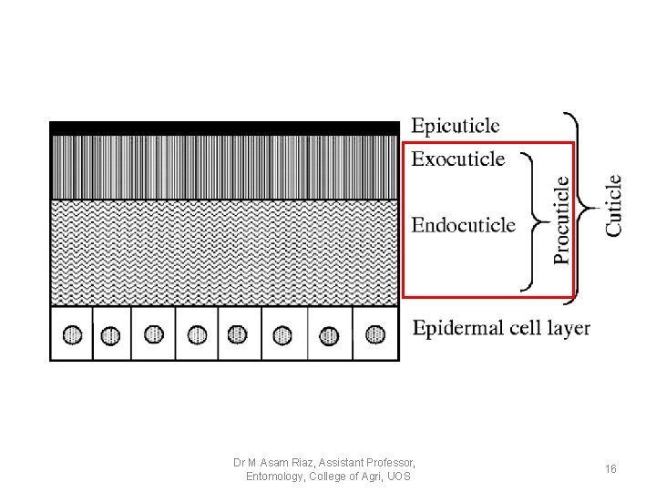
Basic structure of cuticle • It is differentiated into two main regions: 1 - Inner region Pocuticle, characterized by the presence of chitin and forming the block of the cuticle. (200 micrometer) 2 - Outer thin epicuticel, which don’t contain chitin. (1 -4 micrometer) Dr M Asam Riaz, Assistant Professor, Entomology, College of Agri, UOS 8
Dr M Asam Riaz, Assistant Professor, Entomology, College of Agri, UOS 9
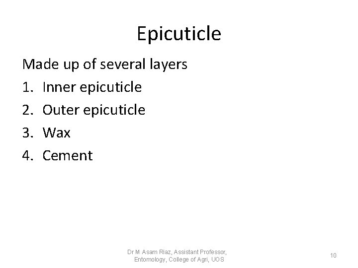
Epicuticle Made up of several layers 1. Inner epicuticle 2. Outer epicuticle 3. Wax 4. Cement Dr M Asam Riaz, Assistant Professor, Entomology, College of Agri, UOS 10
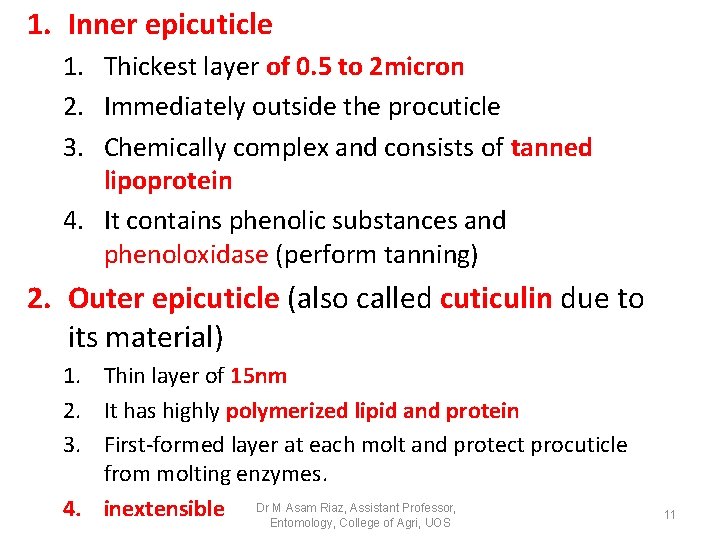
1. Inner epicuticle 1. Thickest layer of 0. 5 to 2 micron 2. Immediately outside the procuticle 3. Chemically complex and consists of tanned lipoprotein 4. It contains phenolic substances and phenoloxidase (perform tanning) 2. Outer epicuticle (also called cuticulin due to its material) 1. Thin layer of 15 nm 2. It has highly polymerized lipid and protein 3. First-formed layer at each molt and protect procuticle from molting enzymes. M Asam Riaz, Assistant Professor, 4. inextensible Dr. Entomology, College of Agri, UOS 11
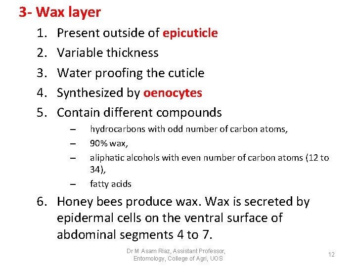
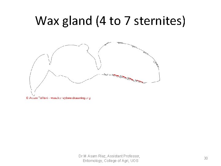
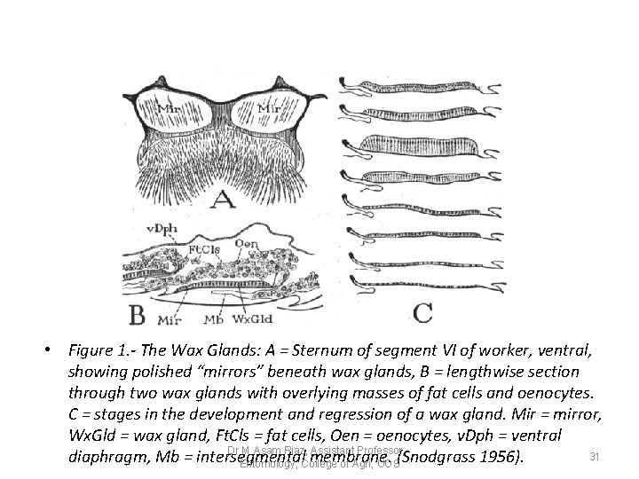
3 - Wax layer 1. 2. 3. 4. 5. Present outside of epicuticle Variable thickness Water proofing the cuticle Synthesized by oenocytes Contain different compounds – – hydrocarbons with odd number of carbon atoms, 90% wax, aliphatic alcohols with even number of carbon atoms (12 to 34), fatty acids 6. Honey bees produce wax. Wax is secreted by epidermal cells on the ventral surface of abdominal segments 4 to 7. Dr M Asam Riaz, Assistant Professor, Entomology, College of Agri, UOS 12
• Wax is used by honey bees to protect themselves against water loss through the integument and in the construction of combs. The major fractions of the cuticular wax were analyzed by gas-liquid chromatography and were shown to be qualitatively similar to those of comb wax (Blomquist et al. 1980). However, the composition of the cuticular wax of the honey bee is quantitatively different from that of the comb wax. Dr M Asam Riaz, Assistant Professor, Entomology, College of Agri, UOS 13
• The major component of the cuticular lipids is hydrocarbon, which comprises 58% of this wax. • In contrast, hydrocarbon comprises only 1317% of the comb wax, and monoester is the largest component (Tulloch 1971). Comb wax is produced by four pairs of glands within the abdomen while cuticular wax is likely produced by epidermal cells of the integument Dr M Asam Riaz, Assistant Professor, Entomology, College of Agri, UOS 14
4 - Cement layer 1. Very thin layer outside most of the wax 2. Consisting of mucopolysaccharide associated with lipids 3. Protect underlying wax 4. Not produced by all insects (e. g. , honey bee) 5. It is secreted by gland cell. Dr M Asam Riaz, Assistant Professor, Entomology, College of Agri, UOS 15
Dr M Asam Riaz, Assistant Professor, Entomology, College of Agri, UOS 16
Procuticle • Chitin commonly comprising 20 -50% of insect dry weight. • Cuticle bound to protein. • the outer part become hard and rigid to form exocuticle, • the inner undifferentiated part is called endocuticle. • Between of the two there maybe a region of hardened, but not fully darkened cuticle (which contain stain) is called mesocuticle. Dr M Asam Riaz, Assistant Professor, Entomology, College of Agri, UOS 17
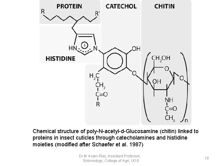
What is Chitin? • Chitin is a polysaccharide made up largely of Nacetylglucosamine residues. • The sugar residues are linked by 1 -4 β linkage, • Adjacent chitin chains are held together by hydrogen bonds to form microfibrils. Dr M Asam Riaz, Assistant Professor, Entomology, College of Agri, UOS 18
Chemical structure of poly-N-acetyl-d-Glucosamine (chitin) linked to proteins in insect cuticles through catecholamines and histidine moieties (modified after Schaefer et al. 1987) Dr M Asam Riaz, Assistant Professor, Entomology, College of Agri, UOS 19
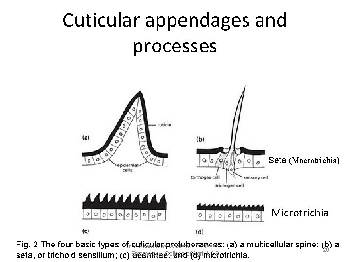
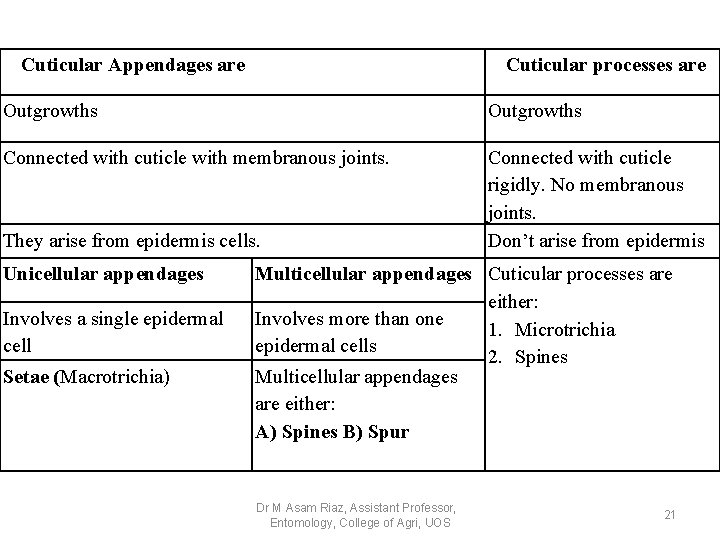
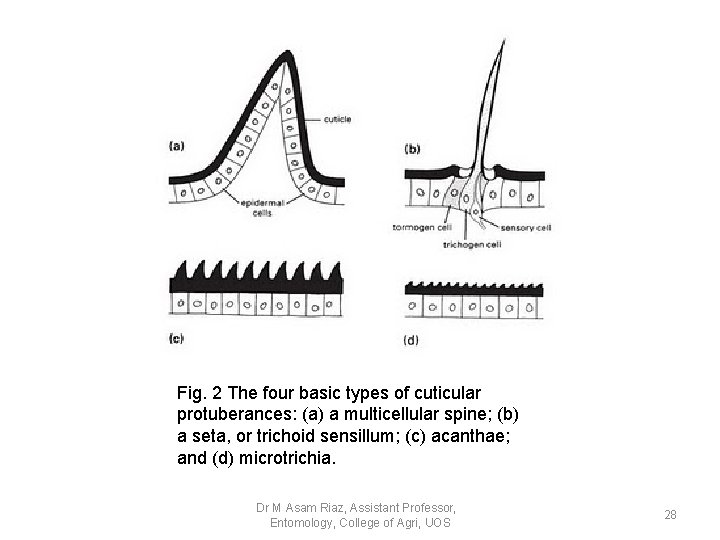
Cuticular appendages and processes Seta (Macrotrichia) Microtrichia Fig. 2 The four basic types of. Dr cuticular M Asam Riaz, protuberances: Assistant Professor, (a) a multicellular spine; (b) a 20 College Agri, microtrichia. UOS seta, or trichoid sensillum; (c) Entomology, acanthae; andof(d)
Cuticular Appendages are Cuticular processes are Outgrowths Connected with cuticle with membranous joints. Connected with cuticle rigidly. No membranous joints. Don’t arise from epidermis They arise from epidermis cells. Unicellular appendages Involves a single epidermal cell Setae (Macrotrichia) Multicellular appendages Cuticular processes are either: Involves more than one 1. Microtrichia epidermal cells 2. Spines Multicellular appendages are either: A) Spines B) Spur Dr M Asam Riaz, Assistant Professor, Entomology, College of Agri, UOS 21
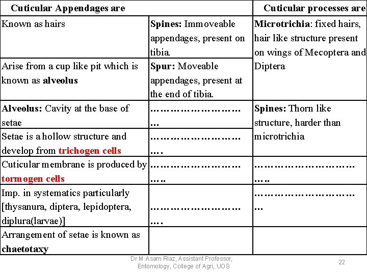
Cuticular Appendages are Cuticular processes are Known as hairs Spines: Immoveable appendages, present on tibia. Arise from a cup like pit which is Spur: Moveable known as alveolus appendages, present at the end of tibia. Alveolus: Cavity at the base of …………… setae … Setae is a hollow structure and …………… develop from trichogen cells …. Cuticular membrane is produced by …………… tormogen cells …. . Imp. in systematics particularly [thysanura, diptera, lepidoptera, …………… diplura(larvae)] …. Arrangement of setae is known as chaetotaxy Dr M Asam Riaz, Assistant Professor, Entomology, College of Agri, UOS Microtrichia: fixed hairs, hair like structure present on wings of Mecoptera and Diptera Spines: Thorn like structure, harder than microtrichia …………… …. . …………… … 22
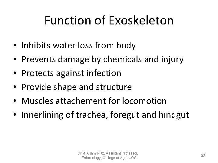
Function of Exoskeleton • • • Inhibits water loss from body Prevents damage by chemicals and injury Protects against infection Provide shape and structure Muscles attachement for locomotion Innerlining of trachea, foregut and hindgut Dr M Asam Riaz, Assistant Professor, Entomology, College of Agri, UOS 23
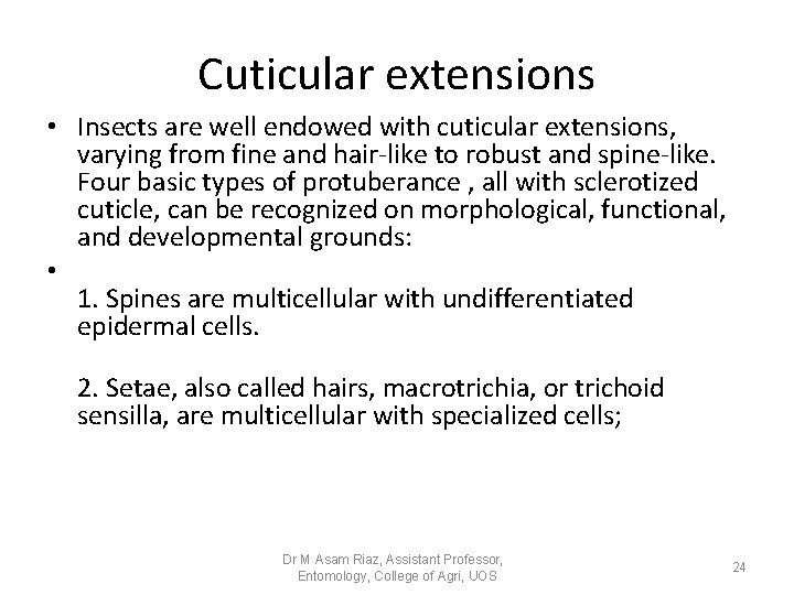
Cuticular extensions • Insects are well endowed with cuticular extensions, varying from fine and hair-like to robust and spine-like. Four basic types of protuberance , all with sclerotized cuticle, can be recognized on morphological, functional, and developmental grounds: • 1. Spines are multicellular with undifferentiated epidermal cells. 2. Setae, also called hairs, macrotrichia, or trichoid sensilla, are multicellular with specialized cells; Dr M Asam Riaz, Assistant Professor, Entomology, College of Agri, UOS 24
• 3. Acanthae are unicellular in origin. 4. microtrichia are subcellular, with several to many extensions per cell Dr M Asam Riaz, Assistant Professor, Entomology, College of Agri, UOS 25
Cuticular extensions Con… Setae sense much of the insect’s tactile environment. Large setae may be called bristles or chaetae, with the most modified being scales, the flattened setae found on butterflies and moths (Lepidoptera) and sporadically elsewhere. Three separate cells form each seta, one for hair formation (trichogen cell), one for socket formation (tormogen cell), and one sensory cell (Fig. 4. 1). Dr M Asam Riaz, Assistant Professor, Entomology, College of Agri, UOS 26
Cuticular extensions Con… • There is no such cellular differentiation in multicellular spines, unicellular acanthae, and subcellular microtrichia. The functions of these types of protuberances are diverse and sometimes debatable, but their sensory function appears limited. The production of pattern, including color, may be significant for some of the microscopic projections. Spines are immovable, but if they are articulated, then they are called spurs. Both spines and spurs may bear unicellular or subcellular processes Dr M Asam Riaz, Assistant Professor, Entomology, College of Agri, UOS 27
Fig. 2 The four basic types of cuticular protuberances: (a) a multicellular spine; (b) a seta, or trichoid sensillum; (c) acanthae; and (d) microtrichia. Dr M Asam Riaz, Assistant Professor, Entomology, College of Agri, UOS 28
Movie before lecture • https: //www. youtube. com/watch? v=f. Zk. Nlv. Kd K 3 g • https: //www. youtube. com/watch? v=BXpwh. U CDzc 8 Dr M Asam Riaz, Assistant Professor, Entomology, College of Agri, UOS 29
Wax gland (4 to 7 sternites) Dr M Asam Riaz, Assistant Professor, Entomology, College of Agri, UOS 30
• Figure 1. - The Wax Glands: A = Sternum of segment VI of worker, ventral, showing polished “mirrors” beneath wax glands, B = lengthwise section through two wax glands with overlying masses of fat cells and oenocytes. C = stages in the development and regression of a wax gland. Mir = mirror, Wx. Gld = wax gland, Ft. Cls = fat cells, Oen = oenocytes, v. Dph = ventral Dr M Asam Riaz, Assistant Professor, 31 diaphragm, Mb = intersegmental membrane. Entomology, College of Agri, UOS(Snodgrass 1956).