OCULAR SYMPTOMATOLOGY DIMINUTION OF VISION Characterization of the


 i. e. rays of")


, bilateral and complete.")


PARTIAL- RELATIVE SCOTOMATA B)COMPLETE-")


 2. Subconscious")


- Slides: 64
OCULAR SYMPTOMATOLOGY
DIMINUTION OF VISION �Characterization of the loss of vision should include: 1. Duration 2. Progression: steadily worsening, improving or static 3. Pattern: constant, intermittent, more for distance or near, episodic or periodic; and 4. Associated symptoms such as pain, redness, watering, photophobia, photopsia, floaters, diplopia, presence of a positive or negative scotoma or peripheral field defect.
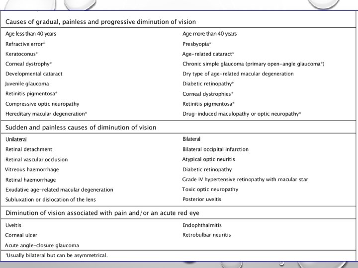
CAUSES OF GRADUAL PAINLESS VISION LOSS • Refractive error • Cataract • ARMD • Refractive error • Chronic (primary) open-angle glaucoma • Diabetic retinopathy • Drugs, toxins or nutritional deficiency
• Hereditary retinal dystrophies • Cerebrovascular disease • Papilledema • Compression of optic nerve or optic pathway
CAUSES OF GRADUAL PAINFUL VISION LOSS • UVEITIS • OPTIC NEURITIS • TRAUMA
SUDDEN PAINLESS DOV • RETINAL DETACHMENT • VIT HAEMORRAGE • CRVO • CRAO • GAINT CELL ARTERITIS • NAION
ANOMALIES OF OCULAR MOTILITY �Disturbances of the extra ocular muscles may manifest as: Eyestrain OR Asthenopia 2. Binocular Diplopia 1.
ASTHENOPIA Weakness or fatigue of the eyes commonly following prolonged close work but may also occur after extended viewing at a distance such watching TV. Aching/burning of eyes • Heaviness of eyelids • Headache • ‘Doubling’ of letters
Seen in: • Insufficiency of convergence • Phorias or other extraocular muscle imbalances • Uncorrected refractive error • Uncorrected refractive correction especially of astigmatism, or early presbyopia.
BINOCULAR DIPLOPIA • Subjective impression of two images of the same object seen by the patient when both eyes are open, but one image disappears on closing either eye • Occurs due to the inability of the two eyes to move together synchronously such that their foveas are both directed towards a target.
• Seen in: Extra ocular muscle paresis • Restrictive squint • Displaced globe •
DISORDERS OF OCULAR SURFACE Ocular irritation • Lacrimation • Photophobia • Red eye •
OCULAR IRRITATION • Sandy or gritty sensation which is generally worse in the morning. • The patient may also complain of tiredness of the eyes or a burning sensation.
LACRIMATION • Reflex increase in the production of tears, as opposed to epiphora, which signifies an overflow of tears due to an obstruction to the outflow of tears. • Caused by irritation of the ocular surface due to the presence of a foreign particle, inflammation, chemical injuries or psychogenic factors.
PHOTOPHOBIA • Discomfort caused by an abnormal sensitivity to ambient light conditions. • Due to pain induced by pupillary constriction and ciliary spasm because of inflammations at the anterior segment, or stimulation of the terminal fibres of the trigeminal nerve in the cornea. • Encountered in patients having abnormalities of the corneal surface or anterior uveitis.
�Mild photophobia 1. Keratoconiunctivitis 2. Posterior uveitis. 3. Some drugs and poisons which dilate the pupil. �True photophobia must be distinguished from 'glare' as well as decreased vision in bright light.
GLARE �Excessive awareness of light could be due to conditions �allow excess light to enter the eye such as aniridia and ocular albinism, �produce excessive irregular scattering of light in the eye such as a posterior sub-capsular cataract.
�Decreased vision in bright light, due to conditions such as posterior subcapsular cataract, congenital cone dystrophy and other central macular disorders may also be mistaken, for photophobia because of the occasionally reported symptom of having to partly close the eyes in bright light.
RED EYE � Prominent symptom 1. Anterior uveitis, 2. Keratitis, and 3. Acute angle-closure glaucoma.
�The distinguishing features between conjunctivitis, iritis and glaucoma are given in the following table:
CONJUNCTIVITIS INFECTIOUS 1. VIRAL 2. BACTERIAL NON INFECTIOUS: 1 ALLREGIC 2. DRY EYE 2. CHEMICAL REACTION 4. FOREIGN BODY KERATITIS INFECTIOUS: 1. FUNAGAL 2. VIRAL 3. BACTERIAL 4. PROTOZOAL I. E ACANTHAMOEBA NON INFECTIOUS: CORNEAL EROSIONS 2. FOREIGN BODY UVEITIS ACUTE ANGLE CLOSURE GLAUCOMA
D/D’S OF THE COMMON CAUSES OF A RED EYE
VISUAL PHENOMENA �Glare �Floaters �Photopsia �Uniocular diplopia �Metamorphopsia �Coloured Halos �Visual Hallucinations �Scintillating scotoma �Coloured vision
FLOATERS CAUSE • Normally transparent vitreous gel liquefies and breaks up, leading to the presence of little particles and fibrous strands floating in the vitreous cavity, with age. • Debris casts shadows onto the retina. COMPLAINTS • Black dots, rings, strands, 'spiderlike' images that are more noticeable against a bright background and 'move' even when the eye is stationary.
FLOATERS • Some form of vitreous degeneration and liquefaction and are usually benign and age related; they are also common at a younger age in myopes. INDICATES • Showers of dots or a sudden increase in their numbers Indicate the formation of a retinal tear • Sometimes the onset of new floaters is secondary to vitreous haemorrhage, often caused by advanced diabetic retinopathy.
PHOTOPSIA • Flashes of light or sensation of flickering lights. COMPLAINTS CAUSE • Vitreous shrinkage or liquefaction, which causes a pull on the vitreoretinal attachments, irritating the retina and causing it to discharge electrical impulses. • These impulses are interpreted by the brain as 'flashes'. • Phenomenon is usually benign and age- related, but could be an indicator of a developing retinal tear or an early retinal detachment.
UNIOCULAR DIPLOPIA • Early senile cataract: • When light is refracted through wedge shaped areas of hydration in immature senile cataract, patient sees multiple images of distant objects such as moon. • Subluxated lens : two images of an object, one through aphakic area and other through phakic zone. • Large iridotomies, if not below the upper lid may also cause shadowing or uniocular diplopia.
UNIOCULAR DIPLOPIA • Folded- over retinal flap in giant retinal tears is a rare cause. • One image is seen by the macula and projected straight ahead. Other image is due to stimulation of the displaced retina and is projected to the point in the visual field normally subserved by that region of the retina.
METAMORPHOPSIA • Patient perceives objects to have an altered, irregular contour or shape. • Reviewed for any changes using an Amsler grid, which tests central 10 degree of vision. • Central serous choroidopathy, ARMD, diabetic Seen in macular edema, macular hole • Micropsia when ordinary, everyday objects look smaller than normal seen in central serous Variant chorioretinopathy due to separation of retinal elements by collection of SRF.
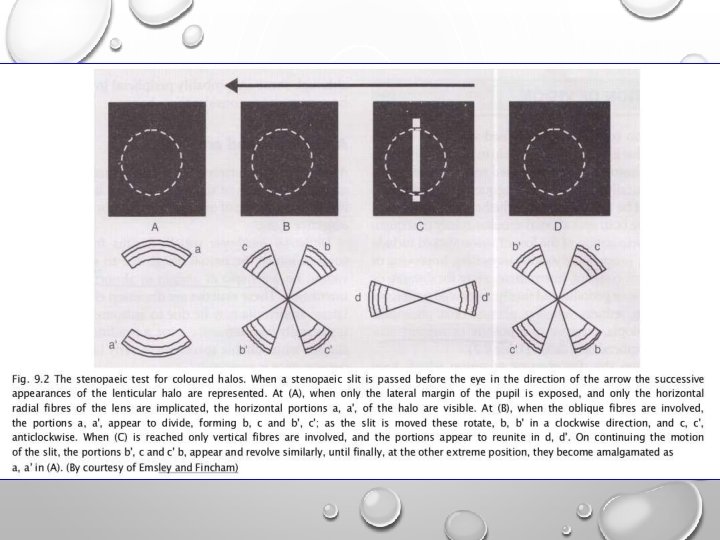
COLOURED HALOS • Rainbow coloured rings around lights at night. • Acute angle closure glaucoma, cataract, corneal Seen in edema, mucus on surface of the conjunctiva. • Occurs due to prismatic dispersion of light bought about these conditions.
COLOURED HALOS • Occurs due to accumulation of fluid in the corneal epithelium & to alterations in the refractive condition of the corneal lamellae. • Halos induced by corneal diseases & early cataractous changes in the lens may be differentiated by the Fincham’s test. • A stenopaeic slit is passed before the eye across the line of vision. glaucomatous halo remains intact but diminished in intensity whereas, a lenticular halo is broken up into segments which revolve as the slit is moved.
VISUAL HALLUCINATIONS • Objects, shapes or lights seen by patients which are not visible to other persons in the vicinity & are fairly Definition specific for disorders involving the cerebral cortex. Types • 1. Unformed visual hallucination • 2. Formed visual hallucination • Unformed hallucinations consist of seeing distorted lights as in flashes & spots, lines, objects, or shapes & are symptoms of lesions affecting the occipital cortex eg. Migraine & AV malformations.
VISUAL HALLUCINATIONS • Formed hallucinations are reported as seeing animals, objects, or people & have specific localizing value, indicating a lesion affecting temporal lobe cortex eg. In temporal lobe tumors & epilepsy. • Formed images could represent misinterpretation of information in brain due to disruption of areas needed to process the information. • Eg. In Charles Bonnet Syndrome, people having a gross diminution of vision ‘see’ detailed images.
�Visual hallucinations can also be due to transitory or chronic abnormalities in the brain, caused by changes like: 1. Electrolyte disturbances 2. High fevers 3. Liver or kidney failure 4. Drugs such as diazepam, alprazolam, LSD
SCOTOMATA �A scotoma is an area of partial alteration in the field of vision consisting of a partially diminished or entirely degenerated visual acuity that is surrounded by a field of normal or relatively well-preserved vision. �Types: 1. Relative scotoma 2. Absolute scotoma
�Relative scotoma: A scotoma in which the visual impairment is not complete. �Absolute scotoma: �Absolute scotoma an area within the visual field in which perception of light is entirely lost.
SCINTILLATING SCOTOMATA � In migraine scintillating scotoma of various kinds occur. � In typical migraine : ◦ Before the attack patient feels unusually well. ◦ Positive scotoma occurs in the field of vision and while obscuring sight it has a peculiar shimmering character. ◦ It gradually increases in size until one half of the field is clouded and fixation point remains relatively clear.
◦ In dark field, bright spots and fortification spectra (teichopsia) i. e. rays of various colors is seen frequently arranged in zigzags. ◦ Homonymous hemianopia usually occurs ( both half fields are affected ) � In other cases : ◦ Whole field becomes clouded but still fixation point is seen momentarily and then becomes obscured until the eyes are moved to fresh spot. ◦ Vision usually clears in quarter of an hour.
◦ Attack is soon followed by violent headache. ◦ Intensified on the side of the head opposite the hemianopic field (hemicrania). ◦ Accompanied by nausea and even sickness ( bilious attack). During the attack numbness in the mouth and tongue, slight aphasia are frequent and copious secretion of urine of low specific gravity.
�Attack occurs periodically but vary in number and severity. �In mild attack and in advanced age the scotoma may occur without the headache or the headache without scotoma. �Migraine is attributed to vasomotor changes in the brain. �Vasodilatation, associated with a feeling of wellbeing, is followed by vasoconstriction, especially in occipital lobes.
�People who suffer ordinary migraine, have attacks in which without any scotoma the headache is followed by partial paralysis of the third nerve (ophthalmoplegic migraine) on the same side as the hemicrania. �Slight ptosis, diplopia and sluggishness of the pupillary reactions continue for some hours and then gradually disappear.
�The paresis is worst and persists longer with succeeding attacks, and eventually sometimes becomes permanent. �Probably most of these cases are not migrainous, but due to some organic nerve lesion such as pressure on the third nerve by a distended artery.
COLOURED VISION • Also known as Chromatopsia , is a rare symptom. • Erythropsia (red vision ) occur in some patients after cataract extraction if the eyes are exposed to bright light. • Objects look red but visual acuity is not affected and no permanent damage results.
COLOURED VISION • Patient should be warned of the possibility of erythropsia as it is somewhat alarming and suggesting of haemorrhage. • On the other hand some patients report an excessive bluish appearance of objects after cataract extraction because blue light was filtered out by the yellowish cataractous nucleus before surgery.
COLOURED VISION • It is also met with in snow blindness. • Occur in some cases during the resolution of optic neuritis when the ensuing atrophy is not complete. • In normal people black print will sometimes suddenly turn deep red owing to strong lateral light entering the eye through the sclera.
AMBLYOPIA & AMAUROSIS �DEFINITION: Partial and complete loss of sight, respectively, in one or both eyes in the absence of ophthalmoscopic or other marked objective sign. ◦ TYPES OF AMBLYOPIA: Unilateral 2. Bilateral 1.
UNILATERAL AMBLYOPIA • Results from physical suppression of retinal image due to sensory deprivation , i. E. Amblyopia or abnormal binocular interaction. • Due to anisometropia, with a unilaterally higher refractive error BILATERAL AMBLYOPIA • Due to bilateral sensory deprivation as in bilateral cataract or corneal opacities or bilateral high refractive error.
Bilateral visual loss due to various exogenous toxins with a normal fundus used to be termed as “Toxic amblyopia”. Now more accurately termed as toxic retinopathies or neuropathies. Occurs in uraemia, meningitis and hysteria.
BILATERAL AMAUROSIS �ONSET: Blindness is sudden or rapid (8 -24 hrs), bilateral and complete. �SEEN IN: 1. Acute nephritis especially complicating pregnancy. 2. After scarlet fever. 3. Chronic renal disease. �FUNDUS: No changes unless there is coincident hypertensive retinopathy.
�Vision improves in 10 -18 hrs and is fully restored in about 48 hrs. �In uraemic amaurosis the pupils are dilated but generally react to light showing that the lower centres are not affected. �The condition is probably due to circulation of toxic material, which acts upon the cells of visual centres. �In cases occuring during pregnancy , there is usually eclampsia.
AMAUROSIS FUGAX �DEFINITION: Transient monocular blindness caused by temporary lack of blood flow either to the brain or retina. �Occurs as a result of emboli from plaques in the carotid artery. �These block an artery for a while and then move on resulting in a loss of vision for the duration of blockage.
• Onset is acute and the episode lasts for several minutes. • Sudden loss may appear like a curtain falling from above or rising from below and vision may be completely absent at the height of the attack. • Recovery occurs in the same pattern.
NIGHT BLINDNESS �Occurs in: 1. Pigmentary retinal dystrophy 2. Xerophthalmia 3. Cirrhosis �Attributed to interference with the functions of the retinal rods.
COLOUR BLINDNESS & ACHROMATOPSIA � TYPES: 1. CONGENITAL 2. ACQUIRED a)PARTIAL- RELATIVE SCOTOMATA B)COMPLETE- DISEASE OF OPTIC Nerve � In most diseases of retina & choroid, changes in colour perception affect mostly the blue end of the spectrum.
�Slight diminution in acuity of perception of these rays is caused normally, owing to their physical absorption, by the increase of amber pigment in the nucleus of the lens (blue blindness). �Seen in sclerosing lenses(black cataract).
�Congenital TOTAL Rare Nystagmus & central scotoma. colour blindness: PARTIAL Seldom discovered Subjects compensate for their defect by attention to shade & texture Spectrum appears grey band like the normal scotopic spectrum, seen with maximum brightness at 510 microns. Caused by a central defect.
COLOUR BLINDNESS �Inherited condition- X-linked recessive �Due to absence of one of the photopigments normally found in the foveal cones. �Red-green cases fall into two chief groups: 1. Protanopes 2. Deuteranopes
� Protanopes: Red end of the spectrum is much less bright than for normal people. Shortened spectrum(red end). � Deuteranopes: Green � Both sensation is defective. have dichromatic vision. � Defects may not be complete & these cases are called protanomalous & deuteranomalous, respectively.
NON-ORGANIC ‘FUNCTIONAL’ VISUAL LOSS �Either due to: 1. Wilful ‘blind’ behaviour (malingering) 2. Subconscious expression of non organic signs & symptoms of defective vision (hysteria).
MALINGERS HYSTERIA Involved with some form of Little or no insight into their financial gain in the form of an infirmity & may sometimes insurance claim, financial display a complete lack of compensation, employement concern over their incapacitating benefits, an excuse to avoid symptoms ( la belle indifference) examinations. • Most common presentation: 1. Decreased visual acuity in one or both eyes. 2. Constricted visual fields
CONDITIONS WHICH PRODUCE VISUAL LOSS WITH A NORMAL FUNDUS