Approach to patient with Arthritis DONE BY TURKI

Approach to patient with Arthritis DONE BY: TURKI ALOTAIBI WALEED ALTALIQI ABDULLAH ALKHAMRI SUPERVISED BY: - PROF. MOHAMMED AL-RUKBAN

Objectives Common causes of arthritis encountered in general practice. What does it mean by arthropathy and arthritis. Highlight on osteoarthritis, Septic arthritis, Gout, Rheumatoid arthritis. Important aspects in History, Clinical examination, Investigations and Management. Red Flags for patient with arthritis. When to refer to specialty clinic.
")
MCQ RA is characterized by which of the following patterns of joint involvement? a) Episodic monoarthritis b) Symmetrical polyarthritis c) Migratory oligoarthritis d) Spondylitis
 MRI b) x-ray")
MCQ The most specific test used in diagnosis of gout: a) MRI b) x-ray c) synovial fluid analysis d) serum uric acid
 S.")
MCQ The most common offending organism in septic arthritis in adults is: a) S. aureus b) Streptococcus pyogenes c) S. pneumoniae d) H. influenzae

MCQ Which one of the following is a characteristic x-ray finding in case of osteoarthritis ? a) chondrocalcinosis b) osteopenia c) Narrowing of joint space d) sequestra

Case Roqaya 42 yo female, saudi book keeper gradually developed painful wrists over 3 months; she consulted the doctor only when the pain and early morning stiffness stopped her from work. Medical history: roqaya’s medical history is unremarkable. Her current medications are iron , Vitamin D and calcium supplement.

Case Physical examination: On examination, both wrists and the metacarpophalangeal joints of both hands were swollen and tender but not deformed. There were no nodules or vasculitic lesions Investigation: On investigation, she was found to have a raised C-reactive protein (CRP) level (27 mg/l) (NR <10) but a normal haemoglobin and white-cell count. A latex test for rheumatoid factor was negative and antinuclear antibodies were not detected.

Case Six months after initial presentation, she developed two subcutaneous nodules on the left elbow; these were small, painless, firm and immobile but not tender. A test for rheumatoid factor was now positive (titre 1/64). X-rays of the hands showed bony erosions in the metacarpal heads. She still had a raised CRP (43 mg/l) but normal serum complement (C 3 and C 4) levels and, she had a biopsy, pannus would have been demonstrable histologically. This woman now had definite X-ray evidence of rheumatoid arthritis and, in view of the continuing arthropathy, her treatment was changed to weekly low-dose methotrexate. This has controlled the arthritis for several years and no further erosions have developed.

Case: A 54 -year-old man complains of severe pain and swelling in his right first toe that developed overnight. He is limping because of the pain and states that this is the most severe pain he has ever had ('even covering my foot with the bed sheet hurts'). He has had no previous episodes. Medical History: His only medication is hydrochlorothiazide for hypertension, He’s an alcoholic.

Case: Physical Examination: There is swelling, erythema, warmth, and tenderness of the right first toe. There is also tenderness and warmth with mild swelling over the mid foot. Investigation: - Deposition of Crystals in synovial fluid among Aspiration. Blood test result revealed Raised Creatine and uric acid. X-ray showing aggressive erosions in PIP of the toe.

case: the patient was treated with colchicine and NSAID’s to control the inflammation and pain. And he was prescribed allopurinol as prophylaxis for controlling his blood uric acid levels. Also he was advised to restrict alcohol intake and reduce meat consumption in his diet. his diuretic was replaced due to it’s contribution to his condition.

Terminology Arthritis: inflammation of the joints. Ankylosis: stiffness of joint. Arthrocentesis: clinical procedure of using a syringe to collect synovial fluid from a joint capsule. Arthroscope: endoscope that's inserted into joint for visual examination.

Overview NHIS 2010 -2012 data analysis showed that 50 million US adults had reported doctor-diagnosed arthritis. In 2007, CDC estimated that 294, 000 U. S. children under age 18 have been diagnosed with arthritis or other rheumatic conditions. A major cause of lost work time and serious disability for many people. Its mainly a disease of adults, but it can also effect children. Treatment of arthritis depends on its type and the main goal of treatment is to reduce the symptoms and improve the quality of life.

Causes Injuries: Leading to degenerative arthritis Abnormal metabolism : Gout Inheritance: Osteoarthritis Infection: Lyme disease Over active immune system : RA and SLE

Diagnostic Approach to Musculoskeletal Pain

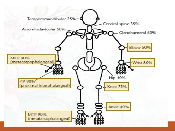
Important aspects in History Duration of Complaints Number of Joints Involved Distribution of Joints Involved Pattern of Involvement Morning Stiffness

Important aspects in History of Joint Swelling. Extra-articular Complaints. Associated Medical Illness. Significant Past History. Family History of Rheumatic Disease.

Importance of Physical Examination Local Warmth Joint effusion Redness Range of Motion Any Deformity

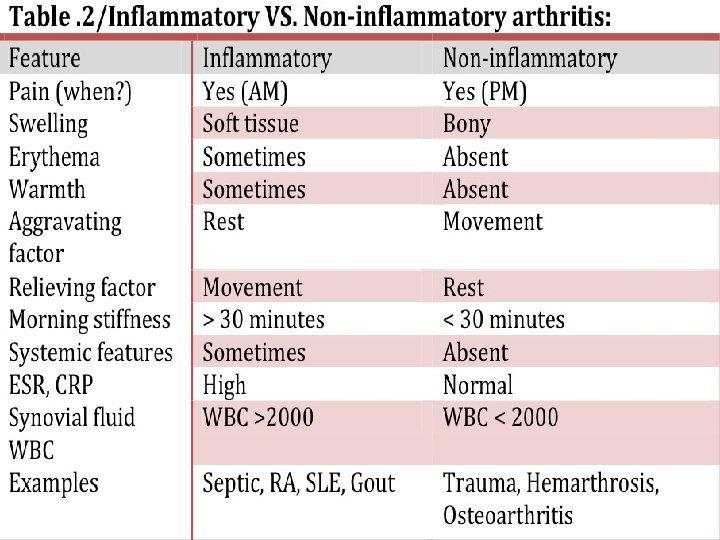
Symptoms 1. Pain Inflammatory joint disease present both at rest and with motion worse at the beginning than at the end of usage. Non-inflammatory pain occurs mainly or only during motion and improves quickly with rest Pain that arises from small peripheral joints more accurately localized than pain arising from larger proximal joints.

Con. 2. Stiffness sensation of tightness when attempting to move joints after a period of inactivity subsides over time Inflammatory arthritis present upon waking typically lasts 30 -60 minutes or longer. Non-inflammatory arthritis experienced briefly (eg, 15 min) upon waking in the morning following periods of inactivity.

Con. 3. Swelling Inflammatory arthritis synovial hypertrophy synovial effusion inflammation of periarticular structures Non-inflammatory arthritis formation of osteophytes synovial cysts Thickening effusions

Con. 4. Limitation of motion structural damage Inflammation contracture of surrounding soft tissues

Con. 5. Weakness result of disuse atrophy Weakness with pain musculoskeletal cause (eg, arthritis, tendonitis)

Temporal pattern of arthritis Abrupt onset symptoms develop over minutes to hours occur in trauma, crystalline synovitis, or infection. Insidious pattern symptoms develop over weeks to months rheumatoid arthritis (RA) and osteoarthritis.

Duration of symptoms: Acute <6 weeks in duration; chronic is 6 or more weeks in duration. Number of involved joints: Monoarthritis - one joint. Oligoarthritis - 2 -4 joints. Polyarthritis -5 or more joints.

Temporal Patterns in Polyarthritis Migratory pattern joints are sequentially affected where, as one joint settles, another becomes inflamed (e. g. , acute rheumatic fever, disseminated gonococcal infection). Intermittent pattern the same joint is involved in different episodes of inflammation, but the joint is quiescent during intervening periods (e. g. , gout). Additive pattern subsequent joints are involved while preceding ones are still inflamed (e. g. RA )

Distribution of affected joints The DIP joints of the fingers involved in psoriatic arthritis, gout, or osteoarthritis spared in RA. Joints of the lumbar spine involved in ankylosing spondylitis spared in RA.

Extra-articular manifestations Constitutional symptoms underlying systemic disorder. include fatigue, fever, and weight loss. Skin lesions SLE, dermatomyositis, scleroderma, Lyme disease, psoriasis and Henoch. Schönlein purpura. Ocular symptoms or signs Episcleritis and Keratoconjunctivitis sicca -RA. Anterior uveitis - ankylosing spondylitis, Conjunctivitis -reactive arthritis

Ocular signs Episcleritis and scleritis Conjunctivitis

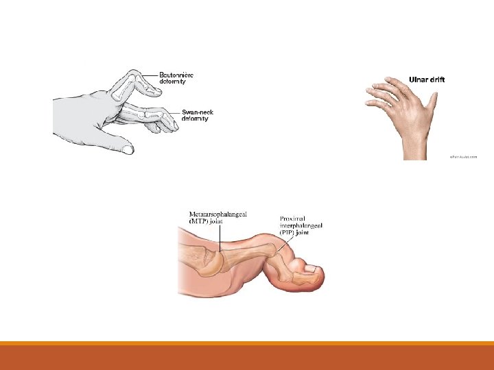
Signs of inflammatory joint disease - Joint effusions - Erythema and warmth - Joint tenderness - Bony overgrowth of the joints (osteophytes) At the DIP joints - Heberden nodes. At the PIP joints are called Bouchard nodes. - Limited range of motion: Crepitus during active or passive range of motion Joint deformity
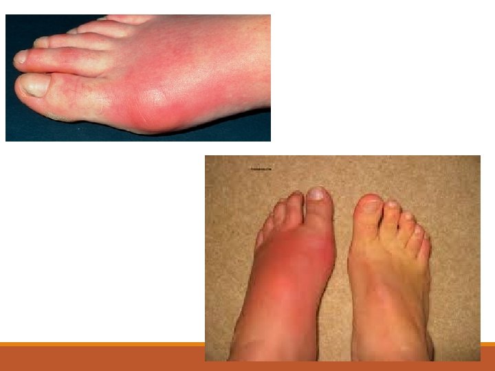

Highlight on Gout Its an Intermittent attacks of acute joint pain due to deposition of uric acid crystals. Usually affect men (10: 1), rare in premenopausal female. Most common joint affected is 1 st MTP joint Prevalence is approximately 20% in patients with a family history of gout.

Gout cont. Causes: Hyperuricemia is the most common cause and it could be because of: 1. Impaired excretion (90%): renal disease, diuretics, NSAID use, and acidosis. 2. Increase production: like chemotherapy, chronic hemolysis, and blood cancers. Risk factors: Alcohol - Dehydration - Urate stones - Diuretics use

Types Acute : common in the late course of untreated gout Chronic : - Chronic tophaceous gout is characterized by collections of solid urate accompanied by chronic inflammatory and often destructive changes in the surrounding connective tissue - often visible and/or palpable - typically not painful or tender

Chronic Tophus of the knee Large tophus and multiple superficial tophi of the knee in patient with chronic uncontrolled gout. Acute Doesn’t have tophi

Pathophysiology

History Mostly involve single joint. Severe pain: Often cannot wear socks. Peak within 24 to 48 hours. Swelling. Redness.

Examination ACUTE GOUT: Fever Mostly involve single joint. Most commonly the first metatarsophalangeal joint "podagra". Severe pain. Erythema. Cellulitis. Chronic Gout: Tophaceous gout. "deposits of monosodium urate crystals in soft tissue

Diagnosis Blood: increase WBC, ESR and Urate X-Ray: Next slide synovial fluid or tophus aspiration with identification of: light microscopy : needle shape crystal compensated polarized light microscopy : positive birefringence with negative elongation

X-ray
")
Criteria Diagnosis from American college of Rheumatology (6 out of 12)

Management Gout is managed in the following 3 stages: Treating the acute attack Providing prophylaxis to prevent acute flares Lowering excess stores of urate to prevent flares of gouty arthritis and to prevent tissue deposition of urate crystals

Pharmacological management of acute gout

Follow up the person 4– 6 weeks after an acute attack of gout has resolved, and: - Check the serum uric acid level. uric acid - Measure their blood pressure and take blood for fasting glucose, blood pressure blood for fasting glucose renal function Consider the need to start prophylactic medication if the person is having two prophylactic medication or more attacks of gout in a year. Colchicine in low doses. Daily NSAID’s.

Rheumatoid Arthritis

Rheumatoid Arthritis A chronic systemic disease primarily of the joints In late stages, deformity and ankylosis develop. - annual incidence of rheumatoid arthritis (RA) has been reported to be around 40 per 100, 000. female: male ration 3: 1 Can present at any age—most common in middle age

Autoimmune disorder - Unknown etiology Genetics – Environmental – Possible infectious component Characterized by: inflammation of the synovial capsule and hyperplasia (“swelling”) swelling autoantibody production (rheumatoid factor and Anti-cyclic citrullinated peptide[ACCP] antinucular antibodies[ANA]). peptide cartilage and bone destruction (“deformity”). deformity Systemic features, including : cardiovascular, pulmonary, psychological, and skeletal disorders.


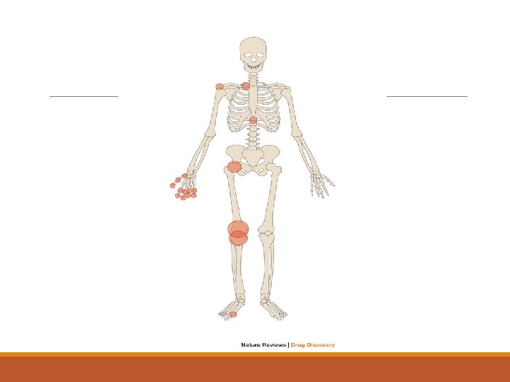
Extra-articular Manifistation

Small vessels Vasculitis Subcutaneous nodule

Episcleritis + scleritis
 OR chronic (6 weeks")
1. Duration of the complaint: Acute (less than 6 weeks) OR chronic (6 weeks or more) History 2. Number of joints involved: rheumatoid arthritis patients have a polyarticular joint involvement. 3. Distribution of Joints Involved: RA has a symmetrical joint involvement. 4. Pattern of involvement: Inflammation persists in involved joints as new ones become affected (Additive). 5. Duration of morning stiffness: Usually morning stiffness last for more than 30 minutes (it can reach one hour). 6. Aggravating and relieving factors: Pain worse after a period of inactivity and relieved by movement. 7. History of joint swelling , Extra-articular complaints Family history

Physical Examination During the physical examination, it is important to assess the following: Stiffness (may improve with heat and active exercise, but they do not prevent the return of stiffness). Tenderness Pain on motion Swelling Deformities (ulnar deviation, boutonniere and swan-neck deformities, hammer toes, and joint ankylosis) Limitation of motion Extra-articular manifestations Rheumatoid nodules (occur in approximately 25% of patients with RA and most commonly found on extensor surfaces [proximal ulna])

Diagnostic criteria

X-Ray

labs RA is a clinical diagnosis; no laboratory test is diagnostic 1. Rheumatoid factor: a. Auto antibodies to the Fc portion of Ig. G Support a diagnosis of Rheumatoid Arthritis but not diagnostic. b. found in 75% to 80% of patients with RA. c. associated with a poor prognosis. d. seen in conditions other than RA like hepatitis C, sarcoidosis, pulmonary fibrosis, and many others. 2. Anti-citrullinated protein antibodies (ACPA): a. These are auto antibodies directed against the body proteins, can be detected by ELISA b. Accuracy (Anti-CCP Assay) Specificity 79%. . Sensitivity 96 -98% c. Diagnosis more accurate when combined with RF.
 a")
Management : -After confirming diagnosis of RA through (history, Physical exams, and investigations) a combination treatment should be started: a. Start DMARD(s): Methotrexate within 3 months to Control symptoms and delay progression of the disease b. Consider NSAIDs (if mild>> To relieve pain and inflammation) c. Consider Local / Low-dose Steroid d. Patient Education e. Physical / Occupational Therapy. f. Referral to rheumatology clinic. g. Follow-UP

 Definition: degeneration of joint cartilage and the underlying bone, most")
Degenerative Arthritis: Osteoarthritis (OA) Definition: degeneration of joint cartilage and the underlying bone, most common from middle age onward. It causes pain and stiffness, especially in the hip, knee, and thumb joints. Risk Factors: genetic predisposition, advanced age, obesity (for knee OA), female, trauma. Signs and Symptoms: localized to affected joints (not a systemic disease). The pain is often insidious, gradually progressive, with an intermittent course.

TYPES OF OSTEOARTHRITIS Idiopathic osteoarthritis — Idiopathic OA can be categorized into localized or generalized forms of the disease. Localized OA most commonly affects the hands, feet, knee, hip, and spine. Other joints are less commonly involved Generalized OA consists of involvement of three or more joint sites.

Secondary osteoarthritis Specific conditions may cause or enhance the risk of developing OA These include: ●Trauma ●Congenital or developmental disorders ●Calcium pyrophosphate dihydrate deposition disease (CPPD) ●Other bone and joint disorders including osteonecrosis, rheumatoid arthritis, gouty arthritis, septic arthritis, and Paget disease of bone ●Other diseases such as diabetes mellitus, acromegaly, hypothyroidism, neuropathic (Charcot) arthropathy, and frostbite.

Pathophysiology: - deterioration of articular cartilage due to local biomechanical factors and release of proteolytic and collagenolytic enzymes OA develops when cartilage catabolism > synthesis loss of proteoglycans and water exposes underlying bone abnormal local bone metabolism further damages joint altered joint function and damage synovitis is secondary to cartilage damage; therefore, may see small effusions in OA

x-ray finding : 1/ joint space narrowing 2/ osteophytes formation 3/ joint destruction 4/ carpometacarpal joint

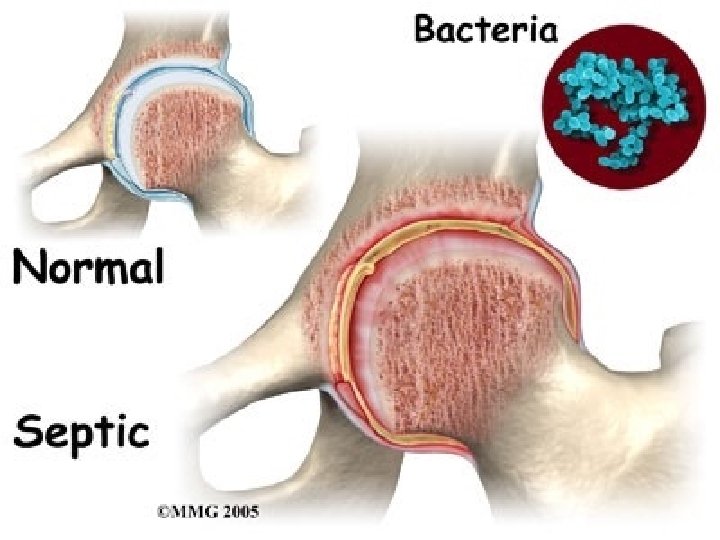
Septic Arthritis Septic arthritis, also known as infectious arthritis, may represent a direct invasion of joint space by bacteria, virus and fungai. It may lead to rapid joint destruction, there for immediate accurate diagnosis is essential. The majority of patients with bacterial septic arthritis will present with acute monoarthritis. The incidence of septic arthritis has been estimated at 2 to 10 cases per 100, 000 in the general population and as high as 30 to 70 cases per 100, 000 in patients with rheumatoid arthritis. The most common mode of spread hematogenous.
 among younger sexually active individuals. Staphylococcus aureus In")
Neisseria gonorrhea (75% of cases) among younger sexually active individuals. Staphylococcus aureus In Adults and children older than 2 years. Pay attention to the following symptoms: -Acute onset of the joint pain. -Previous history of joint disease or trauma. -The presence of extra-articular symptoms. Septic artharitis is a medical emergancy

The most commonly involved joint in septic arthritis is the knee (50% of cases)

A classic presentation for septic arthritis is a patient with feaver who has rigors, an increased leukocyte count, and elevated sedimentation rate. However, none of these is highly sensitive or specific for septic arthritis. In one series: , 40% to 60% of patients with septic arthritis were febrile. 25% to 60% had an elevated leukocyte count. and 60% to 80% had a sedimentation rate greater than 50 mm/hr. Signs and symptoms of infection may be muted in elderly, those who are immunocompromised (especially those with rheumatoid arthritis) and who abuse intravenous drugs.

Investigations of SA Joint Fluid Analysis and Culture: Normal synovial fluid is clear and colorless - Culture results in patients with non gonococcal septic arthritis are almost always positive. - Cultures of the joint fluid in gonococcal infections yield positive results in only about 25 -50% of cases. Blood Cultures: By Obtaining at least 2 sets of blood cultures to rule out a bacteremic origin of the septic joint. Polymerase chain reaction: For detection of bacterial DNA in joint fluid and synovial tissue. Radiography and Ultrasonography: it is most useful in ruling out underlying osteomyelitis caused by the joint infection itself Ultrasonography may be used to diagnose effusions in chronically distorted joints. MRI and CT scanning: More sensitive for distinguishing osteomyelitis , periarticular abscesses and joint effusions.

Synovial Fluid Analysis

Treatment and Management of septic arthritis Medical management of infective arthritis focuses on adequate and timely drainage of the infected synovial fluid, administration of appropriate antimicrobial therapy, and immobilization of the joint to control pain Antibiotic Therapy: In native joint infections, antibiotics usually need to be administered parentally for at least 2 weeks. However, each case must be evaluated independently.

Synovial Fluid Drainage Aspirating the joint 2 -3 times a day may be necessary during the first few days

Joint Immobilization and Physical Therapy - Usually, immobilization of the infected joint to control pain is not necessary after the first few days. - Initial physical therapy consists of maintaining the joint in its functional position and providing passive range-of-motion exercises. - The joint should bear no weight until the clinical signs and symptoms of synovitis have resolved.

Brief: Reactive Arthritis Reactive arthritis is joint pain and swelling triggered by an infection in another part of your body — most often the: Intestines (campylobacter, Salmonella, Shigella and Yersinia) Genitals (Clamidya Trachomatos) or urinary tract. Usually affected parts 1 -joints (knee, feet and ankles). 2 -May affect eyes. 3 -skin. 4 -Urethra. reactive arthritis is sometimes called Reiter's syndrome, which is specific rare type of reactive arthritis. For most people, signs and symptoms of reactive arthritis come and go, eventually disappearing within 12 months.

Symptoms of Reactive Arthritis: 1 - Painful urination and discharge from the penis if there’s inflammation of the urethra(Genitalia). 2 - Diarrhea may occur if the intestines are affected(Bowel). Both followed by arthritis from 4 to 28 days later that usually affects finger, tows, ankles, hips and knee joints.

Treatment: Treat the main infection. NSAIDS for swelling and joint pain. Steroids for swelling. DMARDS for protection of the joints. TNF blockers a new treatment. Physical therapy and exercise. Immuno-suppressant drugs.

Highlights: Juvenile Arthritis What is Juvenile Arthritis? Juvenile arthritis. Also known as pediatric rheumatic disease, pediatric rheumatic diseases that can develop in children under the age of 16 of unknown etiology. Although the various types of juvenile arthritis share many common symptoms, like pain, joint swelling, redness and warmth, each type of JA is distinct and has its own special concerns and symptoms. Some types of juvenile arthritis affect the musculoskeletal system. Juvenile arthritis can also involve the eyes, skin, muscles and gastrointestinal tract. May not involve the joints.
. Considered the most common form of")
Types of Juvenile Arthritis Juvenile idiopathic arthritis (JIA). Considered the most common form of arthritis, JIA includes six subtypes: oligoarthritis, polyarthritis, systemic, enthesitis-related, juvenile psoriatic arthritis or undifferentiated. Juvenile dermatomyositis. , causes muscle weakness and a skin rash on the eyelids and knuckles. Juvenile lupus. Lupus can affect the joints, skin, kidneys, blood and other areas of the body. Juvenile scleroderma. “Hard skin” Kawasaki disease. This disease causes blood-vessel inflammation that can lead to heart complications. Mixed connective tissue disease. This disease may include features of arthritis, lupus dermatomyositis and scleroderma. Fibromyalgia. This chronic pain syndrome is an arthritis-related condition, which can cause stiffness and aching, along with fatigue, disrupted sleep and other symptoms. More common in girls,

Diagnosis & Treatment Diagnosis is made by: careful physical exam. medical history. Any specific tests a doctor may perform will depend upon the type of JA suspected. No specific treatment exist The goal of treatment is to relieve inflammation, control pain and improve the child’s quality of life. Most treatment plans involve a combination of medication, physical activity, eye care and healthy eating.

Juvenile Arthritis Self Care: An important part of JA treatment is teaching the child the importance of how to follow the treatment prescribed by the healthcare team. help the child address the emotional and social effects of the disease. Self management encompasses the choices made each day to live well and stay healthy and happy.

Red Flags in patient with Arthritis 1: Bumps Arthritis sufferers commonly have small lumps on their finger joints. These are actually bone protrusions or bone spurs that tend to result in swelling of the finger joints. Though these are especially common in women, they also tend to run in families, and may not produce any pain.

2: Pain Causing Lack of Sleep While normal aches and pains are common as we get older, persistent pain in the joints that interferes with regular sleep patterns is a warning sign of arthritis. Osteoarthritis pain comes because the cartilage in your joints wears away and can cause your bones to grind against one another. This persistent pain can lead you to lose considerable sleep.

3: Achy Hands If you begin experiencing a loss of fine motor skills, such as an inability to use a fork and knife, this may be a sign of arthritis. Pain in the knuckles and the finger joints, or at the base of the thumb, is very common in osteoarthritis sufferers.

Referral criteria: Patients with suspected inflammatory arthritis should be referred urgently if symptoms have been present for more than six weeks and any of the following apply: 1. swelling is present in two or more joints 2. there is a positive MCPJ or MTPJ squeeze test 3. early morning joint stiffness for more than 30 minutes 4. joint stiffness following periods of immobility 5. the presence of other conditions associated with inflammatory arthritis such as psoriasis, iritis or uveitis, inflammatory bowel disease.

Conclusion Arthritis is a major cause of lost work time and serious disability for many people. Temporal pattern of arthritis could be Abrupt (develop over minutes to hours), or Insidious (develop over weeks to months) Rheumatoid Arthritis is a chronic systemic disease that can be presented with Extra-articular Manifestation
")
MCQ RA is characterized by which of the following patterns of joint involvement? a) Episodic monoarthritis b) Symmetrical polyarthritis c) Migratory oligoarthritis d) Spondylitis
 MRI b) x-ray")
MCQ The most specific test used in diagnosis of gout: a) MRI b) x-ray c) synovial fluid analysis d) serum uric acid
 S.")
MCQ The most common offending organism in septic arthritis in adults is: a) S. aureus b) Streptococcus pyogenes c) S. pneumoniae d) H. influenzae

MCQ Which one of the following is a characteristic x-ray finding in case of osteoarthritis ? a) chondrocalcinosis b) osteopenia c) Narrowing of joint space d) sequestra

Refrences 1. 1 -Prevalence of Doctor-Diagnosed Arthritis and Arthritis-Attributable Activity Limitation United States. 2. National and State Estimates of Childhood Arthritis and Other Rheumatic Conditions. 3 - Clarson LE, Hider SL, Belcher J, Heneghan C, Roddy E, Mallen CD. Increased risk of vascular disease associated with gout: a retrospective, matched cohort study in the UK Clinical Practice Research Datalink. Ann Rheum Dis. 2014 Aug 27. 4 - Dalbeth N, Kalluru R, Aati O, et al. Tendon involvement in the feet of patients with gout: a dual-energy CT study. Ann Rheum Dis 2013; 72: 1545. 5 - Singh JA, Reddy SG, Kundukulam J. Risk factors for gout and prevention: a systematic review of the literature. Curr Opin Rheumatol. 2011 Mar. 23(2): 192 -202. [Medline]. 6 -Mc. Adams-Demarco MA, Maynard JW, Coresh J, Baer AN. Anemia and the onset of gout in a population-based cohort of adults: Atherosclerosis Risk in Communities study. Arthritis Res Ther. 2012 Aug 20. 14(4): R 193. [Medline]. 5. oxford handbook of general oractice, 4 th ed.

Thank You. .
- Slides: 98