DIFFERENTIAL DIAGNOSIS AND TREATMENT OF PATELLOFEMORAL PAIN SYNDROME


 •")


")


 • Success considered")


 •")
 • Pt supine • Examiner fist under the subject’s")


")
 •")


 • 49 patients with PFPS symptoms for at least")


 ages 18 -36")


 • Inclusion criteria: anterior knee pain")
 – TABLE 1")
 – TABLE 2")


 • 20 y/o female BMI 22 – part time student and")


 • Pt A 76 Baseline- 85 follow up,")


- Slides: 81
DIFFERENTIAL DIAGNOSIS AND TREATMENT OF PATELLOFEMORAL PAIN SYNDROME Patrick Mc. Namara SPT University of North Carolina- Chapel Hill
OUTLINE/LEARNING OBJECTIVES • Review of anatomy and sources of knee pain • Listeners will recognize pain provocators in the knee • How PFPS presents in the clinic • Listeners will recognize the cluster of complaints individuals with PFPS often present with. • What deficiencies are common among those who complain of PFPS • Individuals will recognize impairments in strength, running mechanics, and limited ROM in the lower extremities common to PFPS. • Treatment approaches in the literature for PFPS • Individuals will understand what the basic literature behind current PFPS treatment advocates for
COMMON THEMES AND SIGNIFICANT DIFFERENCES IN PFPS
COMMON CHIEF COMPLAINTS • Pain with prolonged sitting, ascending stairs, with squatting (2) • More common among women than men. • Anterior knee pain, especially with weight bearing flexed knee activities. (6) • Vague or diffuse? Does it radiate? Where in relation to patella? • Insidious onset of symptoms • May have already tried and failed conservative therapy (6) • Fulkerson – blunt trauma leads to pain/crepitus in the 70 -120 degree range of flexion. More likely to injure proximal patellar cartilage • History of prolonged activity or newly started weightbearing activity • Is the pain in running with knee flexed or extended? • Lastly ask about instability/giving way
KNEE MUSCULOSKELETAL ANATOMY Adapted from http: //www. webmd. com/pain-management/knee-pain/picture-of-the
OTHER KNEE STRUCTURES Adapted from http: //www. kneeguru. co. uk/KNEEnotes/knee-dictionary/bursa
KNEE BIOMECHANICS Adapted from Lewek 2013
KNEE BIOMECHANICS Adapted from http: //uicptanatomyreviews. blogspot. com/2012/09/knee-handout-from-theres
PATELLAR MECHANICS-WITHOUT YOUTUBE Adapted from http: //www. rosedalenaturalhealth. com/partnering-with-rnhc/articles-2/patellofemoral-pain-syndrome-%E 2%80%9 Crunner%E 2%80%99 s-kne
PATELLAR STABILIZER ANGLES Adapted from Waryasz et al. 2008
PROPOSED PATHOMECHANICS • Once thought that chondromalacia was the prime indicator – recent studies show otherwise • Mal-alignment – of patella and femur at extension • Prevailing theory is that the patella tracks laterally and makes increased contact with the femur • Femoral internal rotation • Femoral adduction • Delayed VMO onset compared to VML (10)
PAIN PROVOCATORS OF THE KNEE DYE ET AL. 1998 Adapted from Dye et al. 1998
DIFFERENTIAL DIAGNOSIS • Plica syndrome • Tibial-femoral OA • Ligamentous (ACL, MCL, LCL, PCL) damage • Meniscal Damage (medial or lateral) • Quad tendinopathy • Neuromas • Patellar stress fracture • Referred pain from lumbar spine or hip joint
CLINICAL PRESENTATION AND POSSIBLE RISK FACTORS • Weakness in: • Knee Extensors – According to recent systematic review – this is the only consistently determined risk factor. (Along with female gender) (16) Measured via isokinetic testing during concentric motion • Hip abductors- an avg of 30% difference from side to side (6, 9) • • Hip eccentric abduction strength (high values) reduces risk of PFPS development • Hip external rotators (6) • Gastrocnemius during functional testing • Knee Flexor Strength- Mixed results Tightness in: • HS, IT band, Gastroc, Soleus, Quadriceps (Piva 2005) • Excessive Q angle – Pooled data by Lankhorst suggests not a reliable factor • Aberrant timing of VMO/VL activation (Cowan et al. ) (PFPS pop v control, VL activated earlier in functional activities) • BMI • Taller individuals? – Mixed results • Quad atrophy – not based on girth measures, based on imaging studies, no ratio btwn VMO/VL
STRENGTH DEFICITS • Robinson et al. – 10 females • • Ireland – 15 females • • At 4 weeks hip group had less pain. Increases in LEFS at 8 weeks and 3 months follow up Barton – 10 case control studies reviewed of EMG of the gluteals during functional tasks • • 24% weaker in hip ER, 26% weaker in hip abduction. IR/Adduction similar to controls Dolak – hip strengthening and quad strengthening groups for 4 weeks then 4 weeks of functional exercises • • 26% weaker in hip abduction and 36% weaker in external rotation Bolgla-18 females • • 29% weakness in hip extension, 23% hip abduction, 14% external rotation compared to contralateral side Evidence for delayed and shorter onset of GMed during stair descent (may lead to adduction during gait) Rathleff – Systematic review/meta-analysis for development of PFPS • No evidence for concentric strength deficits (ER/abduction/IR/extension/adduction/hip flexion) and developing PFPS. • Some evidence for eccentric strength deficits (extension/abduction/ER)
PROPRIOCEPTION DEFICITS • Cross-sectional study with 20 asymptomatic vs 20 PFPS • NWB and WB test for JPS • NWB- knee flexed to 20 or 60 degrees pt. had to replicate • WB- knee flexed to 40 degrees, pt asked to replicate • Relative error- arithmetic difference btwn test/response • Absolute- difference btwn test/response in absolute magnitude • Variable – std deviation from the set of 5 • PFPS patients had greater relative, absolute, and variable errors • No statistical correlation btwn pain and JPS Image adapted from Baker et al.
COLLECTION OF SYMPTOMS • Tyler- 35 patients (29 female, 6 males) • Success considered 1. 5 cm decrease in VAS • 21 patients (26 knees) – successful, 14 patients (17 knees) – unsuccessful • 39/43 had positive Ober test at baseline • 83% that had negative Ober at follow up had successful outcome, 27% that did not have a positive Ober had successful outcome • Normal Ober, Thomas test, and improved hip flexion strength – 93% chance of successful outcome. 2/3 – 75% reached a successful outcome, 27% - for 1/3 and 0% for 0/3. • Piva- compared 30 patients with PFPS to 30 healthy controls • Found no difference in ITB length and hip ER strength • PFPS group has decreased gastroc/soleus length, HS length, quadriceps length • Could discriminate PFPS from healthy controls if grastoc, soleus length decreased and/or hip ER strength
SINGLE LEG SQUAT Levinger et al. 2007
LEVINGER SLS • Subjects asked to perform SLS on affected leg to 45 degrees of knee flexion • Under half reported minor pain • Control subjects – 7. 8+/-4. 2 degrees of frontal plane movement • PFPS subjects – 11. 8+/- 3. 6 degrees of frontal plane movement • Nakagawa et al Cross-sectional study of 4 groups (20 female PFPS, age matched pain free females, males with PFPS, and age matched pain free males) • During SLS – PFPS patients showed significantly more: • Ipsilateral trunk lean • Contralateral Pelvic Drop • Hip adduction • Knee abduction • Female PFPS patients showed less EMG activity of glut med (weakness in eccentric testing of the abductors and ERs) • Bolgla et al 2008 found that females with PFPS did not display altered kinematics (but showed significant hip weakness in abductors/ERs) during stair descent.
BIOMECHANICAL CHANGES AFTER RUNNING • Bazzett-Jones et al 2013 • Prospective study looking at biomechanical changes after exhaustive run in college aged indiviiduals (age 18 -40). All active. • Healthy vs. PFP individuals diagnosed by AT • 38 subjects participated • Strength data – normalized to body mass. Max isometric contractions for hip abduction, ER, hip extension, and lateral trunk flexion. • Did NOT look at knee extension strength! • Peak internal joint moments calculated in stance phase for knee, hip, pelvic, and trunk motions. • Subjects paired for exhaustion protocol on treadmill. RPE and % max HR used
CHANGES NOTED • Hip abduction and ER strength significantly reduced in PFP group but not control after the run. • Hip extension strength decreased in PFP both before (by 19%) and after run. • Anterior pelvic tilt, hip flexion moment, and knee flexion peak angle all increased in PFP group after run compared to control. • Post run PFP group demonstrated more knee valgus than control • Post run PFP significantly increased hip extension moment, but control did not change • PFP group also reported significantly more pain than control and baseline
CAN KINEMATICS CHANGE? • Ferber compared kinematics between 10 PFPS participants and 10 healthy controls • All recreational runners. Studied biomechanics during 20 s of treadmill running at 2. 55 m/s • Performed hip abduction and hip extension with theraband. Performed daily for 3 sets of 10 over 3 weeks. • Knee valgus angle (in gait) and football “variability” were initially reduced but normalized at follow up in the PFPS group. • Saw concurrent decline in VAS pain. • Bell – screened healthy participants, searching for valgus in DLS, corrected with heel lifts. • 10 exercise sessions over 3 weeks to compare exercise and no intervention • Hamstring curls, posterior chain stretches, single leg balance, and star excursion balance techniques used. • Saw decreased valgus in DLS and increased DF ROM with an extended knee
AREAS TO LOOK AT – NOT JUST THE KNEE • Souza et al found that females with PFPS displayed the following during running, drop jump, and step down. • Greater peak Hip IR compared to pain free controls (especially in running) • Less torque produced by abductors and hip extensors • Greater glute max recruitment – even though it was weakened • Glute med EMG activity and hip adduction angle were similar btwn groups
SPECIAL TESTS • Vastus medialis coordination test (Positive Likelihood Ratio – 2. 26) • Patellar apprehension test (Positive Likelihood Ratio – 2. 26) • Eccentric Step Test (Positive Likelihood Ratio – 2. 34) • Taken from a sample of 45 patients complaining of knee pain. Must be independent ambulators and have not undergone a previous knee surgery. Separated into a PFPS and non-PFPS group as diagnosed by an MD. PFPS group patients could not display signs of any other pathology and must report pain with 2 of these: prolonged sitting , squatting, ascending/descending stairs, or kneeling. • A systematic review found similar values, without identifying any tests with better predictive values – that the authors considered valid. • The articles review demonstrated low QUADAS scores.
VASTUS MEDIALIS COORDINATION TEST (3) • Pt supine • Examiner fist under the subject’s knee • Pt extends knee slowly without pressing into or moving off of examiner’s fist. • Pt instructed to achieve full extension • Positive if a lack of coordinated full extension evident • Unable to smoothly extend fully or recruited other muscle groups to achieve full extension
VASTUS MEDIALIS COORDINATION TEST Adapted from Nijs et al. 2006
PATELLER APPREHENSION TEST • Pt supine and relaxed • Examiner pushes the patient’s patella as laterally as possible • Start point – knee at 30 degree flexion • Grasping the leg with other hand at ankle/heel a slow combined hip/knee flexion performed • Lateral glide held throughout the test • Positive test – if it reproduced the pain or apprehension was evident
PATELLAR APPREHENSION TEST Adapted from Nijs et al. 2006
ECCENTRIC STEP TEST • 15 cm step – adjusted to 50% of tibial length • Std demonstration given • Instructions • ‘Stand on the step, put your hands on your hips and step down from the step as slowly and as smoothly as you can” • Repeated using the other leg • Positive test – knee pain reported during test
ECCENTRIC STEP TEST Adapted from Nijs et al 2006
OUTCOME MEASURES • Double blind RCT for conservative tx (quad strengthening vs sham ultrasound) • VAS-usual was the reference standard (looked for change of 1 cm or more in VAS-usual, worst, and with activity. • The Anterior Knee Pain Scale (AKPS) has the highest effect size/most responsive • 7 point change desired • FIQ, global rating of change also studied
WHO BENEFITS FROM EXERCISE THERAPY? • RCT (both groups got advice/information regarding PFPS) • Exercise group got a standardized program (9 treatments over 6 weeks). Static/dynamic quadriceps training, flexibility exercises, and balance work. This became an HEP for 6 weeks. • Participants grouped into symptom duration phases (1 -2 months, 2 -6 months, 6 -12 months, 12+ months) • No significant differences at 3 month and 12 month follow ups on pain, perceived recovery, and functional score. • Trends noted for female sex and longer duration of symptoms (over 6 months). • Could be a result of decreased muscle size/strength due to disuse. • May want to explore other treatment methods (taping/manipulation/orthotics) for shorter durations. • Rathleff- may not see kinematic differences even with symptom reduction.
DYE ET AL 2001 – REHAB CONSIDERATIONS FOR CLINICIANS Adapted from Dye et al 2001
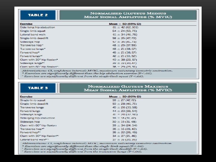
CLAMSHELL EXERCISE • Neutral pelvis • More hip flexion increased glut med activity w/o TFL increase Image adapted from Willcox et al.
CLAMSHELL EMG Image adapted from Willcox et al.
EXERCISES Image adapted from Youdas et al.
YOUDAS
IT BAND STRETCHING Image adapted from Fredericson et al.
PROGNOSTIC FACTORS: QUADRICEPS STRENGTHENING (1) • 49 patients with PFPS symptoms for at least 2 months, aged 15 -50, and no radiographic presence of OA, osteochondritis dissecans, or loose bodies in the knee. • Positive factors: • 1. Smaller knee extensor strength disparity btwn limbs • 2. Less pain in the patella apprehension test • 3. Less patella crepitation
PROGNOSTIC FACTORS CONT. • Negative Factors: • 1. Taller individuals • 2. Older individuals • 3. Development of bilateral symptoms • Noteworthy • 1. X-Ray and MRI changes in affected knee had NO association with 7 year follow up
FUNCTIONAL STABILIZATION PROGRAM • RCT 31 female patients btwn 18 -30 y/o. Quad Strengthening (n=16) vs. Functional Stabilization (n=15). • All rec athletes (3 x per week or more of 30 minutes or more of exercise). • Outcome measures: VAS, LEFS, GRC (Global rating of change), Single leg-triple hop, single leg squat trunk and LE kinematic analysis, trunk muscle endurance, strength of the LE musculature. • Training protocol 3 x per week for 8 weeks. • Quad strengthening – stretching of LE and OKC/CKC exercises • Functional Stabilization – first 2 weeks involved enhancement of motor control. Next 3 weeks focused on trunk strengthening, hip strengthening, motor control improvement. Finally patients educated about GRF and knee flexion angle, exercises progressed. Frontal plane alignment emphasized in neutral.
OUTCOMES • Baseline VAS worst pain 6. 1+/-1. 8 for Quad group, 6. 6+/-1. 1 for Functional Group • At 3 months follow up Quad group (2. 5+/-2. 7), Functional group (0. 9+/-1. 5) • FST group – greater eccentric hip abduction/knee flexion Greater trunk endurance. • Decreased trunk inclination/pelvis depression/hip adduction/knee abduction as well as more pelvic anteversion and hip flexion during a SLS. • Improved on triple hop, while Quad group did not • Both groups – improved eccentric strength of hip adductor, ERs, knee extensors.
EXERCISE SUMMARY • Dolak et al. 2011 found both hip strengthening and quad strengthening led to pain reduction at 8 weeks. Hip strengthening significantly decreased pain at 4 weeks • Kushion et al. found similar VMO: VL ratios for 4 different exercises ( SLRneutral, SLR- ER at hip, Short qrc quad in neutral and short arc quad in hip ER healthy pop).
LUMBAR MANIP CPR • NPRS measured pre and post manip for step up (20 cm step), step down, and squat • During squat test- knee flexion angle at first instance of pain measured • Manip performed, if no cavitation, re-performed. Max of 2 per side • Success defined as 50+% improvement in NPRS, +4 or more on the GRC • Mean NPRS for success group – 80+/-17%, non-success 12+/-28% • Median GRC in success was 5, non-success 1 Image adapted from Iverson et al.
VARIABLES ASSESSED Image adapted from Iverson et al.
CPR ITEMS Image adapted from Iverson et al.
CPR FOR LUMBOPELVIC MANIP PSYCHOMETRICS Image adapted from Iverson et al.
CPR FOOT ORTHOTICS • 42 participants, 28 y/o avg with 3 years of symptoms median • Improvement at 12 week follow up, after fitting for orthotic by PT • 6 visits to modify and fit orthoses. Pre-fabricated ethylene-vinyl acetate orthotic. Have a 6 degree varus wedge and arch support. (PTs would heat mold, wedge, heel raise addition) • Fit was first designed for comfort then pain free function • Success defined as those that reported a “marked improvement” on a 5 point Likert scale (marked improvement, mod improvement, same, mod worse, markedly worse) • Lack et al. found higher FIQ scores, more forefoot valgus, and higher peak reartfoot eversion had good outcomes
CPR ORTHOTIC PSYCHOMETRICS 6/7 individuals that met 3 of the 4 criteria were in the success group Image adapted from Vicenzino et al.
CPR TAPING • 50 active duty soldiers (27 male, 23 female) ages 18 -36 • NPRS for step up (20 cm step), step down, and squat (knee flexion angle measured) • Success considered 50+% improvement on NPRS or a change of +4 or more on the GRC • Mean improvement change in success group – 63. 5+/-24%, GRC 4+/-1. 7 • Mean in non-success group – 30. 6+/-22. 4%, GRC 0. 4+/-1. 8
TAPING METHOD Image adapted from Lesher et al.
CPR TAPING Image adapted from Lesher et al.
GAIT RETRAINING • Rathleff - Retraining runners with high hip adduction peak levels has managed hip pain • Rathleff – high levels of hip IR/adduction in running are risk factors for PFPS development • Teng – compred self-selected, flexed, extended trunk postures in running. • Small but significant (5 -7%) reduction in PFJ stress (force/contact area) with flexed position vs extended position.
RCT MULTIMODAL • Crossley et al 2002 (10) • Inclusion criteria: anterior knee pain with functional activities, insidious onset, positive clinical test (pain with palpation of anterior knee, DLS, or step down) • Exclusion: Other knee pathology (eg ACL tear) or previous surgery on patellofemoral complex, referred pain, or patellar dislocations. • Interventions • • Biofeedback via EMG for VMO • Patellar Taping (anterior tilt medial tilt glide fat pad unloading) Provided in this order until 50% reduction in perceived pain • Gluteal muscle strengthening and HEP • Stretching of hip/knee musculature (deep friction massage to lateral soft tissue, HS, patellar mobs, prone anterior hip stretches) Results • Reduced avg pain, worst pain, and disability after 6 weeks and at 3 month follow up
CROSSLEY (10) – TABLE 1
CROSSLEY (10) – TABLE 2
INTERVENTION CROSSLEY
MODALITIES? • Systematic review by Lake et al. • 1 study looking at iontophoresis, cryotherapy, US, phonophoresis • • 3 studies for NEMS • • • Low quality studies. All included exercise regimens. Could not determine if beneficial 4 studies with EMG biofeedback • One study found short term pain reduction, but in combination with exercise and taping • Useful in retraining the VMO. May or may not help with pain reduction 3 Studies with E-Stim • • Very low quality. Included a strengthening regimen. Unable to determine if any changes as a result of modalities. Short-term pain reduction with high voltage, monophasic pulsed stim 1 study with low-intensity laser • No significant changes found
CASE REPORTS (MASCAL/POWERS) • 20 y/o female BMI 22 – part time student and waitress (Patient A) • 2 traumatic patella dislocations (falls) – failed PT consisting of walking program and quad strengthening X-Ray unremarkable, symptoms worsening x 2 years. • Walking 2 hours or descending 2 flights of stairs set off pain. Wanted to start jogging. • 37 y/o female BMI 27 accounts asst for hospital (Patient B) • Insidious onset. Had fallen 3 x in the last 1. 5 years due to painful buckling. X-rays unremarkable. • “Intermittent throbbing” behind patella, worsened by stair descent. Liked to walk 30 -45 mins several times a week, but was limited by pain. Wanted to walk, dance, and ascend/descend stairs pain free.
CLINICAL EXAM FINDINGS • Clinical tests used to rule out rearfoot, tibiofemoral, hip, and lumbar sources of pain. • No obvious patellar alignment/tracking issues in standing/dynamic movements • Both experienced pain with compression and apprehension test as well as in the 0 -20 degree range of resisted knee extension Image adapted from Mascal et al.
CASE STUDIES CONTINUED • No structural abnormalities found • Gait assessment – “excessive hip adduction, hip IR, and contralateral pelvic drop” • Step down test – significant hip adduction and contralateral pelvic drop valgus at the knee • Interventions • First targeted glut max/med, hip ERs, and abductors (2 -3 sets of 10 -15 reps or 3 x/week and a total of 6 -12 weeks strength • HEP performed 2 x/day
EXERCISE PROGRESSION • Non-weight bearing 0 -6 weeks Image adapted from Mascal et al.
WEIGHT BEARING EXERCISES • “Neutral LE alignment” – ASIS and knee remained over 2 nd toe with hip in 10 degrees of ER. Image adapted from Mascal et al.
FUNCTIONAL TRAINING Image adapted from Mascal et al.
OUTCOMES • Kujala Questionnaire (100=no disability) • Pt A 76 Baseline- 85 follow up, Pt B 70 Baseline – 84 Follow up • Pt A – no pain during 10 hr shift, running 2 -3 miles no pain, up and down stairs no pain • Pt B – no pain up/down stairs, 2/10 pain if substantial time on her feet, 45 minutes of walking with no pain. • Both showed decreased adduction and hip IR. Pt A had biomechanical follow up indicating 2. 6 degrees of ER (baseline 1. 4 degrees IR), 2. 3 degrees hip adduction (8. 7 baseline) and contralateral pelvic drop 1. 1 degree (3. 9 baseline) during a step-down.
QUADRICEPS STRENGTHENING EXERCISES • Quad sets • Leg Press/Shuttle Machine • Leg extensions • Supine Quad Extensions • Squats
QUAD SETS Adapted from http: //www. hep 2 go. com/exercise_editor. php? ex. Id=13450&user. Ref=
SUPINE QUAD EXTENSIONS Adapted from http: //www. hep 2 go. com/exercise_editor. php? ex. Id=7978&user. Ref=0
KNEE EXTENSIONS 45 -90 DEGREES Image adapted from Baldon et al.
LEG PRESS 0 -45 DEGREES Image adapted from Baldon et al.
WALL SQUATS 0 -60 DEGREES KNEE FLEX Image adapted from Baldon et al.
STEP-UPS/DOWNS 20 CM BOX Image adapted from Baldon et al.
SINGLE LEG BALANCE Image adapted from Baldon et al.
TRANSV ABD EX BALL Image adapted from Baldon et al.
TRUNK EXTENSIONS Image adapted from Baldon et al.
THANKS! WHY I’M INTERESTED IN PFPS
REFERENCES • 1. Natri A. , Kannus P. , Jarvinen M. Which Factors predict the long-term outcome in chronic patellofemoral pain syndrome? A 7 -yr prospective follow-up study. (1998) Medicine & Science in Sports & Exercise. 30(11): 1572 -1577 • 2. Cook C, Mabry L, Reiman M, Hegedus E. Best tests/clinical findings for screenings and diagnosis of patellofemoral pain syndrome: a systematic review. (2011) Physiotherapy, doi: 10. 1016/j. physio. 2011. 09. 001 • 3. Nijs J. , Van Geel C. , Van der auwera C. , Van de Velde B. Diagnostic value of five clinical tests in patelllofemoral pain syndrome. Manual Therapy (2006) 11: 69 -77 • 4. Souza R. , Powers C. Differences in Hip Kinematics, Strength, and Muscle Activation in Subjects With and Without Patellofemoral Pain. (2009) JOSPT. 39(1): 12 -19 • 5. Rabelo N. , Lima B. , Curcio A. , Bley A. , Yi L. , Fukuda T. , Costa L. Neuromuscular training and muscle strengthening in patients with patellofemoral pain syndrome: a protocol of randomized controlled trial. BMC Musculoskeletal Disorders. (2014) 15: 157 • 6. Ireland M. , Wilson J. , Ballantyne B. , Davis I. Hip Strength in Females with and without patellofemoral pain. (2003) 33(11): 671 -676 • 7. Crossley K. , Bennell K, Cowan S. , Green S. Analysis of Outcome Measures for Persons with Patellofemoral Pain: Which are Reliable and Valid? Arch Phys Med Rehabill (2004) 85: 815 -822 • 8. Fulkerson J. Diagnosis and Treatment of Patients with Patellofemoral Pain. The American Journal of Sports Medicine (2002) 30(3): 447 -456 • 9. Robinson R. , Nee R. Analysis of Hip Strength in Females Seeking Physical Therapy Treatment for Unilateral Patellofemoral Pain Syndrome. (2007) JOSPT 37(5): 232 -238 • 10. Crossley K. , Bennell K. , Green S. , Cowan S. , Mc. Connell J. Physical Therapy for Patellofemoral Pain : A Randomized, Double-Blinded, Placebo Controlled Trial. (2002) The American Journal of Sports Medicine. 30(6) : 857 -865 • 11. Iverson C. , Sutlive T. , Crowell M. , Morrell R. , Perkins M. , Garber M. , Moore J. , Wainner R. Lumbopelvic Manipulation for the Treatment of Patients with Patellofemoral Pain Syndrome: Development of a Clinical Prediction Rule. (2008) J Orthop Sports Phys Ther. 38(6): : 297 -312 • 12. Dye S. , Vaupel G. , Dye C. Conscious Neurosensory Mapping of the Internal Structures of the Human Knee Without Intraarticular anesthesia. (1998) The American Journal of Sports Medicine. 26(6): 773 - 777 • 13. Waryasz G. , Mc. Dermott A. Patellofemoral pain syndrome (PFPS): a systematic review of anatomy and potential risk factors. (2008) Dynamic Medicine. 7(9): 1 -14 • 14. Dye S. Patellofemoral Pain Current Concepts : AN Overview. (2001) Sports Medicine and Arthroscopy Review • 15. Bazzett-Jones D. , Cobb S. , Huddleston W. , Connor K. , Armstrong B. , Earl-Boehm J. Effect of Patellofemoral Pain on Strength and Mechanics after an Exhaustive Run. (2013) Medicine & Science in Sports and Exercise. 1331 -1339 • 16. Lankhorst N. , Bierma-Zeinstra S. , Van Middelkoo M. Risk Factors for Patellofemoral Pain Syndrome: A systematic Review. (2012) J Orthop Sports Phys Ther. 42(2): 81 -94
REFERENCES • Lankhorst N. , Middelkoop M. , Trier Y. , et al. Can we predict which patients with patellofemoral pain are more likely to benefit from exercise therapy? A secondary exploratory analysis of a randomized controlled trial. (2015) J Orthop Sports Phys Ther. 45(3): 183 -189 • Witvrouw E. , Lysens R. , Bellemans J. , et al. Open versus closed kinetic chain exercises for patellofemoral pain. A prospective, randomized study. (2000) The American Journal of Sports Medicine. 28(5): 687 -694 • Barton C. , Lack S. , Malliaras P. , et al. Gluteal muscle activity and patellofemoral syndrome: a systematic review. (2013) Br J Sports Med. 47: 207 -214 • Rathleff M. , Rathleff C. , Crossley K. , et al. Is hip strength a risk factor for patellofemoral pain? A systematic review and meta-analysis. (2014) Br J Sports Med. 48 • Teng HL. , Powers C. Sagittal plane trunk posture influences patellofemoral joint stress during running. (2014) J Orthop Sports Phys Ther. 44(10): 785 -792 • Ferber R. , Kendall K. , Farr L. Changes in knee biomechanics after strengthening protocol for runners with patellofemoral pain syndrome. (2011) Journal of Athletic Training. 46(2): 142 -149 • Bell D. , Oates D. , Clark M. Two- and 3 -dimensional knee valgus are reduced after an exercise intervention in young adults with demonstrable valgus during squatting. (2013) Journal of Athletic Training. 48(4): 442 -449 • Piva S. , Goodnite E. , Childs J. Strength around the hip and flexibility of soft tissues in individuals with and without patellofemoral pain syndrome. (2005) J Orthop Sports Phys Ther. 35(12): 793 -801 • Baldon R. , Serrao F. , Silva R. , et al. Effects of functional stabilization training on pain, function, and lower extremity biomehcanics in women with patellofemoral pain: a randomized clinical trial. (2014) J Orthop Sports Phys Ther. 44(4): 240 -251 • Lake D. , Wofford N. Effect of therapeutic modalities on patients with patellofemoral pain syndrome: A systematic review. (2011) Sports Health. 3(2): 182 -189 • Youdas J. , Boor M. , Darfler A. , et al. Surface electromyographic anallysis of core trunk and hip muscles during selected rehabilitation exercises in the side-bridge to neutral spine position. (2014) Sports Health. 6(5): 416 -421 • Bolgla L. , Malone T. , Umberger B. , Uhl T. Hip strength and hip and knee kinematics during stair descent in females with and without patellofemoral pain syndrome. Journal of Orthopaedic & Sports Physical Therapy. (2008) 38(1): 12 -18 • Dolak K. , Silkman C. , Mc. Keon J. , et al. Hip strengthening prior to functional exercises reduces pain sooner than quadriceps strengthening in females with Patellofemoral Pain Syndrome: A randomized clinical trial. (2011) Journal of Orthopaedic & Sports Physical Therapy. 41(8): 560 -570 • Tyler T. , Nicholas S. , Mullaney M. , et al. The Role of Hip Muscle Function in the treatment of Patellofemoral Pain Syndrome. (2006) The American Journal of Sports Medicine. 34(4): 630 - 636 • Iverson C. , Sutlive T. , Crowell M. , et al. Lumbopelvic manipulation for the treatment of patients with patellofemoral pain syndrome: development of a clinical prediction rule. (2008) J Orthop Sports Ther. 38(6): 297 - 312 • Crossley K. , Dip G. , Bennell K. , et al. Physical Therapy for Patellofemoral Pain: A randomized, double-blinded, placebo-controlled trial. (2002) The American Journal of Sports Medicine. 30(6): 857 -865 • Vicenzino B. , Collins N. , Cleland J. , et al. A clinical prediction rule for identifying patients with patellofemoral pain who are likely to benefit from foot orthotics: a preliminary determination. (2010) Br J Sports Med. 44: 862 - 866 • Kooiker L. , Van de Port I. , Weir A. , et al. Effects of physical therapist guided quadriceps strengthening exercises for treatment of patellofemoral pain syndrome: a systematic review. (2014) J Orthop Sports Phys Ther. 44(x): xxx- xxx • Verma C. , Krishnan V. Comparison between Mc. Connell Patellar taping and conventional physiotherapy treatment in the management of patellofemoral pain syndrome – a randomised controlled trial. (2012) JKIMSU. 1(2): 95 -104
REFERENCES • Cowan S. , Bennell K. , Crossley K. Physical therapy alters recruitment of the vasti in patellofemoral pain syndrome. (2002) Medicine & Science in Sports & Exercise. 1879 -1885 • Boling M. , Bolgla L. , Mattacola C. , et al. Outcomes of a weight-bearing rehabilitation program for patients diagnosed with patellofemoral pain syndrome. (2006) Arch Phys Med Rehabil. 87: 1428 -1435 • Distefano L. , Blackburn T. , Marshall S. , et al. Gluteal muscle activation during common therapeutic exercises. (2009) J Orthop Sports Phys Ther. 39(7): 532 -540 • Callaghan MJ. , Mc. Carthy CJ. , Oldham JA. Electromyographic fatigue characteristics of the quadriceps in patellofemoral pain syndrome. (2001) Manual Therapy. 6(1): 27 -33 • Kushion D. , Rheaume J. , Kopchitz K. , et al. EMG activation of the vastus medialis oblique and vastus lateralis during four rehabilitative exercises. (2012) The Open Rehabilitation Journal. 5: 1 -7 • Fredericson M. , White J. , Mac. Mahon J. , et al. Quantitative analysis of the relative effectiveness of 3 iliotibial band stretches. (2002) Arch Phys Med Rehabil. 83: 589 -592 • Reiman M. , Bolgla L. , Loudon J. A literature review of studies evaluating gluteus maximus and gluteus medius activation during rehabilitation exercises. (2012) Physiotherapy Theory and Practice. 28(4): 257 -268 • Selkowitz D. , Beneck G. , Powers C. Which exercises target the gluteal muscles while minimizing activation of the tensor fascia lata? Electromyographic assessment using fine-wire electrodes. (2013) J Orthop Sports Phys Ther. 43(2): 54 -64 • Barton C. , Kennedy A. , Twycross-Lewis R. , et al. Gluteal activation during the isometric phase of squat exercises with and without a swiss ball. (2014) Physical Therapy in Sport. 15: 39 -46 • Papadopoulos K. , Noyes J. , Jones J. , et al. Clinical tests for differentiating between patients with and without patellofemoral pain syndrome. (2014) Hong Kong Physiotherapy Journal. 32: 35 -43 • Kettunen J. , Harilainen A. , Sandelin J. , et al. Knee arthoscopy and exercises versus exercise only for chronic patellofemoral pain syndrome: 5 -yearfollow up. (2012) Br J Sports Med. 46: 243 -246 • Lesher J. , Sutlive T. , Miller G. , et al. Development of a clinical predication rule for classifying patients with patellofemoral pain syndrome who respond to patellar taping. (2006) J Orthop Sports Phys Ther. 36(11): 854 -866 • Baker V. , Bennell K. , Stillman B. , et al. Abnormal knee joint position sense in individuals with patellofemoral pain syndrome. (2002) Journal of Orthopaedic Research. 20: 208 -214 • White L. , Dolphin P. , Dixon J. Hamstring length in patellofemoral pain syndrome. (2009) Physiotherapy. 95: 24 -28
DIFFERENTIAL DIAGNOSIS AND TREATMENT OF PATELLOFEMORAL PAIN SYNDROME • • Patrick Mc. Namara Student Physical Therapist: University of North Carolina at Chapel Hill Patellofemoral pain syndrome (PFPS) – commonly known as runner’s knee, is often considered a diagnosis of exclusion. There are common complaints, limitations, and clinical presentations that have been used to properly diagnose PFPS. Once a correct diagnosis is made, PFPS treatment is varied, but recent work is indicating quadriceps strengthening should be a key component of the rehab effort. This presentation will detail current evaluation methods, psychometrics of special tests, efficacy of rehab techniques, and key areas to address. Image adapted from http: //ironstruck. com/runners-knee