Articular syndrome differential diagnosis tactics of GP Associate


 Arthritis-if you have defiguracii or swelling or")


 Spondylosis, Osteochondrosis, Spondylolisthesis;")
; 2.")


 l This disease, which is based on immunotoxic inflammation of the")


 chronic inflammatory joint disease ankylosing axial skeleton (intervertebral, sacroiliac) belonging")

 and radiological evidence of")


 Severe pain in the joints regional limfoadenit Explicit")


 Reiter triad syndromes arthritis • Urogenital (urethritis, cystitis, prostatitis) ёki")
 In joints prevail exudative")

 epidemiological")


 Consulting")
 anamnesis Laboratory and instrumental examination (III")

 echocardiography")


; • Prolonged")


 therapeutic exercise rheumatoid arthritis Symptomatic")
 therapeutic exercise Ankylosing spondylitis Symptomatic")

 Intra-articular injection")
 Gouty arthritis, Unsharp Waiver of")

- Slides: 74
Articular syndrome differential diagnosis, tactics of GP Associate professor: Kh. S. Akhmedov
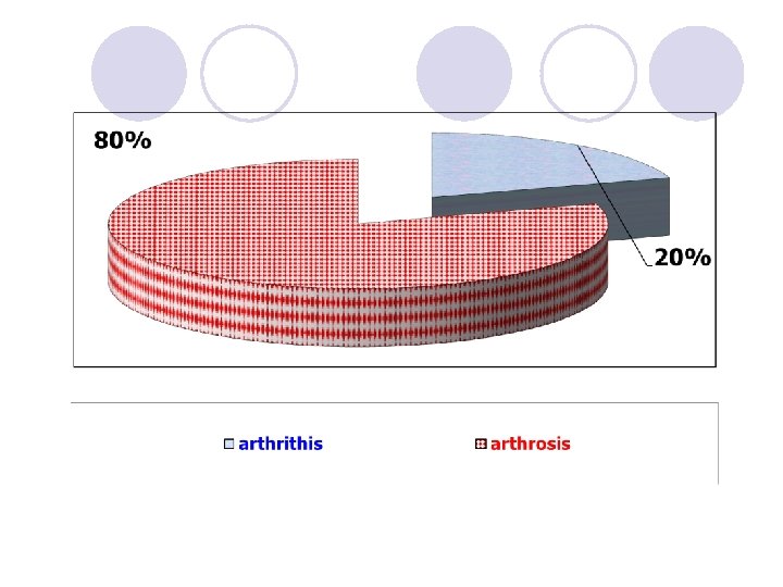
Articular syndrome is simptomakompleks, which is the manifestation of many rheumatic and non-rheumatic diseases, and he found more than 200 diseases Articular syndrome is found in 20 -45% of the cases among the population and in 6 -10% of cases leads to early disability.
Pain over the joint Deformation Disfiguration Hyperemia of acromioclavicular joint Articular syndrome Hyperthermia of acromioclavicular joint Restricted mobility Swelling Ankylosis Morning stiffness Dupuytren Crepitus
l Monoartrit is a manifestation of one joint l Oligoartrit is a manifestation of two or three joints l Polyarthritis-in 3 or more joints
Arthralgia-pain over the joint (without objective changes) Arthritis-if you have defiguracii or swelling or hyperthermia, as well as the pain of Osteoarthritis alone-mechanical pain during movement
The main causes of articular syndrome, which occurs in the form of arthritis Ø Rheumatoid arthritis. Rheumatism. DZST (SLE, SSC, b-HB Syndrome, darmatomiozit, medicine polimialgia, etc. ). Systemic Vasculitis (periarterit, hemorrhagic nodular vasculitis, nespecifič. aortoarterit, etc). Ûvenilnyj arthritis, Bechterew's disease (ankylosing spondylitis).
l Articular syndrome accompanying spondiloartritami: psoriatic arthritis; -illness or Reiter's syndrome; -cron. bowel disease (NSU, b-HB Koruna)-brucellosistuberculosis Microcrystalline arthritis: -gout (primary VA secondary); -chondrocalcinosis;
l Arthritis associated with infectious factors: a. infectious arthritis: -bacterial (stafilo-, strepto-, gonococcal, brucelëz, syphilis, tuberculosis and Mycobacterial etc. ); -viral (rubella, mumps, chicken pox), anti-fungal; -parasitic. B. artritlar: postènterokolitičeskie Reagent (yersiniosis, salmanillëz, shigellosis); -urogenital (except b-nor Reuter); -postnoso-pharyngeal; -after vaccination.
Joint syndrome occurring type of osteoarthritis: 1. DOA (osteartrit) Spondylosis, Osteochondrosis, Spondylolisthesis;
Arthropathy in non-rheumatic diseases: 1. allergic conditions or illnesses (serum or medicinal disease); 2. Metoboličeskie disorders (hemochromatosis, amyloidosis, Hyperlipidemia); 3. Congenital defect in the metabolism of connective tissue (Marfany, syndrome, Mucopolysaccharidosis); 4. Endocrine Disorders (thyroid, pituitary diseases, genital glands, diabetes); 5. Asab tizimi kasalliklari (NDCS, Syringomyelia, treepanža sindromi WA bošķalar); 6. Ķon kasalliklari tizimi (haemophilia, lejkozlar, kasalligi, ëmon sifatli mielon ŭsmalar); Paraneoplastik artropatiâlar (turli sohalarda žojlašgan ëmon sifatli ŭsmalar) 8. Diseases of the nervous system (neurocirculatory asthenia syndrome, Syringomyelia, shoulder-brush, etc. ); 9. other diseases.
Other diseases 1. Polindromnyj rheumatism; 2. Intermitteruûŝij gidroartroz; 3. Joint Chondromatosis; 4. Polychondritis;
Diseases occurring in the form of arthritis
Rheumatism (Rheumatic fever) l This disease, which is based on immunotoxic inflammation of the connective tissue, mainly affecting the cardiovascular system and joints, etiologically related to β - hemolytic streptococcus group A and develops in individuals predisposed to the disease, particularly among those from 7 to 15 years.
Features articular syndrome in rheumatism Changes in the joints are the volatile nature Manifestations of articular syndrome lasts from several hours to several weeks Articular syndrome Can manifest itself in the form of arthralgia and arthritis in the form Predominantly affects medium and large joints Oligoarthritis or polyarthritis Changes in the joints are completely (ie without organic changes) Do not leave: strains contractures ankylosis
Major key manifestations: l l l l Manifestation of 10 -14 days after streptococcal infection carditis chorea annular erythema subcutaneous nodules fever ↑ ESR, ↑ acute phase indicators (ASL -O, α 2, - globulins mukoproteidyr) l Elongation P-Q intervalaa l abdominal syndrome l The development of heart disease
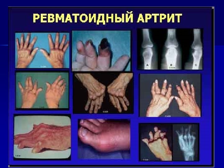
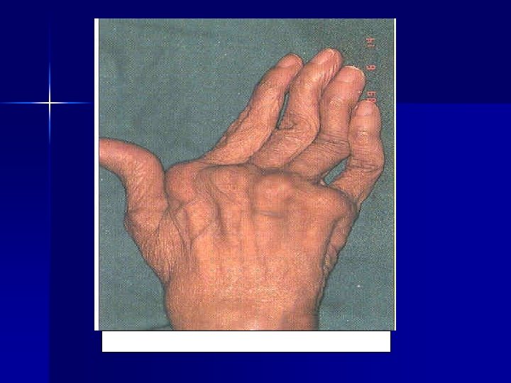
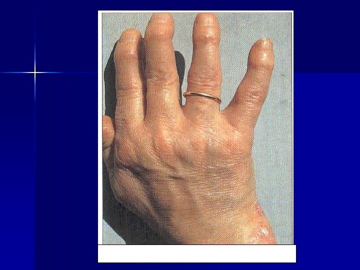
Rheumatoid arthritis It is an autoimmune inflammatory disease of connective tissue with an explicit joint disease, dominated by progressive erosive and destructive-exudative phenomena.
Features articular syndrome in Rheumatoid arthritis Changes joints are sustainable Symmetrical joint involvement Begins with the defeat of the small and medium joints contracture Articular syndrome ankylosis Morning stiffness (at least 30 -60 minutes) Oligoarthritis of polyarthritis Development of deformation - "area walrus", "Leben neck"
Major key manifestations: • Characteristic rentgnography changes (stage 4: osteoparosis, joint space narrowing, and ankylosis uzuratsiya) • Quick regional atrophy of the muscles. • The appearance of painless moving rheumatoid nodules; • Rheumatoid factor (reaction Voler - Rose, latex test) • Visceral (pleurisy, gastritis, liver enlargement, myocarditis, perekardit, amyloidosis kidney polyserositis and others. ) • Hyper α 1, α 2 gammaglobulinemiya Islands; ↑ Subtitle Creactive protein, ↑ ESR
Diffuse diseases of connective tissue Key moments Lack mona etiological factor The similarity of the pathological changes Lesions (signs of inflammation) of arterial vessels Polisindromal flowing DDCT articular syndrome The effectiveness of glucocorticoids, cytotoxic drugs and NSAIDs
Features articular syndrome in SLE Migratory arthralgia or arthritis Symmetrical joint disease Morning stiffness (less than 30 minutes) Transient painless contractures Articular syndrome Often, myalgia and myositis, ossalgiya Predominant involvement of small and medium-sized joints, and sometimes large Deformation and ankylosis are not observed
Leading clinical features that are crucial in the diagnosis of SLE: Ø Ø Ø Lupus "butterfly" discoid lupus Raynaud's syndrome alopecia photosensitization ulceration of the mucous membrane of the nasopharynx (stomatitis) Nephritis, carditis, Polyserositis (pneumonia, pleurisy and (or) pericarditis) Psychosis and (or) seizures with seizures LE-cell test, false-positive Wassermann test Verlgofa syndrome, hemolytic anemia, and (or) leukopenia and (or) thrombocytopenia.
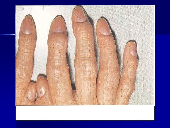
Features articular syndrome in SSc Migratory arthralgia or arthritis Symmetrical joint disease Morning stiffness (less than 30 minutes) Contracture (due to injury myshsch or ligaments) Articular syndrome myositis Predominant involvement of small and medium-sized joints, and sometimes large Deformation and ankylosis are not observed
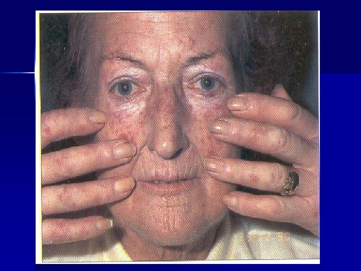
Leading clinical features that are crucial in the diagnosis of SSc: • Skin lesions, "solid" edema, induration, atrophy and pigmentation. Major changes are on the face "maskaobraznaya" and brush. • Raynaud's phenomenon (95%) • Sclerodactyly CREST- syndrome (calcinosis, Raynaud's, esophagitis, sclerodactyly, teleangiektoziya) • Anemia and (or) leukopenia, thrombocytopenia • Osteolysis of the terminal phalanges of hands and feet • Visceral (esophagitis with dysphagia, basal pulmonary fibrosis, macrofocal cardio, true scleroderma kidney) • peripheral hyperpigmentation, trophic changes • Lymphadenopathy, polyserositis, polyneuritis • Increased ESR, hyperproteinemia, hypergammaglobulinemia, RF
Features articular syndrome systemic dermatomyositis polyarthralgia Symmetrical joint disease Contracture - mostly muscle origin Articular syndrome Ankylosis - mostly muscle origin Morning stiffness (less than 30 minutes) The deformation is not observed
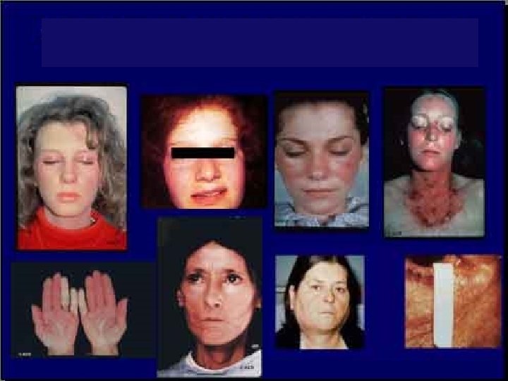
Leading clinical features that are crucial in the diagnosis of SD: l Typical skin changes - periorbital edema and erythema symptom score), telangiectasia l 2. progressive weakness, myalgia, edema, and further muscle atrophy (proximal parts of the limbs) l 3. The muscle biopsy (degeneration, necrosis, basophilia, inflammatory infiltrate, fibrosis); l 4. increase : CPK, aldolase, transaminase l 5. The electromyographic muscle changes. l 6. Calcification; l 7. Dysphagia;
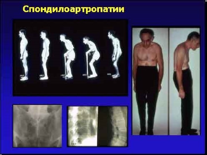
Bekhterev’s disease (Ankylosing Spondylitis) chronic inflammatory joint disease ankylosing axial skeleton (intervertebral, sacroiliac) belonging to the group of seronegative spondyloarthritis. More common in men aged 20 - 40 years.
Clinical forms peripheral form Affects the peripheral joints The central form Lesions of the spine and sacro-ileal compound Spondylitis and defeat sacro-ileal compound Scandinavian form The defeat of the small joints Rizomilic form The defeat of the hip and (or) of the shoulder joint
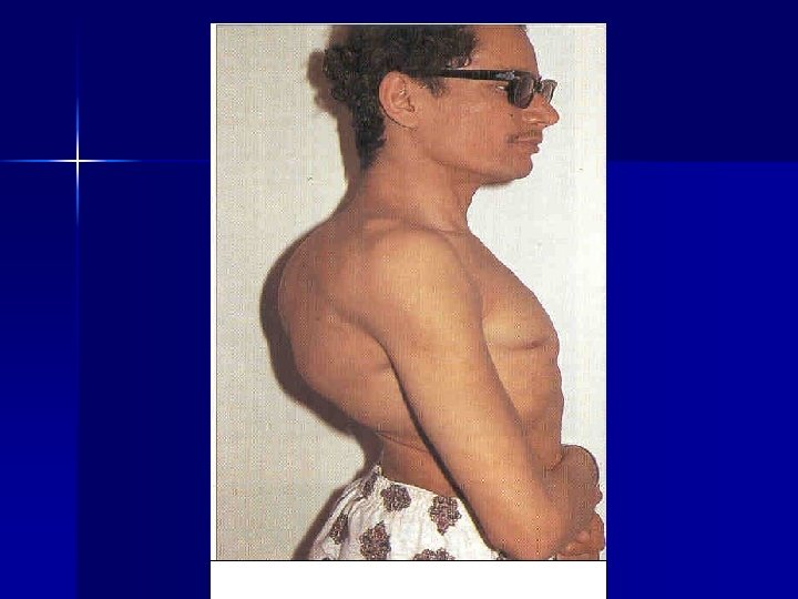
Features articular syndrome in Ankylosing spondylitis Clinical (("+" signs Kushelevsky) and radiological evidence of sacroiliitis Symmetrical joint disease Morning stiffness (at least 30 -60 minutes) Pain in the spine Articular syndrom Restriction of mobility in the spine ("+" Signs Otto Schober, chin-sternum Forestier) Spinal deformity ("board shaped spin", "pose petitioner" flattening of the lumbar lordosis)
Leading clinical features that are crucial in the diagnosis Ankylosing spondylitis: • Early radiographic signs of bilateral sacroiliitis • Radiographic signs in the form of "bamboo trunk" • If affects the internal organs, the aorta, aortic valve insufficiency, pulmonary fibrosis retsediviruyuschee, iritis or iridocyclitis, sometimes renal amyloidosis
Features articular syndrome in Psoriatic arthritis The asymmetric mono-oligoarthritis Frequent loss of the distal interphalangeal joints Unilateral sacroiliitis Articular syndrome Fingers can purchase "sausages-shaped" form The defeat of the joints of the first thumb, early loss of the big toe with a bluish or purple-bluish color; heel pain
Leading clinical signs of key importance in the diagnosis of psoriatic arthritis: • The presence of psoriatic plaques • Psoriasis in the immediate family • Radiographic manifestations: osteolytic process raznoosevym offset bone peri-rest overlay, no near-articular osteoporosis • Clinico-radiological evidence of sacroiliitis
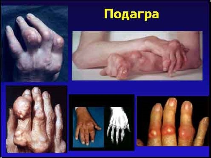
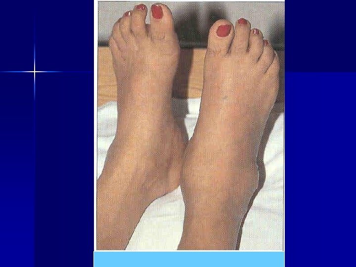
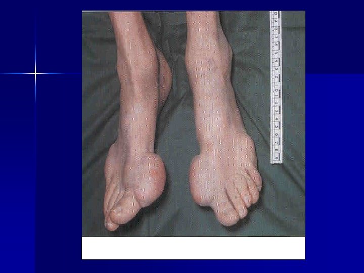
Features articular syndrome in gout A typical attack of arthritis, affecting the first metatarsophalangeal joint of the big toe Long bouts of arthritis is not more than 2 weeks articular syndrome Explicit hyperemia and hyperthermia over the joint during an attack Provoking an attack from taking alcoholic drinks and diet violation Obvious swelling of the joints and sharp pain when touched
Leading clinical features that are crucial in the diagnosis of gout: l Male sex l The presence of obesity l urolithiasis l tophi l giperurekimeya. l sinovial suyuқlikda wa tўқimalarda natriyli siydik acid tuzi kristallarini aniқlanishi. l The absolute sign of gout - detection of urate crystals in synovial fluid or tissue
Arthritides associated with infectious factors Articular syndrome infectious arthritis reactive arthritis Direct joint disease infectious factor Sterile articular syndrome (lack of local infectious) • purulent arthritis • gonorrheal arthritis • tuberculous arthritis • Brucellosis arthritis • Disease (syndrome) Reiter • Yersinia arthritis • Arthritis on a background of nasopharyngeal infection • Dysentery and arthritis salmonellosis
Features articular syndrome in arthritis associated with infectious factors Asymmetric joint disease Chronological Us articular syndrome with infection (infectious process) Basically defeat large joints articular syndrome The defeat of the leg joints monoartrit often, sometimes oligoarthritis Often unilateral sacroiliitis
Infectious arthritis Fever (febrile fever, chills) Severe pain in the joints regional limfoadenit Explicit congestion over the joint purulent arthritis Obvious swelling Explicit hyperthermia over the joint When arthrocentesis turbid and purulent fluid
Often the lesion of the knee and ankle The presence of gonococci microscopy study at discharge from the urethra Early development of contractures, defiguration joint and muscular atrophy Urethritis (with purulent discharge) Hyperemia over the affected joint gonorrhea arthritis swelling of joints In the history of promiscuity
undulating fever Mono oligoarthritis hepatosplenomegaly lymphadenopathy Unilateral sacroiliitis Brucellosis arthritis Bursitis, periarthritis spondylitis epidemiological anamnesis "+" Reaction Wright. Heddelsona
Often defeat one knee, hip, wrist and ankle Hyperemia over the joint Unilateral sacroiliitis In the later stages of the formation of fistulas rachiopathy tuberculous arthritis effusion "+" Tuberculin test Radiographically lobular reorganization of bone trabeculae, destruction and melting of the articular ends of bones, their displacement, subluxation Typical changes on chest radiographs epidemiological anamnesis
reactive arthritis Disease (syndrome) Reiter triad syndromes arthritis • Urogenital (urethritis, cystitis, prostatitis) ёki • Gastroenterology (diarrhea) conjunctivitis chlamydia «+» Reiter's disease «-» Reiter's syndrome
Upper joint hyperemia Staircase shaped type of joint disease ("bottom-up") In joints prevail exudative manifestations Joint pain worse in the morning and evening Disease (syndrome) Reiter Obvious swelling of joints Sometimes ulcerative stomatitis, glossitis, balanitis, keratoderma leukocytosis, accelerated erythrocyte sedimentation rate
Joint syndrome that develops after 1 -3 weeks preceding the intestinal manifestations such as transient diarrhea, cholecystitis, appendicular colic, pain in the right iliac region. often fever Often tendovaginitis, bursitis In the process may involve the acromioclavicular and sternoclavicular joints, the joints of the first fingers and toes Yersinia arthritis Sometimes, episcleritis, conjunctivitis, iritis, myocarditis Characteristically asymmetric joint disease of the lower extremities Unilateral sacroiliitis Detection of antibodies to Yersinia
Intestinal changes (diarrhea with greenish tinge, the presence of blood in the stool) epidemiological anamnesis Sometimes, tenosynovitis, bursitis Severe pain in the joints Dysentery and salmonellosis arthritis The chronological relationship with intestinal manifestations Leukocytosis, accelerated erythrocyte sedimentation rate Unilateral sacroiliitis Serology reaction
Articular syndrome by type ARTHROSIS
DOA Norm cartila ge bone
Commonly affects the knee, hip, and distal interphalangeal joints Pain in the joints to the end of the day, and (or) the first half of the night deformation Periodically - a symptom of "blockade" of the joint Articular syndrome Pain mechanical nature "Starter" pain crepitus
Leading clinical features that are crucial in the diagnosis of DOA: l. Heberden's nodes and Bouchard l. Radiological findings: cystoid restructuring bone structure, joint space narrowing, osteosclerosis of the articular surface, osteophytosis l Sometimes secondary synovitis
TACTICS GPS AT THE ARTICULAR SYNDROME Early diagnosis of the underlying cause (disease) Consulting a specialist (rheumatologist) according to the characteristics of the GP qualifying (I or II category) Implementation of preventive measures, and if necessary medication in conjunction with a rheumatologist Clinical supervision and rehabilitation
1. Early diagnosis of the underlying cause (disease) anamnesis Laboratory and instrumental examination (III and CAT IV) objective inspection
Objective examination localization Special tests defiguratsiya, swelling Examination of the skin funduscopy hyperemia The main reason hyperthermia Pharyngoscope. rhinoscopy anorectal examination deformation Inspection of the penis restriction of mobility "Crunch" Ankylosis, contracture Palpation, percussion auscultation of
Laboratory and instrumental examination If suspected: rheumatism rheumatoid arthritis DZST ECG Serological (revma-test) echocardiography Rentgrafiya joints Voler-Rose or latex test acute phase indicators LE cells skin biopsy Chest X-ray ECG blood creatinine CPK, aldolase, transaminase acute phase indicators coagulation A general analysis of blood and urine
continuation Bekhterev's disease gout Chronic intestinal disease Radiography of the pelvis and spine acute phase indicators Uric acid in the blood creatinine Zimnitskiy renal ultrasound Colonoscopy in the gastrointestinal tract X-rays A general analysis of blood and urine
continuation Arthritides associated with infectious factors DOA Reactions Wright-Heddlsona Bak. posev microscopy or urethral discharge Buck stool cultures RW Serology bak. issledovaniya throat IFA X-ray of the pelvis and spine X-ray of the lungs X-ray of the joints A general analysis of blood and urine
To remember! If the patient is observed • Articular syndrome (especially visceral); • Prolonged fever of unknown origin; • serozity; • Eruptions with manifestations of vasculitis; • Weight Loss; • Raynaud's phenomenon; • The appearance of the seals; Should be deleted DZST
2. Strategy GP Category I Diagnosis and management in terms of PVP or OP Rheumatism, inactive phase DOA uncomplicated gout Arthritis associated with nasopharyngeal infection Category II Early diagnosis and referral to a specialist or to hospital The remaining cases
3. Implementation of preventive measures, and if necessary medication in conjunction with a rheumatologist The antibiotics penicillin (10 -14 days) rheumatism The active phase peace NSAIDs (aspirin) 10 -14 days. At high activity - glucocorticoids) Bitsillino- profilatika (3 -5 years) Bitsillinoprofilaktika (35 years) aminohinolinovogo drugs rheumatism inactive phase Remediation of chronic foci of infection
Basic anti-inflammatory drugs (aminohinolinovogo, gold preparations, kuprenil and others) therapeutic exercise rheumatoid arthritis Symptomatic treatment (NSAID) Glucocorticoids (high activity, rapidly-progressive course and vistserity) Immunosuppressants - cytostatics (high activity, rapidly-progressive course and vistserity) Intra-articular injection (kenalog, diprospan) organ-treatment
Basic anti-inflammatory drugs (aminohinolinovogo, gold preparations, kuprenil and others) therapeutic exercise Ankylosing spondylitis Symptomatic treatment (NSAID) Glucocorticoids (high activity, rapidlyprogressive course and vistserity) Immunosuppressants - cytostatics (high activity, rapidly-progressive course and vistserity) Intra-articular injection (kenalog, diprospan) organ-treatment
Antibiotic therapy Arthritis on background nasopharyngeal infection NSAIDs sanitation
rest NSAIDs Gouty arthritis, acute Rich alkaline water Glucocorticoids (with inefficiency NSAIDs) Intra-articular injection (kenalog, diprospan) diet
diet Urikodepressivnye preparatlar (allopurinol, milurit, tiopurinol et al. ) Gouty arthritis, Unsharp Waiver of alcoholic beverages Uricosuric drugs (anturan, ketazon et al. ) physiotherapy lose weight