Diagnosis and differential diagnosis of rheumatoid arthritis Update

 is a symmetric, inflammatory, peripheral polyarthritis of unknown")


 is")
 can")

/(EULAR) classification criteria focus on features that would")

 Classification Criteria for RA")


![Viral polyarthritis � ●Viral infections such as rubella [12], parvovirus B 19 [13],](https://slidetodoc.com/presentation_image_h/9aef1055c8ebe305fbb899abdf536890/image-25.jpg "Viral polyarthritis � ●Viral infections such as rubella [12], parvovirus B 19 [13],")
 � — Early RA may be difficult to distinguish")


 can sometimes be mistaken for RA in")
 � The diagnosis is established by")

 can be confused with RA in the middle aged or")


 should be suspected")


- Slides: 48
Diagnosis and differential diagnosis of rheumatoid arthritis Update By Dr Vongsinh Phothisanesak Heard of Rheumatology Internal Medicine Lao PDR (All topics are updated as new evidence becomes available and our peer review process is complete. Literature review current through: Nov 2016. ( This topic last updated: Jan 22, 2016. )
INTRODUCTION 1 � Rheumatoid arthritis (RA) is a symmetric, inflammatory, peripheral polyarthritis of unknown etiology. It typically leads to deformity through the stretching of tendons and ligaments and destruction of joints through the erosion of cartilage and bone. If it is untreated or unresponsive to therapy, inflammation and joint destruction lead to loss of physical function, inability to carry out daily tasks of living, and difficulties in maintaining employment.
Introduction � RA: an autoimmune disease in which the normal immune response is directed against an individual’s own tissue, including the joints, tendon, bones, resulting in inflammation and destruction of these tissues � Characterized by persistent inflammatory synovitis leading to cartilage damage, bone erosions, joint deformity and disability
� Age : any age � Female: 3: 1 � Pattern of joint involvement: Polyarticular Oligoarticular Monoarticular � Morning joint stiffness> 1 hr � Small joints of hand feet are typically involved
Clinical manifestations � Articular � Extra-articular
Incidence of joint involvement in RA � MCP and PIP of hand MTP of feet 90% � Knees, ankles, wrists � Shoulder � Elbows � Acromio-clavicular and SC joints � Others : cervical spine 80% 60% 50% 30% � Thoracolumbar, sacroiliac and DIP of hand were not common
Extra- articular manifestations � Present in 30 -40% � Consitutional symptoms � Rheumatoid nodules � Hematological normocytic normochromic anemia leucocytosis thrombocytopenia � Felty’s syndrome: chronic nodular RA splenomegaly neutropenia
� Respiratory ; pleural effusion, pneumonitis, pleuro- pulmonary nodules, ILD � CVS: asymtomatic pericarditis, pericardial effusion, cardiomegaly � Rheumatoid vasculitis: mononeuritis multiplex, cutaneous ulceration, digital gangrene, visceral infarction � CNS: peripheral neuropathy, cord compression from spine subluxation, entrapment neuropathies � Eye: kerato-conjunctivitis sicca, episcleritis, scleritis
INTRODUCTION 2 � Early recognition and treatment with disease- modifying antirheumatic drugs (DMARDs) is important in achieving control of disease and prevention of joint injury and disability. However, in patients with early disease, the joint manifestations are often difficult to distinguish from other forms of inflammatory polyarthritis. The more distinctive signs of RA, such as joint erosions, rheumatoid nodules, and other extraarticular manifestations, are seen primarily in patients with longstanding, poorly controlled disease but are frequently absent on initial presentation.
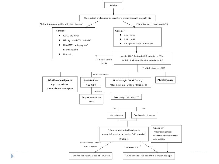
Our diagnostic criteria of RA � — The diagnosis of rheumatoid arthritis (RA) can be made when the following clinical features are all present: � � ●Inflammatory arthritis involving three or more joints. (See "Clinical manifestations of rheumatoid arthritis", section on 'Symptoms and physical findings'. ) � ●Positive rheumatoid factor (RF) and/or anti-citrullinated peptide/protein antibody (such as anti-cyclic citrullinated peptide [CCP])) testing. (See "Biologic markers in the diagnosis and assessment of rheumatoid arthritis", section on 'Anti-citrullinated peptide antibodies'. ) � ●Elevated levels of C-reactive protein (CRP) or the erythrocyte sedimentation rate (ESR). (See "Biologic markers in the diagnosis and assessment of rheumatoid arthritis", section on 'Erythrocyte sedimentation rate'. ) � ●Diseases with similar clinical features have been excluded, particularly psoriatic arthritis, acute viral polyarthritis, polyarticular gout or calcium pyrophosphate deposition disease, and systemic lupus erythematosus (SLE). (See 'Differential diagnosis' below. ) � ●The duration of symptoms is more than six weeks. � These criteria are consistent with the 2010 American College of Rheumatology (ACR)/European League Against Rheumatism (EULAR) classification criteria for RA. (See '2010 ACR/EULAR criteria' below. ) � � The diagnosis of RA may also be made in some patients who do not meet all of our criteria.
ACR cricteria of Rhumatoid
CLASSIFICATION CRITERIA � — The 2010 (ACR)/(EULAR) classification criteria focus on features that would identify patients at an earlier stage of disease than would the previously used criteria that had been last revised in 1987. The 1987 ACR criteria were formulated to distinguish patients with established rheumatoid arthritis (RA) from patients with other defined rheumatic diseases; the 2010 ACR/EULAR criteria for RA focused on identifying the factors, among patients newly presenting with undifferentiated inflammatory synovitis, which could allow for the identification of patients for whom the risk of symptom persistence or structural damage is sufficient to be considered for intervention with disease-modifying antirheumatic drugs (DMARDs). (American College of Rheumatology (ACR) European League Against Rheumatism (EULAR) )
2010 ACR/EULAR criteria � � � ●Number and site of involved joints • 2 to 10 large joints (from among shoulders, elbows, hips, knees, and ankles) = 1 point • 1 to 3 small joints (from among the metacarpophalangeal joints, proximal interphalangeal joints, second through fifth metatarsophalangeal joints, thumb interphalangeal joints, and wrists) = 2 points • 4 to 10 small joints = 3 points • Greater than 10 joints (including at least 1 small joint) = 5 points ●Serological abnormality (rheumatoid factor or anti-citrullinated peptide/protein antibody) • Low positive (above the upper limit of normal [ULN]) = 2 points • High positive (greater than three times the ULN) = 3 points ●Elevated acute phase response (erythrocyte sedimentation rate [ESR] or Creactive protein [CRP]) above the ULN = 1 point ●Symptom duration at least six weeks = 1 point In addition to those with the criteria above, which are best suited to patients with newly presenting disease, the following patients are classified as having RA:
ACR/EULAR (2010) Classification Criteria for RA
� ●Patients with erosive disease typical of RA with a history compatible with prior fulfillment of the criteria above � ●Patients with longstanding disease, including those whose disease is inactive (with or without treatment) who have previously fulfilled the criteria above based upon retrospectively available data � 1987 ACR criteria — It is important to recognize that RA has been defined in virtually all clinical trials of drugs for RA initiated from 1987 through 2010 based upon the criteria developed and validated by the ACR (previously the American Rheumatism Association) in 1987 (table 1) [9, 10]. A patient was classified as having RA if at least four of these seven criteria were satisfied; four of the criteria must have been present for at least six weeks: morning stiffness, arthritis of three or more joint areas, arthritis of the hands, and symmetric arthritis. Rheumatoid factor (RF) was included as a criterion, but anti-cyclic citrullinated peptide (CCP) antibody testing was not available at that time. The other two criteria were rheumatoid nodules and radiographic erosive changes typical of RA, but these are generally not present in the early stages of disease. � � Thus, while these criteria were very good at separating inflammatory from noninflammatory arthritis, the major drawback of the 1987 criteria has been their insensitivity in identifying some patients with early disease who subsequently develop typical established RA [10]. On the other hand, the criteria did not require any exclusions, and patients could initially fulfill the diagnostic criteria but occasionally
Labs � RF: 85% of patients with RA over the first 2 years become RF+ � RF: have poor prognosis with high titres, more extra articular manifestation
Causes of positive test for RF � RA � Sjogrens syndrome � Vasculitis � SLE � Chronic liver disease � Infections: TB, bacterial endocarditis, leprosy, syphylis, malignancies…… � Old ages ( 5% woman aged above 60)
Anti-CCP � Sensitivity 65% and specificity 95% � Can predictive of erosion disease � Disease severity � Radiologic progression � Poor functional outcomes
Others labs � Elevated ESR, CRP � Thrombocytosis � Leucocytosis � ANA- 30% � Inflammatory synovial fluid � hypoalbuminemia
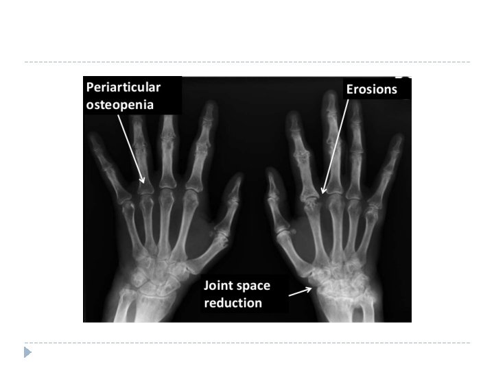
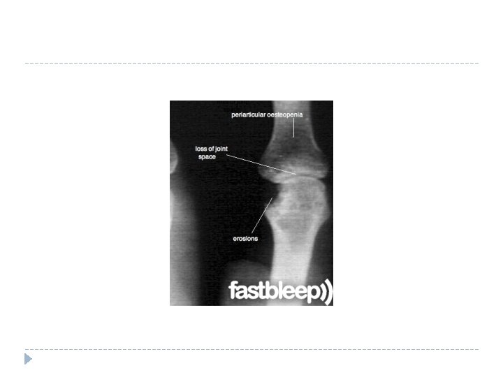
Radiologic features � Peri-articular osteopenia � Uniform symmetric joint space narrowing � Marginal subchondral erosions � Joints subluxations � Joint destruction � Collapse � Ultrasound : detects early soft tissue lesions � MRI: greatest sensitivity to detect synovitis and marrow changes
C- spine subluxation
DIFFERENTIAL DIAGNOSIS � — A variety of conditions must be considered in the differential diagnosis of rheumatoid arthritis (RA). Features of some disorders that are included in the differential diagnosis of RA are shown in the table (table 2). (See "Evaluation of the adult with polyarticular pain". )
Viral polyarthritis � ●Viral infections such as rubella [12], parvovirus B 19 [13], and hepatitis B virus (HBV) can cause an acute polyarthritis syndrome that may be mistaken for the inflammatory polyarthritis of RA. However, the syndrome is usually short-lived, lasting only from a few days to several weeks, and rarely beyond six weeks. Hepatitis C virus (HCV) can cause a polyarthritis or oligoarthritis in a minority of patients, but is more commonly associated with arthralgias.
Systemic lupus erythematosus (SLE) � — Early RA may be difficult to distinguish from the arthritis of systemic lupus erythematosus (SLE), Sjögren's syndrome, dermatomyositis (DM), or overlap syndromes such as mixed connective tissue disease. In contrast with RA, these disorders are generally characterized by the presence of other systemic features, such as rashes, dry mouth and dry eyes, myositis, or nephritis, and by various autoantibodies not seen in RA. Additionally, the relative responses of the erythrocyte sedimentation rate (ESR) and C-reactive protein (CRP) can be less well-correlated with each other in other diseases, particularly SLE, than in RA. Whereas both are commonly raised in RA, the CRP is often normal or only minimally elevated in patients with active SLE even when the ESR is elevated.
●Jaccoud's arthropathy � Jaccoud's arthropathy occurs in up to 5 to 10 percent of patients with Sjögren's syndrome or SLE and can also occur in sarcoidosis [21]. (See "Musculoskeletal manifestations of systemic lupus erythematosus" and "Sarcoid arthropathy". ) � � The joint deformities of Jaccoud's arthropathy are not caused by destruction of joints but by loosening and lengthening of periarticular structures and tendons. The ulnar drift or swan neck deformities caused by this disorder resemble RA superficially but can be distinguished by the fact that they are "correctable" on physical examination: fingers with these deformities can be moved manually back into normal alignment. In addition, radiographs in Jaccoud's arthropathy rarely reveal the cartilage loss, erosions, or cysts that are typical of longstanding RA.
Palindromic rheumatism � Palindromic rheumatism is characterized by episodes of joint inflammation sequentially affecting one to several joint areas for hours to days, with symptom-free periods that may last from days to months. Some patients presenting with this syndrome eventually develop a welldefined rheumatic disease, the most common being RA (ranging from 28 to 67 percent); some of the remaining patients develop SLE and other systemic disorders [22, 23]. Patients with anti-CCP antibodies appear more likely to progress to definite RA [24, 25]. Close follow-up and specific serologic evaluation can help distinguish among these disorders. (See "Clinical manifestations of rheumatoid arthritis", section on 'Palindromic rheumatism'. )
●Fibromyalgia � Pain, rather than stiffness or swelling, is the dominant symptom of the two common disorders, hypermobility syndrome and fibromyalgia. Although the hypermobility syndrome and fibromyalgia can both bear superficial resemblances to RA due to the presence of polyarthralgia, there are important distinguishing features:
Reactive arthritis and arthritis of IBD — Reactive arthritis often presents as a monoarthritis or oligoarthritis in large joints, such as the knees, and RA may occasionally present in this fashion as well [28]. The physical signs of both reactive arthritis and RA can be identical in the knees. (See "Reactive arthritis". ) � � The following findings on history, physical examination, or other assessments are more consistent with reactive arthritis than RA: � � ●History of recent urethritis or enteric infection � ●Asymmetric pattern of joint involvement � ●Symptoms or signs of enthesopathy (inflammation at the site where a tendon inserts into a bone, eg, the insertion point of the Achilles tendon into the heel) � ●Keratoderma blenorrhagica or circinate balanitis (see "Reactive arthritis" and "Reactive arthritis", section on 'Extraarticular signs and symptoms') � ●Radiologic evidence of sacroiliitis and/or spondylitis � ●The presence of human leukocyte antigen (HLA)-B 27 �
Lyme arthritis � � Lyme arthritis, a late manifestation of Lyme disease, occurs primarily in individuals who live in or travel to Lyme disease-endemic areas. Lyme arthritis is characterized by intermittent or persistent inflammatory arthritis in a few large joints, especially the knee. The most commonly involved joints, after the knee, are the shoulder, ankle, elbow, temporomandibular joint, and wrist. Migratory arthralgias without frank arthritis may occur during early localized or early disseminated Lyme disease. (See "Musculoskeletal manifestations of Lyme disease". ) The diagnosis of Lyme arthritis can usually be made by serologic testing, which should be performed in patients presenting with undiagnosed inflammatory arthritis in endemic areas. In addition, several clinical features help distinguish Lyme arthritis from RA. Unlike RA, for example, involvement of the small joints of the hands and feet is uncommon in patients with Lyme arthritis. Furthermore, many, but not all, patients with Lyme arthritis will describe an antecedent history of erythema migrans or other early disease manifestations. (See "Musculoskeletal manifestations of Lyme disease", section on 'Laboratory testing'. )
Psoriatic arthritis � Psoriatic arthritis can be difficult to distinguish from RA because a symmetric polyarthritis can be observed in both disorders [29]. We generally make the diagnosis of psoriatic arthritis in such patients who also have psoriasis and are seronegative for RF and anti-CCP. However, we diagnose RA in those with a symmetric polyarthritis who are seropositive for at least one of these antibodies, since skin psoriasis is so common. However, serologic testing and skin findings may not be informative, since patients with RA may not have RF or CCP antibodies (eg, seronegative RA) and the joint symptoms of psoriatic arthritis may precede the onset of skin disease by many years. Such patients should be evaluated and monitored for other signs more characteristic of psoriatic arthritis, such as nail changes or enthesitis; occasional patients exhibit overlapping features of both disorders.
Polymyalgia rheumatica — � Polymyalgia rheumatica (PMR) can sometimes be mistaken for RA in patients presenting with more limited arthritis over the age of 50 who are seronegative or only have a low RF titer. Unlike RA, PMR is usually associated with marked myalgias in the shoulders and hips, and joint involvement tends to be milder, more limited, and more often asymmetric. � � Stiffness is thus more axial in PMR and more likely to be described as difficulty getting out of bed, while stiffness in the small joints of the hands and other involved joints predominates in RA, in which difficulty buttoning clothing is more likely to be reported. However, similar complaints to RA may be present in patients with PMR with synovitis affecting the small joints in the hands. �
Crystalline arthritis — Crystalline arthritis (gout and pseudogout) � The diagnosis is established by the finding of urate or calcium pyrophosphate crystals, respectively, in synovial fluids. The presence of tophi on physical examination, the detection of serological markers of RA, and the characteristic appearance of gouty erosions are also useful in distinguishing RA from polyarticular gout. (See "Clinical manifestations and diagnosis of gout" and "Clinical manifestations and diagnosis of calcium pyrophosphate crystal deposition (CPPD) disease". )
Infectious arthritis � Infectious arthritis is usually monoarticular, but polyarthritis can occur. The diagnosis is established by culturing the pathogen from the synovial fluid or from the blood. Patients with septic arthritis may or may not appear toxic on examination, depending upon the stage of their infection, the presence of medications that can mask infection (eg, glucocorticoids), and other clinical variables. Peripheral blood leukocytosis with a left shift is common but not invariably present.
Osteoarthritis � Osteoarthritis (OA) can be confused with RA in the middle aged or older patient when the small joints of the hands are involved. However, different patterns of clinical involvement usually permit the correct diagnosis. The following are examples (see "Clinical manifestations and diagnosis of osteoarthritis"): � ●OA of the fingers typically affects the distal interphalangeal joints and is frequently associated with Heberden's nodes in this area. In contrast, RA typically affects the MCP and proximal interphalangeal (PIP) joints and is not associated with Heberden's nodes. � ●The carpometacarpal joint of the thumb is typically involved in OA. � ●Swelling of the joints is hard and bony in OA. In contrast, soft, warm, boggy, and tender joints are typical of RA.
management
Goals of management � Relieving pain � Preventing damage/ disability � Patient education about the disease � Physical therapy for stretching and range of motion exercises � Occupational therapy for splints and adaptive devices � Treatment should be started early and should be individualised
Treatment modalities for RA � NSAIDs � Steroids � DMARDs � Immunosuppressive therapy � Biological therapies � surgery
Conventional DMARDs � Anti malarial � Methrotraxate � Sulfasalazin � Gold salt � ……
immunosuppressive � Recommendation in � Loss of effectiveness with conventional DMARDs � Toxicity from DMARDS…. . � Including: azathioprine, cyclosporin, leflunomide and cyclophosphamide
Biologic in RA � Anti TNF agents � IL 1 receptors antagonist � IL 6 receptors antagonist � Anti CD 20 antibody � T cell costimulatory inhibitors
How to monitors treatment in RA � Duration of morning stiffness � Tender joints count � Swollen joints count � Observe global assessment � Patient global assessment for pain… � ESR, CRP. � Patients should be observed for 6 months before declaring a DMARDs ineffectiveness
surgical � Persistent synovitis involving the dorsal compartments of the wrist can lead to extensor tendon sheath rupture resulting in severe disability of hand function � Nerve entrapment � Removal of symptomatic rheumatoid nodule � Total joint arthroplasties, Knee, hip, wrist, elbow…
SUMMARY AND RECOMMENDATIONS 1` � � � � ●Rheumatoid arthritis (RA) should be suspected in the adult patient who presents with inflammatory polyarthritis. The initial evaluation of such patients requires a careful history and physical examination, along with selected laboratory testing to identify features that are characteristic of RA or that suggest an alternative diagnosis. (See 'Evaluation for suspected RA' above and 'Differential diagnosis' above. ) ●The following components of the medical evaluation are helpful in making a clinical diagnosis of RA, both for the identification of characteristic findings and for the exclusion of other diagnoses (see 'Evaluation for suspected RA' above): • A thorough medical history, with particular attention to joint pain, stiffness, and associated functional difficulties • A complete physical examination to assess for synovitis, limited joint motion, extraarticular disease manifestations, and signs of diseases included in differential diagnosis • Basic and selected laboratory testing, including assays for acute phase reactants (erythrocyte sedimentation rate [ESR] and C-reactive protein [CRP]), rheumatoid factor (RF), anti-cyclic citrullinated peptide (CCP) antibodies, and antinuclear antibodies (ANA) • Selected imaging studies, including bilateral radiographs of the hands, wrists, and feet • Arthrocentesis, if there is diagnostic uncertainty. ●The diagnosis of RA can be made in a patient with inflammatory arthritis involving three or more joints, positive RF and/or anti-citrullinated peptide/protein antibody, disease duration of more than six weeks, and elevated CRP or ESR, but without evidence of diseases with similar clinical features. (See 'Our diagnostic criteria' above. )
SUMMARY AND RECOMMENDATIONS 2 � � � � � ●RA may also be diagnosed in patients without all of the classic findings of disease. This includes patients with seronegative RA, those with clinically quiescent disease, and those with recent onset RA. Such patients have findings/clinical features that are generally consistent with those described as meeting the American College of Rheumatology (ACR)/European League Against Rheumatism (EULAR) classification criteria for RA. (See 'Patients not meeting above criteria' above. ) ●The 2010 classification criteria for RA were developed primarily for the identification for research purposes of patients with RA who are at high risk of persistent symptoms and joint injury unless treated with disease-modifying antirheumatic drugs (DMARDs). These criteria have replaced the 1987 criteria, which were based only upon patients with established disease. (See 'Classification criteria' above. ) ●The differential diagnosis of RA includes multiple disorders that can generally be distinguished clinically or by limited laboratory testing, based upon a combination of the following features (see 'Differential diagnosis' above): • Limited duration (eg, in viral arthropathy) • The presence of other diseases (eg, in psoriatic arthritis or arthritis of inflammatory bowel disease [IBD]) • The pattern of joint involvement and other symptoms (eg, in psoriatic arthritis, spondyloarthropathy, or polymyalgia rheumatica [PMR]) • The presence of systemic features (eg, in systemic lupus erythematosus [SLE] or dermatomyositis [DM]) • Diagnostic laboratory tests associated with other conditions (eg, specific autoantibodies in SLE, synovial fluid crystals in gout or calcium pyrophosphate disease) • Relatively high specificity of anti-CCP antibodies for RA Use of Up. To. Date is subject to the Subscription and License Agreement.
REFERENCES � � � � Whiting PF, Smidt N, Sterne JA, et al. Systematic review: accuracy of anticitrullinated Peptide antibodies for diagnosing rheumatoid arthritis. Ann Intern Med 2010; 152: 456. Nishimura K, Sugiyama D, Kogata Y, et al. Meta-analysis: diagnostic accuracy of anti-cyclic citrullinated peptide antibody and rheumatoid factor for rheumatoid arthritis. Ann Intern Med 2007; 146: 797. Finckh A, Liang MH. Anti-cyclic citrullinated peptide antibodies in the diagnosis of rheumatoid arthritis: bayes clears the haze. Ann Intern Med 2007; 146: 816. Lee DM, Schur PH. Clinical utility of the anti-CCP assay in patients with rheumatic diseases. Ann Rheum Dis 2003; 62: 870. Luime JJ, Colin EM, Hazes JM, Lubberts E. Does anti-mutated citrullinated vimentin have additional value as a serological marker in the diagnostic and prognostic investigation of patients with rheumatoid arthritis? A systematic review. Ann Rheum Dis 2010; 69: 337. Klareskog L, Catrina AI, Paget S. Rheumatoid arthritis. Lancet 2009; 373: 659. Aletaha D, Neogi T, Silman AJ, et al. 2010 Rheumatoid arthritis classification criteria: an American College of Rheumatology/European League Against Rheumatism collaborative initiative. Arthritis Rheum 2010; 62: 2569.