Pre Cancerous Lesions of the Skin Mucosa Neirita


- outdoor work/hobby fair skin,")


 in situ. Progress to Bowen carcinoma (invasive SCC)")


- Slides: 31
Pre- Cancerous Lesions of the Skin & Mucosa Neirita Hazarika
ACTINIC KERATOSES Aka solar keratosis, or senile keratosis Precursor lesions of cutaneous squamous cell carcinoma (SCC). Consists of proliferations of atypical epidermal keratinocytes , that may progress to invasive SCC The overall risk of progression to invasive SCC is (5 -10)%.
ACTINIC KERATOSES Risk factors cumulative UV radiation exposure (most important)- outdoor work/hobby fair skin, red or blond hair, blue eyes (Fitzpatrick type 1). age, immunosuppression, prior history of non-melanoma skin cancer. certain genetic syndromes, namely albinism, xeroderma pigmentosum,
ACTINIC KERATOSES Site: sun-exposed areas, balding scalp, head, neck, forearms, dorsal hands, dorsal legs in women asymptomatic, pruritus, burning or stinging pain, bleeding. 2 - to 6 -mm, erythematous, flat, rough, gritty or scaly papule more easily felt than seen. against a background of photodamaged skin
ACTINIC KERATOSES A punch biopsy with depth upto mid-reticular dermis Common indications for a biopsy 1. Rapidly enlarging lesions, 2. bleeding or ulceration, 3. Evidence of inflammation, 4. strong induration, 5. lesions extending beyond 1 cm of size, 6. resistance to treatment.
ACTINIC KERATOSES DD seborrheic keratosis, arsenical keratosis melanocytic nevi, Senile lentigo, cutaneous lupus erythematosus Established AKs chronic course lesions persist, spontaneously regress, or progress to invasive SCCs. Spontaneous resolution range from 20% to 30% limiting sun exposure, use of sunscreen promote regression.
PROGRESSION TO INVASIVE SCC each AK : potential to progress into SCC (5 -10% in reality) Direct cancerous invasive transformation of basaloid atypical keratinocytes is the most common mechanism of disease progression In situ SCC AK I basal, suprabasal AK II lower two-thirds AK III > two-thirds SCC
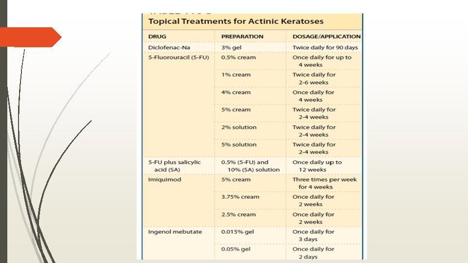
ACTINIC KERATOSES Treatment treat entire actinically damaged areas
PREVENTION of AK SUNSCREENS : Broad-spectrum against UVB and UVA, minimum SPF 30 RETINOIDS: Systemic retinoids in preventing nonmelanoma skin cancer, Ak only effective while taking them; limited by systemic toxicities Oral nicotinamide: doubtful
BOWEN DISEASE squamous cell carcinoma (SCC) in situ. Progress to Bowen carcinoma (invasive SCC) - 5% of cases. Etiologic factors include 1. UV radiation, 2. arsenic, 3. previous therapy with psoralen and UVA radiation (PUVA), 4. immunosuppression, 5. exposure to ionizing radiation, 6. infection with HPV(16, 18, 31, 34, 35, 54, 58, 61, 62, and 73)
BOWEN DISEASE Slow growing, usually asymptomatic. erythematous plaques with irregular, clearly demarcated borders. Surface scaly, crusted, hyperkeratotic. measure up to several centimeters.
Bowen Disease Clinical variants pigmented, intertriginous, periungual, and subungual BD. HPE full-thickness epidermal atypia with large, round cells and possible adnexal involvement.
Differential Diagnosis of Bowen Disease
Treatment of Bowen Disease Surgical &Destructive therapies Topical Therapies 1. Excision 1. 5 -Fluorouracil 2. Mohs micrographic surgery 3. Curettage with or without 4. Electrosurgery 5. Cryosurgery Nonsurgical Ablative Therapies 1. Photodynamic(PDT) 2. Laser ablation 3. Chemoablation : TCA 4. Radiation therapy
ARSENICAL KERATOSES result from chronic exposure to arsenic potential to become squamous cell carcinoma (SCC) punctuate, keratotic, yellow papules overlying pressure points Palms and soles.
HPV ASSOCIATED EPITHELIAL PRECANCEROUS LESIONS
BOWENOID PAPULOSIS BP is a precancerous condition of the genitalia infection with high-risk HPV, (16, 18, and 33) young to middle-aged sexually active, M>F multiple red to brownish flat papules on the penis or vulva.
BOWENOID PAPULOSIS location on the glans penis, prepuce, and penis in males, around the labia minora and majora in females. BP : transitional state between genital warts and in situ SCC. Benign, clinical course: spontaneous regression, persistence, rarely transformation into BD and invasive SCC (1% to 2. 6%). DD lichen planus, condylomata acuminata, erythroplasia, molluscum contagiosum, and seborrheic keratoses.
Treatment of Bowenoid Papulosis Local destructive Topical 1. curettage with or without electrosurgery 1. Tretinoin, 2. CO 2 -laser 3. Cidofovir 3. neodymium: YAG laser, 4. Imiquimod 2. 5 -FU, 4. cryosurgery 5. excision. HPV-specific vaccination for types 6, 11, 16, and 18 in prevention: ? ?
EPIDERMODYSPLASIA VERRUCIFORMIS inherited skin condition Loss-of-function mutations of the genes EVER 1 and EVER 2 High local susceptibility to infection with HPV, commonly types 5 and 8. Factors contributing to pathogenesis 1. host genetic background 2. HPV infection, 3. UV exposure 4. immunosuppression
EPIDERMODYSPLASIA VERRUCIFORMIS develop skin lesions early in life clinical presentation 1. numerous thin, pink, flat-topped papules and plaques that resemble verrucae planae ; knees, elbows, and trunk. 2. widespread scaly, erythematous, or hypopigmented macules and flat papules resemble tinea versicolor High risk to develop AK, BD, invasive SCC in future Sun avoidance, sun-protective measures, regular follow up, screening of family members topical 5% imiquimod cream and retinoids – mixed results
EPIDERMODYSPLASIA VERRUCIFORMIS
POTENTIALLY MALIGNANT DISORDERS OF THE ORAL CAVITY LEUKOPLAKIA white lesion of the oral mucosa that cannot be rubbed off or characterized by any other definable lesion or known disease. most common potentially malignant lesion of the oral mucosa potential to become oral SCC is 0. 2% to 3. 4%.
LEUKOPLAKIA Etiologic factors for leukoplakia 1. Chronic chemotoxic exposure, Tobacco is the strongest risk factor: either as smoke or by chewing 2. mechanical trauma with poor oral hygiene ill-fitting prosthesis, chronic cheek biting, 3. HPV (16 and 18) 4. alcohol consumption
Differential Diagnosis of Oral Leukoplakia 1. Tobacco-associated lesion 7. Frictional lesion 2. Candida-associated lesion 8. Oral white sponge nevus 3. Leukoedema 9. Oral hairy leukoplakia 4. Lichen planus 10. Verrucous carcinoma 5. Lupus erythematosus 11. Squamous cell carcinoma 6. Habitual cheek biting
LEUKOPLAKIA treatment OMMISSION 1. consumption of tobacco products, alcohol, bethel nuts, 2. chronic mechanical trauma Surgical excision is the first treatment of choice. recurrence after surgical excision : cryotherapy or CO 2 -laser the patient should be followed closely
ERYTHROPLAKIA a red macule or patch on a mucosal surface that cannot be categorized as any other known disease entity caused by inflammatory, vascular, or traumatic factors. commonly seen with leukoplakia least common of all oral potentially malignant lesions greatest potential to become oral squamous cell carcinoma Males Risk factors tobacco products, alcohol consumption.
ERYTHROPLAKIA asymptomatic, solitary, erythematous macule or patch. sharply demarcated from the surrounding pink mucosa, surface is smooth and homogeneous in color
ERYTHROPLAKIA - DD Erythematous candidiasis Atrophic lichen planus Chronic allergic contact dermatitis Lupus erythematosus Chronic mechanical trauma Pemphigus Thermal or mechanical injury Cicatricial pemphigoid Squamous cell carcinoma Kaposi sarcoma • High potential for malignant transformation : early treatment. • Surgery or excision with CO 2 - laser: treatment of choice
THANK YOU